Implications
There is intensifying concerns about the increasing rates of antimicrobials misuse and the decreasing endogenous antioxidant capacity of the weaning pig that influence its lifelong performance potential in commercial swine production. Therefore, confirming the efficacy of the nutritional alternative ingredients such as spermine that used therapeutically and prophylactically to reduce the negative effects of weaning is imperative. The present study shows that spermine promotes the antioxidant defense in organisms. Hence, the results of this study may make it possible to define the spermine as a natural dietary antioxidant in pig feeding that can promote weaning endogenous health.
Introduction
Spermine is a polycationic low-molecular weight molecule with multiple cellular and physiological functions in almost all tissues and cells (Pegg, Reference Pegg2014). One of its major functions is to protect cells from oxidative damage by directly reacting with oxidative metabolites or indirectly interacting with antioxidative enzymes. Supplementation with appropriate doses of spermine partially counteract the metabolite changes of amino acid and lipid metabolism induced by oxidative stress (Liu et al., Reference Liu, Fang, Yan, Jia, Zhao, Chen, Wu and Wang2014). Meanwhile, spermine has positive effects on the attenuation of oxidative stress response by scavenging free radicals and ameliorating lipid peroxidation in jejunum (Cao et al., Reference Cao, Liu, Fang, Wu, Jia, Zhao, Chen, Wu, Wang and Cai2015). Furthermore, spermine can also protect rat liver mitochondria from lipid peroxidation and protein oxidation by scavenging the hydroxyl radical (Sava et al., Reference Sava, Battaglia, Rossi, Salvi and Toninello2006). However, the effects of spermine supplementation and extended supplementation on the antioxidant properties of pig liver have not been studied to the best of our knowledge. When pigs are subjected to oxidant stresses, lipid peroxidation and protein oxidation in the muscle systems largely determine meat quality. Dietary supplementation with arginine (as an upstream product of spermine) can increase the antioxidative capacity and ameliorate stress in the longissimus muscle of finishing pigs (Ma et al., Reference Ma, Lin, Jiang, Zheng, Zhou, Yu, Cao, Wang and Fang2010). Nevertheless, information is not available regarding the effects of spermine supplementation and extended supplementation for improving oxidative stability in the longissimus dorsi of pigs.
We recently reported that dietary spermine can enhance the antioxidant defense systems of serum in pigs by increasing the enzymatic antioxidant and non-enzymatic antioxidant activities (Fang et al., Reference Fang, Liu, Cao, Wu, Jia, Zhao, Chen, Wu and Wang2016). An increase in enzyme activity is frequently accompanied by gene expression changes in the organism. However, whether spermine supplementation and duration of spermine supplementation could change the expression of antioxidant enzymes genes as well as the underlying molecular mechanism in pigs are still unclear. In addition, the expression of several genes for antioxidant enzymes are regulated by a pivotal regulator, NF-E2 p45-related factor 2 (Nrf2). The Nrf2 is the primary target of Kelch-like ECH-associated protein 1 (Keap1), which acts as a negative regulator of Nrf2 (Uruno and Motohashi, Reference Uruno and Motohashi2011). The Nrf2–Keap1 system is recognized as an important cellular defense mechanism against oxidation in animals (Motohashi and Yamamoto, Reference Motohashi and Yamamoto2004). However, the relationship of spermine supplementation and its duration to Nrf2–Keap1 signaling pathway in the liver and longissimus dorsi of pigs remains largely unknown and warrants further investigations. Previous research on the lipoic acid-stimulated detoxification stress response elucidated the mechanism of the mammalian target of rapamycin (mTOR) mediation in nuclear Nrf2 accumulation (Shay et al., Reference Shay, Michels, Li, Kong and Hagen2012), but the mechanism by which spermine supplementation and its duration regulate mTOR translation is generally unknown.
The present study is part of a larger research project that involves the determination of various effects of spermine supplementation on the intestinal development and serum antioxidant status in piglets (Fang et al., Reference Fang, Liu, Cao, Wu, Jia, Zhao, Chen, Wu and Wang2016). This study aimed to further investigate the effects of spermine supplementation and extended supplementation on the antioxidant status and gene expression of antioxidant-related signaling molecules in the liver and longissimus dorsi of piglets.
Material and methods
Animal experiments
Animal maintenance and experimental treatments were performed in accordance with the guidelines of the National Research Council’s Guide for the Care and Use of Laboratory Animals, and approved by the Animal Care and Use Committee of Sichuan Agricultural University. A total of 80 Duroc×(Landrace×Yorkshire) piglets with an average BW (BW) of 3.3±0.03 kg were acclimatized to the experimental conditions with basic formula milk from 2 days to 12 days in individual metabolic cages with trough and water tank (0.8×0.7×0.4 m). The piglets were randomly grouped into eight, with 10 replicates in each of the following groups: C-7h, C-3d, C-6d, C-9d, S-7h, S-3d, S-6d and S-9d. The S-7h, S-3d, S-6d and S-9d groups were fed with basic formula milk and had sufficient nutrient intake supplemented with spermine (0.4 mmol/kg BW) once a day for 7 h or 3, 6 and 9 days. The other four control groups, namely, C-7h, C-3d, C-6d and C-9d, were fed with the same amount of formula milk as spermine pigs and supplemented with normal saline in pairs. The optimum dosage of spermine and the extended time of spermine supplementation in this work were set according to prior research (Cheng et al., Reference Cheng, Li, Xing, Guo and Li2006). The basic formula milk powder was formulated according to a previous study (Fang et al., Reference Fang, Liu, Cao, Wu, Jia, Zhao, Chen, Wu and Wang2016), and the milk formula contained cow’s milk-based powder (58%), whey protein concentrate (25%), casein (5.7%), coconut oil (10.0%), CaH2PO4 (0.1%), choline chloride (0.1%), amino acids (0.5%), minerals and vitamins (0.6%).
Sample collection
Immediately after slaughtering, the liver and longissimus dorsi tissue samples (about 10 g) of each piglet were quickly obtained, snap-frozen in liquid nitrogen and then stored at −80 °C until further analysis.
Measurement of antioxidative parameters
Liver and longissimus dorsi malondialdehyde (MDA) content was analyzed with the thiobarbituric acid reaction, and the protein carbonyl (PC) content was calculated from the peak absorbance at 370 nm (Baltacioğlu et al., Reference Baltacioğlu, Akalin, Alver, Değer and Karabulut2008). The antisuperoxide anion (ASA) and the antihydroxyl radical (AHR) were measured according to the protocol in a previous study (Jiang et al., Reference Jiang, Zheng, Zhou, Liu and Feng2009). The catalase (CAT) activity , the total superoxide dismutase (T-SOD) activity, the glutathione (GSH) content, as well as the glutathione peroxidase (GPx) and glutathione S-transferase (GST) activities were determined as described by Che et al. (Reference Che, Xuan, Hu, Liu, Xu, Fang, Lin, Xu, Wu and Zhang2015). The total antioxidant capacity (T-AOC) activity was estimated according to Miller et al. (Reference Miller, Rice-Evans, Davies, Gopinathan and Milner1993). All antioxidant-related indices were assayed with the corresponding diagnostic kits purchased from Nanjing Jiancheng Bioengineering Institute (Nanjing, China).
RNA extraction and quantitative real-time polymerase chain reaction analysis
The total RNA of the liver and longissimus dorsi were extracted with the TRIzol Reagent (Takara, Dalian, China) according to the manufacturer’s protocol. The quality of RNA samples were checked by 1.0% agarose gel electrophoresis (Beckman DU-800; Beckman Coulter, CA, USA). Subsequently, total RNA was reverse transcribed into cDNA by the real-time reverse transcription–polymerase chain reaction with the PrimeScriptTM RT reagent (Takara) by following the manufacturer’s instructions. For quantitative real-time PCR, specific primers for the β-actin, SOD1, CAT, GPx1, GST, glutathione reductase (GR), Nrf2, Keap1, mTOR, ribosomal p70 S6 kinase (S6K1), and eukaryotic initiation factor 4E (eIF4E)-binding protein 1 (4EBP1) genes were designed with Primer Express Software (version 3.0; Applied Biosystems, Foster City, CA, USA) and synthesized by the TaKaRa Biotechnology Company (Takara). The primer sequences of the corresponding genes and the optimal annealing temperatures are summarized in Supplementary Material Table S1. All real-time PCR reactions were performed with the SYBR® Green I PCR Reagent Kit and a real-time PCR instrument (ABI 7900HT; Applied Biosystems). The reaction mixture (8 µl) contained 4 µl of SYBR Premix Ex Taq II with the ROX reference dye, 0.8 µl of cDNA, 1.6 µl of primer pairs (10 µM each of the forward and reverse primers) and 1.6 µl of ddH2O. The thermal cycler conditions for PCR were: prerun at 95°C for 10 s, 42 cycles of denaturation at 95°C for 10 s, annealing at 58°C for 35 s, and extension at 72°C for 15 s. The expression results were analyzed by the
 $${\rm 2}^{{{\minus}\Delta \Delta C_{t} }} $$
method as described by Livak and Schmittgen (Reference Livak and Schmittgen2001).
Statistical analysis
All data were analyzed by two-way ANOVA for the 2×4 factorial experimental design using the GLM procedure of SPSS 22.0 (SPSS Inc., Chicago, IL, USA). The statistical model used included the main effects of spermine doses (0 or 0.4 mmol/kg BW), treatment time (7 h or 3, 6 or 9 days) and spermine dose×treatment time interaction. If a significant treatment effect of their interaction was observed, the significant difference between the treatments was identified separately by using Tukey’s test. All results were expressed as mean, and P<0.05 was used to assess statistical significance. Pearson’s correlation coefficients among the data were determined using the correlation analysis of SPSS.
Results
Antioxidant indicators in the liver
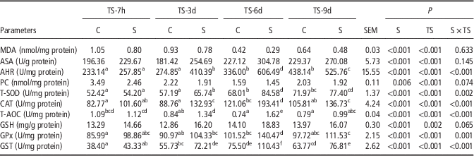
Table 1 presents the antioxidant indicators in the liver. Both spermine dose and treatment time significantly increased the ASA, AHR, T-SOD, CAT, T-AOC, GPx and GST activities, as well as the GSH content, but decreased the MDA and PC content in liver (P<0.05). There were significant interactions between spermine dose and treatment time on AHR, T-SOD, CAT, T-AOC, GPx and GST activities in liver (P<0.05). Moreover, the AHR, T-SOD, CAT and GST activities were lowest in the S-7 group compared with the S-3, S-6, and S-9 groups after extending the spermine supplementation (P<0.05). When the treatment time was prolonged, the AHR, T-SOD, CAT, T-AOC, GPx, and GST activities were gradually increased, particularly after extending spermine supplementation for 6 days. The S-6 group showed highest AHR, T-SOD, CAT, T-AOC, GPx and GST activities compared with the other groups (P<0.05). The AHR and T-SOD activities significantly increased with the extended duration of spermine supplementation (9 days) relative to 7 h or 3 days (P<0.05).
Effects of spermine and its duration extension on the antioxidant status of liver in piglets

TS-7h=treatment time with spermine for 7 h; TS-3d=treatment time with spermine for 3 days; TS-6d=treatment time with spermine for 6 days; TS-9d=treatment time with spermine for 9 days; C=control; S=0.4 mmol/g BW of spermine; MDA=malondialdehyde; ASA=antisuperoxide anion; AHR=antihydroxyl radical; PC=protein carbonyl; T-SOD=total superoxide dismutase; CAT=catalase; T-AOC=total antioxidant capacity; GSH=glutathione content; GPx=glutathione peroxidase; GST=glutathione S-transferase; SEM=standard error of the means.
a,b,c,d,e,fMean values within the same row having different superscript letters differ significantly at P<0.05 (n=10).
Antioxidant indicators in longissimus dorsi
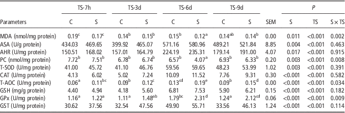
Table 2 presents the antioxidant indicators in the longissimus dorsi. Both spermine dose and treatment time significantly increased ASA, AHR, T-SOD, CAT, T-AOC, GPx and GST activities, as well as the GSH content, but decreased the MDA and PC content in the longissimus dorsi (P<0.05). There were effects of the interaction between spermine dose and treatment time on T-AOC and GPx activities, as well as MDA and PC content (P<0.05). Unlike other activity observed after 3, 6 and 9 days of spermine supplementation, after 7 h of spermine supplementation, the MDA content were highest (P<0.05). T-AOC and GPx activities gradually increased after 3, 6 and 9 days of supplementation. By contrast, the MDA and PC content gradually decreased. Meanwhile, extension of spermine administration for 6 days produced the highest T-AOC activity, but the lowest MDA and PC content relative to the other treatment groups (P<0.05). Time extension of spermine administration (9 days) significantly increased T-AOC and GPx activities relative to the levels observed in spermine administration for 7 h or 3 days (P<0.05).
Effects of spermine and its duration extension on the antioxidant status of longissimus dorsi muscle in piglets

TS-7h=treatment time with spermine for 7 h; TS-3d=treatment time with spermine for 3 days; TS-6d=treatment time with spermine for 6 days; TS-9d=treatment time with spermine for 9 days; C=control; S=0.4 mmol/g BW of spermine; MDA=malondialdehyde; ASA=antisuperoxide anion; AHR=antihydroxyl radical; PC=protein carbonyl; T-SOD=total superoxide dismutase; CAT=catalase; T-AOC=total antioxidant capacity; GSH=glutathione content; GPx=glutathione peroxidase; GST=glutathione S-transferase; SEM=standard error of the means.
a,b,c,d,eMean values within the same row having different superscript letters differ significantly at P<0.05 (n=10).
mRNA levels of antioxidant-related parameters in the liver
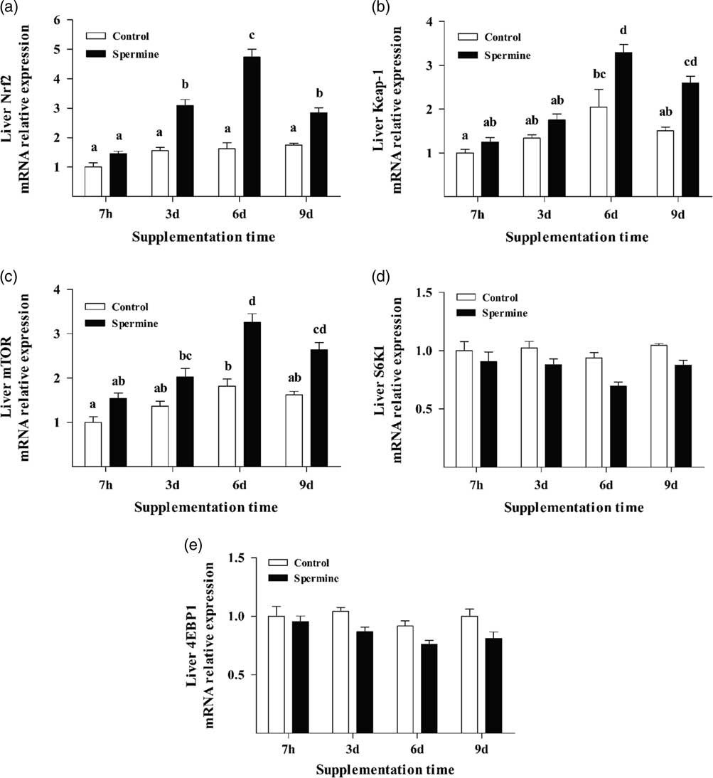
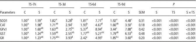
The mRNA levels of different genes in the liver are shown in Table 3 and Figure 1. The SOD1, CAT, GPx1, GST, GR, Nrf2, Keap1 and mTOR mRNA levels in the liver were significantly increased; the S6K1 mRNA level was decreased in response to spermine dose and treatment time (P<0.05), and significant interaction between spermine dose and treatment time were shown for SOD1, CAT, GPx1, GST, GR, Nrf2, Keap1 and mTOR mRNA levels (P<0.05). Moreover, the SOD1, CAT, GPx1, GST, GR, Nrf2, Keap1 and mTOR mRNA levels were gradually increased by extending spermine supplementation to 3, 6 and 9 days (P<0.05). The SOD1, CAT, GPx1, GR and Nrf2 mRNA levels were the lowest after extending spermine administration to 7 h, relative to that observed at 3, 6 and 9 days (P<0.05). By contrast, extension of spermine supplementation for 6 days revealed the highest SOD1, CAT, GR, Nrf2, Keap1 and mTOR mRNA levels compared with the 7 h and 3 days treatment groups. The SOD1, GPx1, Keap1 and mTOR mRNA levels were increased in the group supplemented with spermine for 9 days (S-9 v. S-7, S-3; P<0.05).
Effects of spermine and its duration extension on the relative mRNA expression levels of the NF-E2-related nuclear factor 2 (Nrf2) (a); Kelch-like ECH-associated protein 1 (Keap1) (b); mammalian target of rapamycin (mTOR) (c); ribosomal p70 S6 kinase 1 (S6K1) (d); and eukaryotic initiation factor 4E (eIF4E)-binding protein 1 (4EBP1) (e) in the liver of the piglets. The values are means, with their standard errors represented by vertical bars (n=6), and mean values with different letters (a, b, c, d) are indicated the significant different among treatments (P<0.05).

Effects of spermine and its duration extension on the antioxidant-related gene expressions of liver in piglets

TS-7h=treatment time with spermine for 7 h; TS-3d=treatment time with spermine for 3 days; TS-6d=treatment time with spermine for 6 days; TS-9d=treatment time with spermine for 9 days; C=control; S=0.4 mmol/g BW of spermine; SOD1=total superoxide dismutase 1; CAT=catalase; GPx1=glutathione peroxidase 1; GST=glutathione S-transferase; GR=glutathione reductase; SEM=standard error of the means.
a,b,c,d,eMean values within the same row having different superscript letters differ significantly at P<0.05 (n=6).
mRNA levels of antioxidant-related parameters in the longissimus dorsi
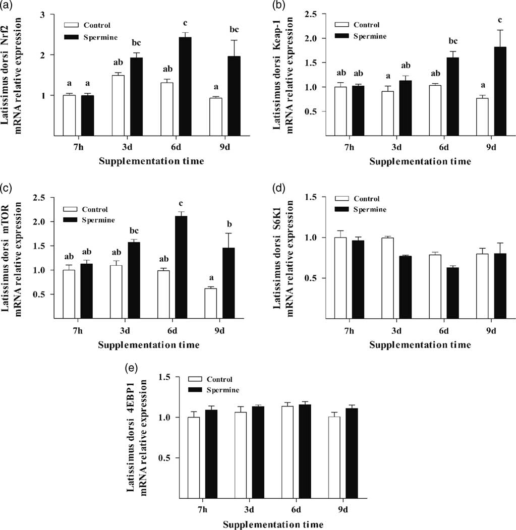
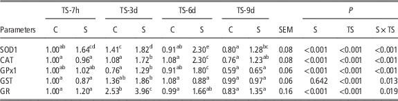
As shown in Table 4 and Figure 2, both spermine dose and treatment time revealed significantly increased SOD1, CAT, GPx1, GR, Nrf2 and mTOR mRNA levels in longissimus dorsi (P<0.05). There were significant interactions between spermine dose and treatment time on SOD1, CAT, GPx1, GST, GR, Nrf2, Keap1 and mTOR mRNA levels (P<0.05). Moreover, the CAT and Nrf2 mRNA levels were lowest after extending spermine administration to 7 h relative to those observed at 3, 6 and 9 days (P<0.05). Extension of spermine administration for 3 days significantly increased GST and GR mRNA levels relative to the levels observed after treatment for 7 h or 9 days (P<0.05). By extending spermine supplementation to 6 days, the SOD1, CAT and GPx1 mRNA levels were increased relative to the values of the other treatment groups (P<0.05). The SOD1 and Keap1 mRNA levels were increased in the group supplemented with spermine for 9 days (S-9 v. S-7, S-3; P<0.05).
Effects of spermine and its duration extension on the relative mRNA expression levels of the NF-E2-related nuclear factor 2 (Nrf2) (a); Kelch-like ECH-associated protein 1 (Keap1) (b); mammalian target of rapamycin (mTOR) (c); ribosomal p70 S6 kinase 1 (S6K1) (d); and eukaryotic initiation factor 4E (eIF4E)-binding protein 1 (4EBP1) (e) in the longissimus dorsi muscle of the piglets. The values are means, with their standard errors represented by vertical bars (n=6), and mean values with different letters (a, b, c) are indicated the significant different among treatments (P<0.05).

Effects of spermine and its duration extension on the antioxidant-related gene expressions of longissimus dorsi muscle in piglets

TS-7h=treatment time with spermine for 7 h; TS-3d=treatment time with spermine for 3 days; TS-6d=treatment time with spermine for 6 days; TS-9d=treatment time with spermine for 9 days; C=control; S=0.4 mmol/g BW of spermine; SOD1=total superoxide dismutase 1; CAT=catalase; GPx1=glutathione peroxidase 1; GST=glutathione S-transferase; GR=glutathione reductase; SEM=standard error of the means.
a,b,c,d,eMean values within the same row having different superscript letters differ significantly at P<0.05 (n=6).
Discussion
Weaning stress is often accompanied by increased susceptibility to disease, metabolic disorders, and the impaired structure and function of numerous organs because of the poor antioxidant system (Campbell et al., Reference Campbell, Crenshaw and Polo2013). The liver, which has high oxygen consumption (Nemeth et al., Reference Nemeth, Baird and O’Farrelly2009), acts as a crucial innate physical barrier that is responsible for filtering of potentially destructive antigens before they reach the body. The longissimus dorsi has low oxygen consumption, whereas protein and lipid oxidation may lead to decreased meat quality such as reduced tenderness and juiciness. Therefore, maintenance of the antioxidant status of the liver and longissimus dorsi are critical for pigs.
Effects of spermine supplementation and extension of its administration on the antioxidant status of the liver and longissimus dorsi
Oxidative stress can lead to overproduction of reactive oxygen species (ROS). The superfluous ROS can cause lipid peroxidation and protein oxidation in organisms. MDA is a key product of lipid oxidation; and PC is frequently used as a direct marker of oxidative modification in proteins (Uchida and Kawakishi, Reference Uchida and Kawakishi1993; El-Sheikh and Khalil, Reference El-Sheikh and Khalil2011). The current study showed that the MDA and PC content were decreased with spermine supplementation in the liver and longissimus dorsi, thereby indicating that spermine can suppress lipid peroxidation and protein oxidation in tissues. The protective effects against lipid and protein oxidative damage are generally related to the decreased interaction with toxic oxygen species, such as the superoxide anion radical (O2 −) and hydroxyl radical (OH−). The O2 − and OH− radicals are strongly involved in ROS-oxidative damage (Kohen and Nyska, Reference Kohen and Nyska2002), and the total scavenging capacity against O2 − and OH− radicals are measured by two indexes: the ASA and the AHR activities. The present study showed that the ASA and AHR activities in the liver and longissimus dorsi was significantly improved with spermine supplementation, thereby indicating their improved free radical-scavenging ability. The results obtained in the present study are similar with previous studies, which showed that spermine can mitigate oxidative damage by decreasing MDA content and scavenging O2 − and OH− radicals in suckling animals (Cao et al., Reference Cao, Liu, Fang, Wu, Jia, Zhao, Chen, Wu, Wang and Cai2015; Fang et al., Reference Fang, Liu, Cao, Wu, Jia, Zhao, Chen, Wu and Wang2016).
Organisms possess the ability to protect themselves from ROS via antioxidant defense systems involving non-enzymatic and enzymatic antioxidants. T-AOC is often adopted as a comprehensive indicator of the redox status to reflect the overall cellular endogenous antioxidative capability in the whole body (Ren et al., Reference Ren, Yin, Liu, Yu, Li, Yang, Li and Wu2012). GSH is a non-enzymatic antioxidant that is ubiquitous in mammals, with important roles in several detoxification reactions as the first line of the antioxidant defense system (Yang et al., Reference Yang, Fang, Hong, Wang, Zhou and Lin2013). In the present study, spermine supplementation significantly increased the T-AOC activity and GSH content in the liver and longissimus dorsi. This finding suggests that spermine supplementation can protect the organs from oxidative stress by enhancing the total antioxidant capacity and the non-enzymatic antioxidant system. In addition, the increased levels of GSH may be associated with its increased synthesis or decreased degradation. In mammals, the maintenance of GSH homeostasis is regulated by several glutathione-dependent enzymes, such as GPx and GST, which are the line of enzymatic antioxidant defense that have cytoprotective roles in eliminating reactive electrophilic chemical compounds and facilitate their excretion from the oxidative metabolism in cells (Baez et al., Reference Baez, Seguraaguilar, Widersten, Johansson and Mannervik1997; Matés et al., Reference Matés, Pérez-Gómez and Castro1999). Notably, the GPx and GST activities in all tissues were improved by spermine administration. Correlation analysis obtained in our experiment declared that GSH content was positively associated with the GPx activity (r liver=+0.951, P liver<0.01; r longissimus dorsi=+0.876, P longissimus dorsi<0.01) and GST activity (r liver=+0.838, P liver<0.01; r longissimus dorsi=+0.874, P longissimus dorsi<0.01), indicating that the improvement of GPx and GST activities in the liver and longissimus dorsi by spermine addition may be attributed to the increased regeneration of GSH.
Similar with the antioxidant defense system in GSH, the CAT and T-SOD activities also have complementary catalytic activity with GPx. CAT and SOD are two important endogenous antioxidant enzymes that can cooperatively convert organic hydroxyl radicals into water and O2 (Winston and Di Giulio, Reference Winston and Di Giulio1991; David et al., Reference David, Munaswamy, Halappa and Marigoudar2008). In the present study, the CAT and T-SOD activities in all organs, which had similar patterns as compared with the ASA and AHR activities were substantially higher in the spermine group than the control group, as evidenced by the positive association between CAT activity and ASA activity (r liver=+0.945, P liver<0.01; r longissimus dorsi=+0.951, P longissimus dorsi<0.01) and AHR activity (r liver=+0.891, P liver<0.01; r longissimus dorsi=+0.951, P longissimus dorsi<0.01), as well as the positive relation between T-SOD activity and ASA activity (r liver=+0.852, P liver<0.01; r longissimus dorsi=+0.984, P longissimus dorsi<0.01) and AHR activity (r liver=+0.974, P liver<0.01; r longissimus dorsi=+0.974, P longissimus dorsi<0.01). A significant increase in the activities of both enzymes suggests that the spermine-enhanced O2 − and OH− radical scavenging abilities may be ascribed to the improved SOD and CAT activities, and these results conform with those observed in the serum from our previous study (Fang et al., Reference Fang, Liu, Cao, Wu, Jia, Zhao, Chen, Wu and Wang2016).
Notably, the investigated antioxidant-related parameters in this study showed similar results, 6-day spermine supplementation increased the level of AHR, T-AOC, GPx, GST, CAT and T-SOD as compared with the three other durations of spermine treatment in liver. Similar results may be explained by the fact that spermine has a time-dependent effect on inducing antioxidant function in the liver. This observation is consistent with the results of a previous study, which indicated that spermine exerts a time-dependent effect on facilitating ileum growth in piglets (Fang et al., Reference Fang, Liu, Cao, Wu, Jia, Zhao, Chen, Wu and Wang2016). However, the relationship between the time-dependent effect of spermine and the antioxidant capacity of organs is largely unknown. The mechanism underlying the mediation of spermine in the antioxidant defense in liver and longissimus dorsi of piglets needs further investigation. Moreover, the enhanced activities of antioxidant enzymes may be a consequence of increased gene expression. Consequently, we determined the influence of spermine supplementation and extended spermine administration on the mRNA levels of antioxidant enzymes in the liver and longissimus dorsi of suckling piglets.
Effects of spermine supplementation and extension of spermine administration on antioxidant enzyme gene expression in liver and longissimus dorsi muscle
The present study showed that dietary spermine upregulated the mRNA levels of SOD1 and CAT in the liver and longissimus dorsi. These results were similar to the increased activities of both enzymes as observed in our study. Correlation analysis indicated that the mRNA level of SOD1 was positively correlated with the T-SOD activity in the liver (r liver=+0.727, P liver<0.01), as well as a positive relation between CAT1 gene expression and CAT activity (r liver=+0.766, P liver<0.01). These findings indicated that the influences of spermine supplementation on the activities of antioxidant enzymes partially occurred at the level of gene transcription. To support the above-mentioned proposition, this study showed that the physiological levels of spermine increased the GPx1 and GR mRNA levels in liver and longissimus dorsi, and correlation analysis exhibited that GPx1 mRNA level was positively associated with GPx activity (r liver=+0.745, P liver<0.01) in liver, which might partly explain the increased GPx1 and GST activities in the spermine administration group. These findings confirmed that increasing the antioxidant enzymes activities is partially attributed to spermine, which upregulated the gene expression of antioxidant enzymes. This response could be an adaptive mechanism against oxidant stress in piglets. This result agrees with the previous report that arginine can significantly elevate the mRNA levels of SOD1, CAT and GPx1 in muscles (Wang et al., Reference Wang, Liu, Feng, Jiang, Kuang, Jiang, Li, Tang and Zhou2015). Furthermore, our study demonstrated that the mRNA levels of SOD1, CAT, GPx1, GST, and GR in the liver increased with the duration of spermine supplementation. In particular, SOD1 and CAT mRNA levels were higher after 6 days than in the other spermine supplementation groups in the liver and longissimus dorsi. A similar trend was observed in the enzymatic activities of T-SOD and CAT. Therefore, extended spermine supplementation is necessary to enhance the gene transcription of antioxidant enzymes in the suckling piglets. In addition, the upregulation of these antioxidant enzymes may be attributed to the activation of intracellular signaling pathways in mammals. Thus, we explored the effects of spermine supplementation and extended spermine supplementation on the gene expression of these related signaling molecules in the liver and longissimus dorsi.
Effects of spermine supplementation and extended spermine administration on the gene expression of Nrf2 and TOR signaling molecules in the liver and longissimus dorsi
Previous studies confirmed that Nrf2 is a transcriptional regulator of a cellular antioxidant pathway by activating the gene expression of a group of antioxidant enzymes, such as GSTs and NAD(P)H quinone oxidoreductase (Kobayashi and Yamamoto, Reference Kobayashi and Yamamoto2005). In the present study, we observed that the mRNA levels of Nrf2 in the liver and longissimus dorsi were upregulated with dietary spermine and Nrf2 gene transcription had substantial association with SOD1 mRNA level (r liver=+0.973, P liver<0.01), CAT1 mRNA level (r liver=+0.970, P liver<0.01), GPx1 mRNA level (r liver=+0.782, P liver<0.05), GST mRNA level (r liver=+0.771, P liver<0.05), and GR mRNA level (r liver=+0.963, P liver<0.01) in the liver, thereby suggesting that spermine can increase the liver antioxidant enzyme gene expression by partially upregulating Nrf2 gene transcription in suckling piglets. This finding is consistent with previous reports, which suggested that Nrf2 mRNA levels can enhance the SOD and GPx gene expression in rats (Singh et al., Reference Singh, Rangasamy, Thimmulappa, Lee, Osburn, Brigeliusflohé, Kensler, Yamamoto and Biswal2006; Jung et al., Reference Jung, Hong, Zheng, Lee and Hong2009). In addition, our results are similar with a study on the enhanced mRNA levels of Nrf2 as the arginine concentration is increased to the optimum level (Wang et al., Reference Wang, Liu, Feng, Jiang, Kuang, Jiang, Li, Tang and Zhou2015). Keap1 is a Nrf2-binding protein that can negatively regulate Nrf2 activity by facilitating Nrf2 degradation via the proteasome and inhibiting Nrf2 nuclear translocation in the cytoplasm (Shibata et al., Reference Shibata, Kokubu, Gotoh, Ojima, Ohta, Yamamoto and Hirohashi2008; Qiang, Reference Qiang2013). In the present study, we revealed that 0.4 mmol/kg BW spermine supplementation significantly increased the mRNA levels of Keap1 in the liver and longissimus dorsi. Correlation analysis showed that the mRNA level of Keap1 was positively related to the mRNA level of Nrf2 in organs (r liver=+0.889, P liver<0.01; r longissimus dorsi=+0.792, P longissimus dorsi<0.05). This observation suggests that spermine may enhance the gene expression of antioxidant enzymes and Nrf2 by upregulating the Keap1 mRNA levels. The effect of spermine supplementation on Nrf2 and Keap1 expression might contribute to the TOR signaling pathway because TOR is an upstream signaling molecule of Nrf2 and can regulate Nrf2 expression (Shay et al., Reference Shay, Michels, Li, Kong and Hagen2012).
mTOR is a serine/threonine (Ser/Thr) protein kinase that integrates signals related to several cellular processes, such as cell growth and proliferation, growth factor stimulation, energy status and amino acid utilization (Rhee and Bae, Reference Rhee and Bae2015). Our results showed that treatment with spermine obviously increased the mTOR mRNA level in both tissues, thereby clearly indicating that the mTOR-mediated pathways were stimulated by spermine. Correlation analysis showed that the mRNA level of mTOR was positively related to the Nrf2 gene transcription (r liver=+0.941, P liver<0.01; r longissimus dorsi=+0.927, P longissimus dorsi<0.01), indicating that the translational system may be involved in the steady-state Nrf2 levels. Moreover, our data showed that S6K1 and 4EBP1 expression were decreased in the liver. S6K1 expression was also decreased in the longissimus dorsi. S6K1 and 4EBP1 are two major downstream effector molecules of the mTOR pathway, and these genes were identified in transcripts that encode proteins for cell stress response and survival (Zhou et al., Reference Zhou, Liu, Liu, Zhang, Zhang, Liu, Zhang, Xu, Liu and Huang2014). The downregulation of S6K1 and 4EBP1 expressions in the liver and longissimus dorsi fit with the interpretation that Nrf2 accumulation may be a result of the alterations in the mTOR pathway caused by spermine. Nevertheless, the underlying mechanism of the relationship between spermine and the Nrf2 and mTOR pathways remains largely unknown and requires further investigation.
In the current study, extended spermine supplementation for 6 days resulted in higher Nrf2, Keap1 and mTOR mRNA levels in comparison with those observed after 7 h, and 3 and 9 days of spermine treatment in liver. This finding supports that spermine can exert simultaneous modulation of Nrf2, Keap1 and mTOR expressions in a time-dependent manner. These changes show a similar trend to the changes in antioxidant enzymes activities and antioxidant enzymes gene expressions, elucidating that extension of spermine administration for 6 days can promote a better effect on induction of Nrf2 and mTOR signaling pathways than the three other extension times.
In conclusion, the current study has shown that dietary spermine and extended spermine supplementation effectively promoted the antioxidant defense in the liver and longissimus dorsi by inhibiting lipid peroxidation, enhancing free radical-scavenging ability and improving non-enzymatic and enzymatic antioxidant capacities. The elevation of antioxidant enzymes activities by spermine supplementation and extended spermine supplementation were partly attributed to the upregulation of antioxidant enzyme gene expression, and the gene expression of antioxidant enzymes was regulated by signaling molecules, such as Nrf2, Keap1, mTOR, S6K1 and 4EBP1. This work describes a partial molecular mechanism for improving the antioxidant capacity by spermine intake and extended spermine supplementation in piglets. Therefore, spermine might be a source of dietary antioxidant agents and have significant effects on production.
Acknowledgments
The authors thank the Natural Science Foundation of China (31301986), the Academy of Kechuang Feed Industry in Sichuan and Specific Research Supporting Program for Discipline Construction in Sichuan Agricultural University (to G. Liu) for financial support. The authors would like to thank their teammates for kind assistance.
Supplementary material
To view supplementary material for this article, please visit https://doi.org/10.1017/S1751731117002737