Introduction
The main goal of assisted reproductive techniques (ARTs) is to ensure the birth of a single healthy, full-term baby, which can be achieved by transferring a single euploid embryo (Nuñez-Calonge et al., Reference Nuñez-Calonge, Santamaria, Rubio and Moreno2024). In recent years, selecting the best embryo for transfer has been a major challenge in in vitro fertilization (IVF). Several scoring systems have been developed for the assessment of embryo viability, taking into account cleavage rate, fragmentation percentage, multinucleation, presence of cytoplasmic irregularities and blastocyst development (Nuñez-Calonge et al., Reference Nuñez-Calonge, Santamaria, Rubio and Moreno2024; Youngster et al., Reference Youngster, Shvaikovsky, Avraham, Strassburger, Esti Kasterstein, Maslansky, Gat, Yerushalmi, Gidoni, Hourvitz and Kedem2024).
A blastomere containing more than one nucleus is defined as a multinucleated blastomere (MNB), and its incidence has been reported to range from 15 to 40% (Sun et al., Reference Sun, Chen, Yang, Yi, Liu and Ou2018). The presence of multinucleation has been associated with poor embryo development and unfavourable ART outcomes (Aguilar et al., Reference Aguilar, Rubio, Munoz, Pellicer and Meseguer2016). However, in recent years, further research performed by the application of time-lapse monitoring systems and advances in preimplantation genetic screening for aneuploidy (PGD-A) suggests that multinucleation may represent previously unknown levels of complexity and diversity of the embryonic cell division cycle (Coticchio et al., Reference Coticchio, Lagalla, Taggi, Cimadomo and Rienzi2024). A study showing that the majority of multinucleated (MLN) embryos were genetically abnormal reported that 74.5% of MLN embryos had chromosomal abnormalities compared to 32.3% of non-MLN embryos, suggesting that MLN embryos should not be recommended for transfer in IVF cycles (Ambroggio et al., Reference Ambroggio, Gindoff, Dayal, Khaldi, Peak, Frankfurter and Dubey2011).
Anti-Müllerian hormone (AMH) is a peptide growth factor produced by granulosa cells of primary, secondary, pre-antral and early antral follicles in female fetuses after 36 weeks of gestation and is a member of the Transforming Growth Factor-beta (Amanda Souza Setti et al., Reference Setti, Braga, Guilherme, Iaconelli and Borges2023). AMH is considered a good marker for quantitative assessment of ovarian response to stimulation during ART cycles (La Marca et al., Reference La Marca, Sighinolfi, Radi, Argento, Baraldi, Artenisio, Stabile and Volpe2010).
However, the relationship between AMH, oocyte and embryo quality and outcome of ART is still controversial.
Although the mechanism of multinucleation has not been clearly established, there are studies reporting a higher incidence of MLN embryos in GnRH agonist cycles, cycles with male factor-induced infertility and cycles with a higher number of oocytes (Desai et al., Reference Desai, Goldberg, Austin and Falcone2018; Fragouli et al., Reference Fragouli, Alfarawati, Spath, Jaroudi, Sarasa, Enciso and Wells2013; Figueira et al., Reference Figueira, Setti, Braga, Iaconelli and Borges2010). In our study, we have aimed to investigate the relationship between serum AMH levels and MLN embryos, one of the parameters indicating embryo quality in intracytoplasmic sperm injection (ICSI) cycles.
Materials and methods
This retrospective cohort study included female participants aged < 44 years who were undergoing fresh ICSI cycles. The study was conducted between January 2018 and December 2020. The study participants were chosen by scrutinizing the medical records of the patients attending the IVF Clinic at Ankara City Hospital. The conduct of the study was approved by the institutional review board (E1-20-1234/2020). Informed consent was obtained from all the patients attending the clinic. The outcomes of 1153 fresh ICSI cycles were analysed. Patients with missing serum AMH measurements, those diagnosed with genetic disorders, and those with a history of recurrent pregnancy loss were excluded from the study. In line with our institutional clinical protocol, all ART procedures at our centre were performed using ICSI. A total of 888 patients were included in the study. Cycles with at least one MLN yield were defined as the study group (Group 1) (n = 237) and cycles without MLN yield as the control group (Group 2) (n = 651).
Patients’ ages, body mass indexes, causes of infertility, spermiograms, baseline E2, follicle-stimulating hormone (FSH), luteinizing hormone (LH) levels, antral follicle counts, ovulation induction protocols, total number of oocytes retrieved, M2 oocyte and 2PN embryo counts, presence of MLN embryos and clinical pregnancy results were evaluated. Indications for ICSI were unexplained infertility, male factor infertility, diminished ovarian reserve (DOR), multiple indications (male factor + female factor infertilities) and other causative factors as polycystic ovary syndrome, endometriosis and tubal pathologies. According to the ESHRE 2023 guideline, unexplained infertility is diagnosed in couples who fail to conceive after at least 12 months of regular, unprotected intercourse, despite normal findings on standard fertility evaluations. These evaluations include confirmation of ovulatory cycles, tubal patency and a normal uterine cavity in the female partner (aged ≤ 40 years), as well as normal semen parameters and testicular function in the male partner, in line with WHO reference criteria. The diagnosis is made by exclusion, following a thorough assessment of both partners. Cases of oligoasthenospermia with total progressive motile sperm count less than 5 million per cc in spermiogram were defined as male factor infertility. DOR was defined as a serum AMH value of < 1 ng/ml.
Long agonist, microdose or antagonist protocols were used depending on the clinical features and indications. The initial doses were determined based on antral follicle counts, female age and baseline FSH levels. The subsequent doses were adjusted according to the degree of follicle maturation and serum estradiol levels.
The GnRH antagonist protocol was based on the administration of recombinant FSH or human menopausal gonadotropin from day 2 of a spontaneous menstrual cycle. Then daily subcutaneous injectable doses of 0.25 mg cetrorelix (Cetrotide®, Merck Serono, Istanbul, Turkey) were administered from the day of the stimulation cycle when the first follicle reached 12 mm in size. These treatments continued till the day of human chorionic gonadotropin (hCG) administration. Micro-dose flare-up protocol was started with an oral contraceptive pill on day 1 of the previous cycle, which was maintained for 21 days. On the second day of withdrawal bleeding, subcutaneous injections of leuprolide acetate (50 mg bid) (Lucrin®; Abbott Lab, Istanbul, Turkey) were initiated. Human menopausal gonadotropin (HMG) therapy was begun on cycle day 3. Treatment with leuprolide acetate and HMG was maintained until the day of hCG administration. In the long agonist protocol, leuprolide acetate (Lucrin®) at daily doses of 1 mg was started. After the onset of ovarian suppression, a maintenance dose of leuprolide acetate (0.5 mg/d) and subcutaneous injections of recombinant FSH or human menopausal gonadotropin were administered until the day of hCG. Final follicular maturation was triggered with the administration of human chorionic gonadotropin (HCG) or recombinant HCG (rHCG alpha) when at least three leading follicles reached a mean diameter of ≥ 18 mm. Transvaginal oocyte retrieval was scheduled 34–38 h later. The luteal phase was supported by daily applications of vaginal progesterone gel (Crinone®; Merck Serono, Istanbul, Turkey) starting one day after oocyte pick-up.
Denudation time was defined as the time interval between oocyte retrieval and the initiation of ICSI. Although a standardized laboratory protocol was followed, variations in denudation time occurred due to differences in patient volume and workflow on retrieval days. Each case was recorded individually, and the denudation time was analysed as a continuous variable. ICSI was performed to the mature oocytes. Mature oocyte ratio was calculated as M2 oocyte/total oocyte ratio. Normal fertilization was identified by the presence of two pronuclei (2PN) at the time of fertilization assessment, 16–19 h after ICSI. Cleavage stage embryos were evaluated on day 2 or 3 for blastomere number, fragmentation, multinucleation and symmetry in accordance with ESHRE/ALPHA consensus guidelines. Multinucleation was defined as the presence of more than one clearly identifiable nucleus within a single blastomere under high-magnification bright-field microscopy (Figure 1). Embryos exhibiting 2–8 blastomeres were included, and at least one MNB was sufficient for classification. Micronucleation, vacuoles or nuclear-like inclusions were not considered as true nuclei. Evaluations were performed by trained embryologists, and embryos with ambiguous morphology were excluded to ensure consistency. Embryo transfer (ET) was performed on day 2 to day 5 using a soft catheter, based on embryo development and clinical indications. Embryos exhibiting multinucleation – defined as the presence of more than one clearly distinguishable nucleus in a single blastomere – were not selected for transfer, as per our institutional embryo selection policy. Clinical pregnancy was determined by the presence of a gestational sac and detection of fetal cardiac activity by transvaginal ultrasound at 6 weeks of gestation. The relationship between the presence of at least one multinucleated (MLN) embryo in a cycle and serum AMH levels was analysed.
(A) Embryo with single nucleus in a blastomere. Arrow indicates nucleus. Bar = 50μm. (B) Embryo with multinucleated blastomere. Arrow indicates nucleus. Bar = 250μm.

Statistical analysis
SPSS 11.5 programme was used to analyse the data obtained from the electronic file system. Mean ± standard deviation and median (minimum–maximum) values were used as quantitative variables and number of patients and percentages were used as qualitative variables. The Mann–Whitney U test was employed to determine whether there was a difference between the categories of the qualitative variables with two categories in terms of the quantitative variables, since the assumptions of normal distribution were not met. Chi-square and Fisher’s exact tests were used to examine the relationship between two qualitative variables. Univariate and multivariate logistic regression analyses were used to determine the risk factors affecting the dependent qualitative variable. The level of statistical significance was accepted as p < 0.05.
Results
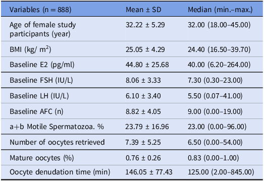
A total of 888 patients with a mean age of 32.22 ± 5.29 years were included in the study. The demographic characteristics and laboratory values of the patients are shown in Table 1. The number (%) of patients with unexplained infertility (n: 189; 21.3%), male factor infertility (n: 312; 35.2%), DOR (n: 268; 30.2), other causative factors (n: 55; 6.1%) and more than one indication (n: 64; 7.2%) were as indicated. As induction protocols, agonist protocol (n: 637; 2%), (n: 755; 86.6%), antagonist protocol and microdose protocol (n: 546; 2%) were used. The number (%) of patients with AMH levels < 1 ng/ml (n: 275; 31.0%), 1–3.5 ng/ml (n: 406; 45.7%), 3.5–5 ng/ml (n: 100; 11.3%) and > 5 ng/ml (n: 107; 12.0%) was as indicated.
Clinical and laboratory data of our patients (numerical identifiers)

Abbreviations: BMI = body mass index; E2 = estradiol; FSH = follicle-stimulating hormone; LH = luteinizing hormone; AFC = antral follicle cell.
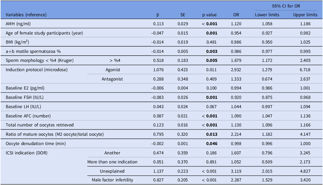
Univariate logistic regression analysis was performed, and when the results were evaluated, variables including AMH, baseline FSH and antral follicle cell (AFC) levels, female age, motile sperm ratio, sperm morphology, total number of oocytes retrieved and denudation time were found to be significant risk factors. So they were included in the multivariate logistic regression analysis. As a quantitative variable, AMH was found to be a significant risk factor regarding its effects on the presence of multinucleation per se as assessed by logistic regression analysis (p < 0.001). It was determined that one unit increase in AMH levels increases the risk of the presence of multinucleation by 1,120 times. According to our study, the likelihood of detecting MLN embryos increased as AMH levels increased. This suggests that higher systemic AMH levels may be linked to an increased likelihood of early cleavage-stage nuclear abnormalities. Similarly, Alexopoulou et al. reported that patients with poor ovarian response had lower AMH levels and exhibited a lower frequency of multinucleation in four-cell stage embryos (Alexopoulou et al., Reference Alexopoulou, Pinborg, Budtz-Jørgensen and Zedeler2019).
Also, it was determined that the likelihood of MLN embryo formation decreased as patients aged. However, as the percentage of motile sperms increases and sperm morphology improves, the likelihood of formation of MLN embryos decreases. As the basal FSH level increased, the likelihood of MLN embryos decreased, while the likelihood of detecting MLN embryos increased as the baseline number of antral follicles increased. As the total number of retrieved oocytes increased, the likelihood of detecting MLN embryos increased. In addition, the risk of multinucleation decreased in line with prolongation of denudation time (Table 2).
Results of univariate logistic regression analysis performed for the presence of multinucleation

Abbreviations: β = beta coefficient; SE = standard error; OR = odds ratio; BMI = body mass index; E2 = estradiol; FSH = follicle-stimulating hormone; LH = luteinizing hormone; AFC = antral follicle cell; DOR = diminished ovarian reserve. Bold text indicates statistical significance (p < 0.05).
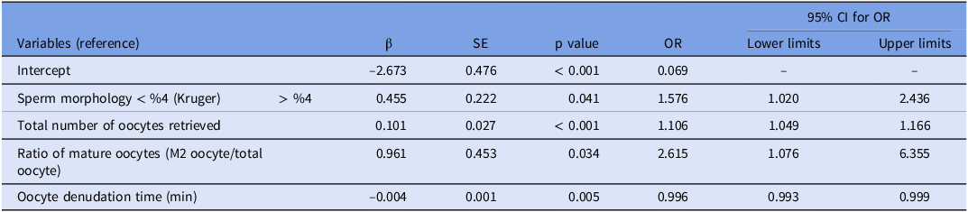
According to the results of multivariate logistic regression analysis, the presence of morphological abnormalities of spermatozoa, the total number of oocytes retrieved and denudation time were found to be statistically significant factors related to fertility potential (p = 0.041, p < 0.001 and p = 0.055), respectively. According to Kruger’s strict criteria for sperm morphology < 4%, the likelihood of MLN increased 1.576 times. One unit increase in the total number of oocytes retrieved increased the likelihood of MLN embryos by 1.106 times, and one unit increase in the rate of mature oocytes also increased the likelihood of MLN embryos by 2.615 times, while a 1-min increase in denudation time decreased the likelihood of MLN embryos by 0.996 times (Table 3).
Results of multivariate logistic regression analysis performed for the presence of multinucleation

Abbreviations: β = beta coefficient; SE = standard error; OR = odds ratio.
The relationship between the MLN embryo/total embryo ratio and clinical pregnancy outcomes was evaluated. A statistically significant difference was found between the groups with and without clinical pregnancy in terms of this ratio (p = 0.010). In cycles that resulted in clinical pregnancy, the mean MLN embryo/total embryo ratio was 0.34 ± 0.18, whereas in cycles without clinical pregnancy, the mean ratio was 0.47 ± 0.30. These findings indicate that an increased MLN embryo/total embryo ratio within a cycle is associated with a reduced likelihood of achieving clinical pregnancy (Table 4). Notably, in accordance with our institutional policy, embryos identified as multinucleated were not selected for transfer; therefore, the observed effect may reflect an indirect association between the MLN embryo/total embryo ratio and overall embryo quality and implantation potential.
Comparisons between hCG positivity and clinical pregnancy rates in terms of MLN embryo/total embryo ratios

Abbreviations: hCG = human chorionic gonadotropin.
Discussion
ART provides great opportunities for infertile patients to have a child. One of the most critical points to be considered here is embryo selection. One of the markers used for embryo selection is the presence of MNBs. In our study, any MLN embryos were not detected in 73.3% (n = 651), and at least one MLN embryo was detected in 26.7% (n = 237) of the cycles. It has been reported in the literature that the incidence of multinucleation varies between 15 and 40% (Sun et al., Reference Sun, Chen, Yang, Yi, Liu and Ou2018). In our patient group, the incidence of multinucleation was correlated with the literature.
According to the results of logistic regression analysis, the most striking outcome of our study was that, as a quantitative variable, serum AMH level was found to be a significant independent risk factor for the presence of multinucleation (p < 0.001). While AMH is widely used as a biomarker of ovarian reserve, it reflects the cumulative activity of granulosa cells across the cohort of growing follicles. The finding that multinucleation was observed in only one or a few embryos within a cycle suggests that local, follicle-specific differences in oocyte quality or AMH expression may exist. Unfortunately, our study did not include intrafollicular AMH measurements or granulosa cell gene expression analyses, which limits our ability to infer mechanistic pathways.
However, several hypotheses may explain the link between high-AMH levels and multinucleation. It has been proposed that elevated AMH levels – often associated with high antral follicle counts and robust ovarian response – could be indicative of oocytes that have undergone accelerated or dysregulated maturation under supraphysiologic hormonal conditions. Sayme et al. suggested that increased AMH levels may be associated with a higher incidence of multiple pronuclei formation due to maturational asynchrony or spindle abnormalities, which may lead to defective meiosis II completion or abnormal fertilization (Sayme et al., Reference Sayme, Kljajic, Krebs and Maas2020). These cytoskeletal or chromosomal instabilities could plausibly contribute to the formation of MNBs.
Taken together, these observations suggest that while AMH is a reliable marker of ovarian reserve quantity, elevated levels in some patients may signal a subset at risk for qualitative deficiencies in oocyte competence. This highlights the importance of individualized ovarian stimulation protocols, particularly in high-AMH responders, to mitigate potential risks to oocyte or embryo quality. Further mechanistic studies – particularly those incorporating follicle-level profiling, time-lapse imaging (TLI) and spindle integrity assessment – are warranted to better understand the underlying biology linking AMH and early embryonic nuclear abnormalities.
Studies have reported the presence of a correlation between multinucleation and higher total ovarian stimulation dose required, number of oocytes aspirated (Royen et al., Reference Royen, Mangelschots, Vercruyssen, Neubourg, Valkenburg, Ryckaert and Gerris2003; Ambroggio et al., Reference Ambroggio, Gindoff, Dayal, Khaldi, Peak, Frankfurter and Dubey2011) and advanced female age (Balakier et al., Reference Balakier, Sojecki, Motamedi and Librach2016; Hashimoto et al., Reference Hashimoto, Nakano, Yamagata, Inoue, Morimoto and Nakaoka2016). However, Talbot et al. found no correlation between the percentages of MLN embryos and female age, BMI, number of aspirated oocytes, fertilization method or cause of female infertility (Talbot et al., Reference Talbot, Alexopoulou, Kallemose, Freiesleben, Nielsen and Zedeler2022).
In our study, age, baseline FSH and AFC levels and total number of oocytes retrieved were found to be significant risk factors according to the results of univariate logistic regression analysis (p < 0.001, p = 0.001, p < 0.001, p < 0.001 and p < 0.001, respectively). According to the results of multivariate logistic regression analysis, we found that the total number of oocytes retrieved conveys significance for the presence of multinucleation (p < 0.001). One unit increase in the total number of oocytes retrieved increased the risk of multinucleation by 1.106 times.
Multinucleation is an abnormality occurring during the division of embryos and is correlated with increased rates of aneuploidy and chromosomal abnormalities (Sun et al., Reference Sun, Chen, Yang, Yi, Liu and Ou2018). The same study also found that the number of MLN embryos was higher in patients with lower baseline FSH levels, lower doses of gonodotropins use, lower LH levels on the day of HCG triggering and in cases where greater number of oocytes could be retrieved.
In our study, we found that the likelihood of detecting MLN embryos decreased with increasing age and baseline FSH levels, and the probability of finding MLN embryos increased with increasing baseline AFC counts and total number of oocytes retrieved. We have also revealed that all of these parameters we searched for were correlated with AMH levels. As the women aged, the number of antral follicles and AMH levels decreased with resulting decline in the number of MLN embryos. As baseline FSH levels increase, AMH levels decline, presumably in line with the reduction in the number of MLN embryos. However, higher AMH levels may be associated with higher baseline AFC counts and increased total number of oocytes retrieved, which may enhance the formation of MLN embryos. According to our study, although different theories have been proposed in the literature to explain the formation of MLN embryos, we believe that many parameters associated with multinucleation may be directly related to AMH levels.
The presence of MNBs has been associated with poor embryo development and unfavourable ART outcomes. MLN embryos are often accompanied by irregularly sized blastomeres and moderate or severe fragmentation, which are indicative of impaired developmental potential (Sun et al., Reference Sun, Chen, Yang, Yi, Liu and Ou2018). Desch et al. reported that embryos containing MNBs are unsuitable for implantation and live birth, even if they proceed to blastocyst formation (Desch et al., Reference Desch, Bruno, Luu, Barberet, Choux, Lamotte, Schmutz, Sagot and Fauque2017). Moreover, a study based on known implantation data from fresh cleavage-stage ETs demonstrated a negative association between day 2 multinucleation and live birth rates (Coticchio et al., Reference Coticchio, Lagalla, Taggi, Cimadomo and Rienzi2024; Sayed et al., Reference Sayed, Reigstad, Petersen, Schwennicke, Hausken and Storeng2022). In our study, multinucleation was evaluated specifically at the cleavage stage (day 2 or day 3), when these abnormalities are considered to be more clinically relevant. We believe that early detection of multinucleation may help identify embryos with compromised implantation potential, even if further development appears morphologically normal. The mean MLN embryo/total embryo ratio in the group with clinical pregnancy was 34 ± 0.18%, while the mean MLN embryo/total embryo ratio in the group without clinical pregnancy was 47 ± 0.30%. According to our study results, as the percentage of MLN embryos in a cycle increased, the occurrence rate of clinical pregnancy decreased. According to our study, as the percentage of MLN embryos in a cycle increased, both chemical and clinical pregnancy rates decreased. In correlation with the literature, our study showed that MLN embryos were associated with poor ART outcomes. There are reports of decreased implantation (Ergin et al., Reference Ergin, Çalişkan, Yalçinkaya, Öztel, Çökelez, Özay and Özörnek2014; Desch et al., Reference Desch, Bruno, Luu, Barberet, Choux, Lamotte, Schmutz, Sagot and Fauque2017), clinical pregnancy (Ergin et al., Reference Ergin, Çalişkan, Yalçinkaya, Öztel, Çökelez, Özay and Özörnek2014) and/or live birth rates (Desch et al., Reference Desch, Bruno, Luu, Barberet, Choux, Lamotte, Schmutz, Sagot and Fauque2017; Barberet et al., Reference Barberet, Bruno, Valot, Antunes-Nunes, Jonval, Chammas, Choux, Ginod, Sagot, Soudry-Faure and Fauque2019) for MLN embryos at the two-cell stage. Since embryo transplantation applied at the cleavage stage makes it impossible to distinguish the potential of the embryo to develop into a blastocyst from its implantation potential, it is necessary to pay attention to the diverse potential of embryos of multinucleated subgroups (Aguilar et al., Reference Aguilar, Rubio, Munoz, Pellicer and Meseguer2016; Desch et al., 2017; Talbot et al., Reference Talbot, Alexopoulou, Kallemose, Freiesleben, Nielsen and Zedeler2022).
In a study investigating the relationship between sperm morphology and multinucleation, a significantly higher number of MLN embryos were detected in the group of patients undergoing male factor-induced IVF. As a concluding remark, the authors emphasized the increased probability of immature spermatozoa development in oligospermic cases, which may induce the formation of centrosomal defects (Manandhar et al., Reference Manandhar, Schatten and Sutovsky2005). Similarly, Sun et al. demonstrated that certain chromosomal polymorphisms in sperm, which are more frequently encountered in oligozoospermic men, are independently associated with the formation of MLN embryos, highlighting the genetic and structural contributions of the sperm to early embryonic division fidelity (Sun et al., Reference Sun, Chen, Yang, Yi, Liu and Ou2018). In our study, univariate logistic regression analysis revealed that percentages of motile sperms and sperm morphologic abnormality were found to be significant risk factors (p = 0.003, p = 0.005, respectively) and were included in the multivariate logistic regression analysis. According to the results of multivariate logistic regression, morphologic abnormalities of spermatozoa were seen at a significantly higher rate (p = 0.041). Accordingly, it was determined that as the percentage of motile spermatozoa increased, the probability of MLN embryos decreased, and according to Kruger’s strict criteria, if the presence of spermatozoa having normal morphology is less than 4%, then the risk of multinucleation increases. According to Kruger’s criteria, in our study, the presence of < 4% spermatozoa increased the risk of MLN embryos by 1.576 times. According to our study, not only oocyte quality but also sperm quality may be associated with the development of MLN embryos.
The study has some limitations. The main one is that it was a retrospective study. In addition, the study was performed with static evaluation. TLI technology was not used, which may have reduced the actual incidence of multinucleation. Another limitation is the absence of receiver operating characteristic (ROC) curve analysis to determine potential cut-off values for AMH, FSH, patient age or sperm count in predicting MLN embryo formation. While these variables were assessed as continuous data, future prospective studies incorporating ROC analysis may help establish clinically useful threshold values. Lastly, fluorescent nuclear staining (e.g., DAPI) was not applied due to ethical and legal restrictions in handling human embryos. This may have limited the ability to definitively distinguish true nuclei from nuclear-like vacuoles, although precautions were taken to minimize misclassification during high-magnification light microscopy.
Conclusion
Multinucleation is defined as a condition where a blastomere has more than one nucleus, and it has been associated with the presence of MLN embryos, poor embryo development and adverse ART outcomes. Detection of MLN embryos is critical to achieve higher rates of pregnancy and live births, and although many mechanisms have been proposed for the presence of multinucleation in the embryo, its exact mechanism has not yet been revealed. Parameters that can predict MLN embryo formation prior to treatment are crucial for achieving higher pregnancy rates and live births. As an endpoint, our results showed the relationship between AMH levels and multinucleation and its effect on embryo quality and pregnancy rates. According to our data, serum AMH levels can be used as a predictive marker for MLN embryos. This is the first study to describe the association between serum AMH levels and MLN embryos. We believe that our study will be a pioneer for the conduct of more comprehensive clinical studies in the future.
Acknowledgements
None
Competing interests
The authors declare none.
Funding
This research received no specific grant from any funding agency, commercial or not-for-profit sectors.