1. Introduction
Diabetic retinopathy (DR) is one of the most common complications of diabetes, which is the main cause of the visual defect in working-age people worldwide. Almost all patients with type 1 and more than 60% of patients with type 2 diabetes develop retinopathy (Nentwich & Ulbig, Reference Nentwich and Ulbig2015). All this time, microvascular lesions have long been regarded as central to the pathogenesis of DR. In recent years, however, many studies showed that retinal neurovascular units have undergone pathological changes before the vascular injury. Especially, retinal ganglion cells (RGCs) death and retinal microglial cells (RMC) activation are regarded as two early features in DR (Rübsam et al., Reference Rübsam, Parikh and Fort2018). Neuroinflammation has been implicated in the pathogenesis of DR. Thus, DR is a microvascular disease as well as chronic inflammation and retinal neurodegeneration (Altmann & Schmidt, Reference Altmann and Schmidt2018). MG activation is often seen in brain diseases, but the role of RMC in DR is rarely addressed. Therefore, in this review, we will summarize the different roles of RMC in early DR to provide some new insights.
2. Origin and function of MG
One century ago, MG was described firstly by Río Hortega, which he called these cells “the third element of nerve centers” and later termed “microglia” (Del Rio-Hortega Bereciartu, Reference Del Rio-Hortega Bereciartu2019). And these cells represent 5–12% in the central nervous system (CNS). Generally, as the macrophages of CNS, MG derives from hematopoietic stem cells (Chan et al., Reference Chan, Kohsaka and Rezaie2007; Saijo & Glass, Reference Saijo and Glass2011). Unlike other ocular macrophages, RMC arises from the Yolk Sac precursors has been confirmed (O’Koren et al., Reference O’Koren, Yu, Klingeborn, Wong, Prigge, Mathew, Kalnitsky, Msallam, Silvin, Kay, Bowes Rickman, Arshavsky, Ginhoux, Merad and Saban2019). Hence, the retina is a well-structured organ within CNS, amoeboid and mesodermal myeloid progenitors enter the retina during development and differentiate into ramified parenchymal MG in the adult retina (Chan et al., Reference Chan, Kohsaka and Rezaie2007; Tay et al., Reference Tay, Carrier and Tremblay2019).
Once the tissue is matured, the MG population is maintained in the retina throughout the entire life span where they compose the resident immune cells (Reu et al., Reference Reu, Khosravi, Bernard, Mold, Salehpour, Alkass, Perl, Tisdale, Possnert, Druid and Frisén2017). In the adult retina, ramified MG are mainly distributed in the plexiform layers, ganglion cell layer (GCL), and never fiber layer (NFL) (Madeira et al., Reference Madeira, Boia, Santos, Ambrósio and Santiago2015). In the healthy retina, MG is required for normal retinal growth, neurogenesis, controlling development, vascular formation, aging, and retinal function. They interact with other neurons, glia, and endothelial cells by secreting growth factors, cytokines as well as neuroprotective anti-inflammatory mediators (Altmann & Schmidt, Reference Altmann and Schmidt2018). However, MG is double-edged.
In the normal retina, MG is located in the plexiform layers where they exhibit elaborate ramified processes responsible for immune surveillance of the retina (Karlstetter et al., Reference Karlstetter, Scholz, Rutar, Wong, Provis and Langmann2015). Noxious insults such as oxidative stress, hypoxia, inflammation, or inherited mutations trigger MG reactivity manifested by amoeboid morphology, increased proliferation, and migration to the sites of injury (Sierra et al., Reference Sierra, de Castro, Del Río-Hortega, Rafael Iglesias-Rozas, Garrosa and Kettenmann2016, Masuda et al., Reference Masuda, Shimazawa and Hideaki2017). And sustained microglial inflammatory responses can instigate severe alterations in retinal integrity, and aggravate neuronal demise (Zhao et al., Reference Zhao, Zabel, Wang, Ma, Shah, Fariss, Qian, Parkhurst, Gan and Wong2015). This is the flip side of MG in the retina, which recent strategies have been shown to suppress microglial inflammatory responses in the retina and prevent neuronal cell death.
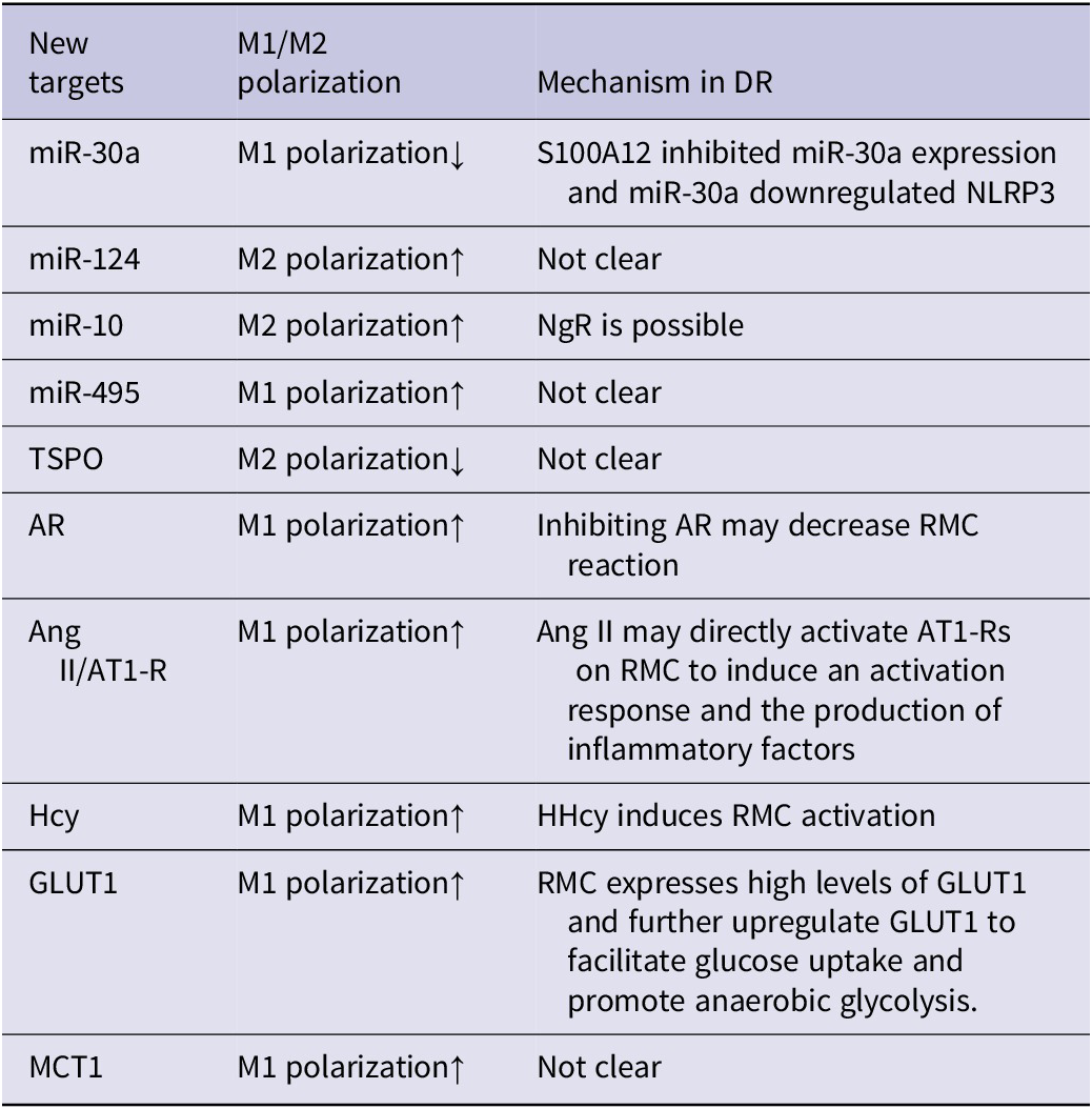
Some new targets associated with RMC polarization

3. The polarization of MG
For a long time, resident ramified MG was described as quiescent cells, waiting for their activation, but now it is known that ramified MG contributes to tissue and neuronal homeostasis (Dereki et al., Reference Dereki, Cronk and Kipnis2013; Hugh Perry & Teeling, Reference Hugh Perry and Teeling2013; Kierdorf & Prinz, Reference Kierdorf and Prinz2017). Once the stimulus is felt, MG would rapidly change their morphology, gene expression, and functional performance upon any threat to tissue homeostasis, acquiring and activated phenotype (McKimmie et al., Reference McKimmie, Roy, Forster and Fazakerley2006), which the procession could be called MG polarization. In DR, RMC is assumed to be the first detector of early signals of hyperglycemia. Located between vasculature and neurons of the retina, RMC activation is often responsible for early harm of the beginning stages of DR (Sorrentino et al., Reference Sorrentino, Allkabes, Salsini, Bonifazzi and Perri2016).
Upon activation, ramified MG undergoes a series of stereotypic, morphological, phenotypic, and functional changes (Streit et al., Reference Streit, Graeber and Kreutzberg1989). Firstly, MG exists in two separate morphological states, namely “resting” and “activated” microglia (Kettenmann et al., Reference Kettenmann, Kirchhoff and Verkhratsky2013). During activation, RMC changes from ramified appearance to ameboid-like. Then, there are roughly two phenotypes of MG. In vitro experiments demonstrate that microglial cells develop either a classic pro-inflammatory M1 or an alternative anti-inflammatory and pro-healing M2 polarization phenotypes (Su et al., Reference Su, Yi, Xu and Zhang2015; Arroba et al., Reference Arroba, Alcalde-Estevez, García-Ramírez, Cazzoni, de la Villa, Sánchez-Fernández, Mellet, García Fernández, Hernández, Simó and Valverde2016). In vivo, MG appears to acquire intermediate phenotypes, whose ultimate function relies on the combination of different polarization markers ranging from M1 to M2 (Lampron et al., Reference Lampron, ElAli and Rivest2013; Arroba et al., Reference Arroba, Alcalde-Estevez, García-Ramírez, Cazzoni, de la Villa, Sánchez-Fernández, Mellet, García Fernández, Hernández, Simó and Valverde2016).
Generally, M1 is induced by the prototypical Th-1 cytokines interferon-gamma (IFN-c) or lipopolysaccharide (LPS). After activating, M1 phenotypes mainly secrete pro-inflammatory cytokines such as tumor necrosis factor (TNF-α), IL (interleukin)-12, IL-23, IL-1β, IL-6, and chemotactic factors as well as induce cytotoxic mediators such as reactive oxygen species and nitric oxide (NO). By contrast, M2 is usually induced by the Th-2 cytokines, IL-4 or IL-10, and IL-13, which express high levels of anti-inflammatory cytokines, etc. (Martinez et al., Reference Martinez, Helming and Gordon2009; Arroba & Valverde, Reference Arroba and Valverde2017). The distinction between M1 and M2 microglia is hazy and MG can adapt their phenotypes to meet demands (Graeber et al., Reference Graeber, Li and Rodriguez2011). Therefore, the unbalance of MG polarization is a key reason for neuroinflammation.
4. RMC polarization and DR
The involvement of inflammation in DR was developed out of initial studies reporting that diabetic patients under treatment with salicylates for rheumatoid arthritis had a lower-than-expected incidence of this complication of diabetes (Powell & Field, Reference Powell and Field1964). While recently, neurodegeneration was confirmed to occur in human diabetes even before the development of clinical stages of DR. RMC is the heart of this possess (Simo & Hernandez, Reference Simo and Hernandez2014) and has a dual phenotype and nature. The study of Arroba has shown that in db/db mice, an early stage of diabetes is characterized by the coexistence of pro-inflammatory and anti-inflammatory cytokines in circulation but, as long as diabetes progresses, this balance is disrupted in favor of a proinflammatory milieu (Arroba & Valverde, Reference Arroba and Valverde2017). Therefore, the M1/M2 responses are closely related to the activation and polarization of RMC (Arroba & Valverde, Reference Arroba and Valverde2017). In the health retina, RMC releases neuroprotective and anti-inflammatory factors. However, in response to prolonged pro-inflammatory stimuli, the chronically activated RMC can lead to neuroinflammation by releasing a plethora of cytokines. In a word, in the early stage of DR, hyperglycemia trigger RMC polarization, and M1/M2 is unbalanced.
Several animal models have been used to study the activation of RMC in diabetes and have confirmed the phenotypic changes mentioned herein (Kinuthia et al., Reference Kinuthia, Wolf and Langmann2020). In the streptozotocin (STZ)-induced diabetic rat, MG changes their morphology from a ramified to an amoeboid form and increases in number within the outer plexiform layers (OPL) and outer photoreceptor layer suggesting that activated microglia are highly proliferative and migratory (Zeng et al., Reference Zeng, Ng and Ling2000). Moreover, in the db/db mouse model of type 2 diabetes, activated RMC was found in the inner nuclear layer and switched from an anti-inflammatory phenotype during early disease toward a pro-inflammatory phenotype in the late stage (Arroba et al., Reference Arroba, Alcalde-Estevez, García-Ramírez, Cazzoni, de la Villa, Sánchez-Fernández, Mellet, García Fernández, Hernández, Simó and Valverde2016).
DR includes nonproliferative diabetic retinopathy (NPDR) and proliferative diabetic retinopathy (PDR). Similarly, clinical studies have confirmed microglia activation in the retina of both NPDR and PDR patients whereby, in NPDR microglia migrated into the plexiform layers and proliferated whereas, in PDR, microglia soma were highly hypertrophied and assembled around ischemic regions (Zeng et al., Reference Zeng, Green and Tso2008; Kinuthia et al., Reference Kinuthia, Wolf and Langmann2020). These indicated that the activation of RMC is a specific feature of DR.
5. New targets associated with RMC polarization
5.1. miRNAs
MicroRNAs (miRNAs) are universal regulators of differentiation, activation, and polarization of MG in normal and diseased CNS (Ponomarev et al., Reference Ponomarev, Veremeyko and Weiner2013), such as DR. A growing body of evidence describes the role of miRNAs in microglial cells under defined stimuli, which induce polarized phenotypes (M1/M2) (Graff et al., Reference Graff, Dickson, Clay, McCaffrey and Wilson2012; Freilich et al., Reference Freilich, Woodbury and Ikezu2013; Cobos Jimenez et al., Reference Cobos Jimenez, Bradley, Willemsen, van Kampen, Baas and Kootstra2014). Here, we are going to summarize some miRNAs regulating MG polarization.
A recent study shows that miR-30a regulates S100A12-induced RMC activation and inflammation by targeting NLRP3 (Dong & Wang, Reference Dong and Wang2019). In the study, they found S100A12 induces RMC activation via regulating NLRP3 activation. But miR-30a downregulate NLRP3 expression by directly targeting its 3′-UTR in RMC, which could be suppressed consistently by S100A12. These results indicate miR-30a may decrease inflammation induced by S100A12 and play an important role in DR.
Commonly, miR-124 is the most abundant miRNA expressed in neurons (Mishima et al., Reference Mishima, Mizuguchi, Kawahigashi, Takizawa and Takizawa2007). Lots of researches demonstrated that miR-124 determines the phenotype of MG in the CNS that exhibited properties of the M2 macrophages (Ponomarev et al., Reference Ponomarev, Maresz, Tan and Dittel2007, Ponomarev et al., Reference Ponomarev, Veremeyko, Barteneva, Krichevsky and Weiner2011). In previous studies of DR, it has been found miR-124 regulates Amador-glycated albumin-induced RMC activation and inflammation by targeting Rac1 (Dong et al., Reference Dong, Xu, Shi and Lu2016). It demonstrates that miR-124 could modulate RMC polarization and thus become an effective target for DR therapy. However, the specific mechanism needs to be further studied.
In the study that we know of, we find that upregulation of Nogo receptor (NgR) expression induces apoptosis of RGCs in diabetic rats (Li et al., Reference Liu, Zuo, Liu, Wang, Hou, Fu and Han2014). Moreover, NgR knockdown attenuates DR in STZ-induced diabetic rats (Guo & Li, Reference Guo and Li2017). These results suggest that NgR may play a role in the progression of DR. According to recent research, in spinal cord injury, miR-10 could target NgR to modulate MG polarization and the secretion of inflammatory cytokines (Wang et al., Reference Wang, Zhan, Zhou, Wang and Chen2018). Overexpressed miR-10 could downregulate the expression of NgR, promoted M2 polarization, and inhibited inflammatory cytokine secretion. Although the expression of miR-10 has not been reported in kinds of literatures, it may become a possible view in the study of DR.
Growing evidence has proved that macrophages are vital in obesity and insulin-resistant in type 2 diabetes (T2D) (Kraakman et al., Reference Kraakman, Murphy, Jandeleit-Dahm and Kammoun2014), which has been demonstrated that M1 is associated with insulin resistance and M2 is related to insulin sensitivity (Cucak et al., Reference Cucak, Grunnet and Rosendahl2014). Moreover, miR-495 has been discovered to be involved in the metabolism and immune response in the human body. A recent study found miR-495 could promote the transformation of macrophages into M1 and inhibit M2 (Hu et al., Reference Hu, Tong, Deng, Zheng and Lu2019). Also, miR-495 could inhibit the expression of its target gene FTO, aggravate insulin resistance and adipose tissue inflammation in T2D mice. In the previous study of DR, we know the miR-495 expression in RGCs was increased in response to high glucose (HG) (Zhang et al., Reference Zhang, Yuhong and Feng2018). However, whether inhibiting miR-495 in DR could delay its progress by inhibiting RMC polarization remains to be further investigated.
5.2. Translocator protein
Translocator protein (TSPO), an 18-kDa protein that expresses on the outer mitochondrial membrane, has been reported as a marker for microglial activation (Thackeray et al., Reference Thackeray, Hupe, Wang, Bankstahl, Berding, Ross, Bauersachs, Wollert and Bengel2018). In CNS, TSPO is present both in neurons and activated glial cells (Kuhlmann & Guilarte, Reference Kuhlmann and Guilarte2000; Maeda et al., Reference Maeda, Higuchi, Inaji, Ji, Haneda, Okauchi, Zhang, Suzuki and Suhara2007). The glial upregulation of TSPO is a major hall marker of neurodegenerative disease (Girard et al., Reference Girard, Liu, Adams, Lacroix, Sinéus, Boucher, Papadopoulos, Rupprecht, Schumacher and Groyer2012). The study of Karlstetter (Karlstetter et al., Reference Karlstetter, Nothdurfter, Aslanidis, Moeller, Horn, Scholz, Neumann, Weber, Rupprecht and Langmann2014) has shown that TSPO is highly expressed in reactive retinal microglia and BV-2 cells stimulated with LPS. They also demonstrated that TSPO is expressed in reactive RMC and modulates microglial inflammation and phagocytosis.
PPAR-γ is a ligand-activated transcription factor that binds to DNA-specific PPAR response elements and regulates the transcription of its target gene. It has demonstrated that TSPO could modulate IL-4-induced M2 polarization via the PPAR-γ pathway. TSPO inhibits the M2 polarization and trophic factors release through the PPAR-γ pathway (Zhou et al., Reference Zhou, Ji and Chen2020). In DR, whether TSPO can interfere with RMC activation via this pathway remains to be further investigated.
Moreover, macroglia and microglia could also interact with each other. The study has shown that TSPO is acutely and specifically upregulated in RMC in separate mouse models of retinal inflammation and injury. Concomitantly, its endogenous ligand, diazepam-binding inhibitor (DBI), is upregulated in the macroglia of the mouse retina. The study of Wang (Wang et al., Reference Wang, Wang, Zhao, Ma, Rodriguez, Fariss and Wong2014) found DBI-TSPO signaling occurs as a form of macroglia–microglia interaction that is activated during retinal inflammation and injury and serves to limit the magnitude of microglial activation responses by negative regulation. These results indicate that TSPO could also regulate RMC activation in this way, but its specific roles in DR remain to be determined.
5.3. Aldose reductase
Aldose reductase (AR), best studied as an enzyme of the polyol pathway, is involved in ocular complications of diabetes, such as DR (Obrosova & Kador, Reference Obrosova and Kador2011; Hampton et al., Reference Hampton, Schwartz, Brantley and Flynn2015). The study of Chang in 2014 (Chang et al., Reference Chang, Ponder, LaBarbera and Petrash2014) showed that aldose reductase inhibitors (ARIs) or genetic deficiency substantially reduced LPS-induced cytokine secretion from macrophages and RMC, and also reduced the activation of MMP-9 and attenuated LPS-induced cell migration. Moreover, their further study found that AR mediates RMC activation, which the overexpression of AR elevates RMC activation in the retina (Kun-Che et al., Reference Kun-Che, Shieh and Petrash2016).
Besides, in response to inflammatory stimulation, RMC migrates from the OPL to the subretinal space where they release cytokines, which cause retinal damage (Ma et al., Reference Ma, Zhao, Fontainhas, Fariss and Wong2009). Therefore, suppressing RMC activation may be a potential strategy to protect the retina. The study of Chang in 2016 and 2019 has found the treatment of mice with ARIs attenuated both RMC activation and its migration (Kun-Che et al., Reference Kun-Che, Shieh and Petrash2016, Reference Kun-Che, Shieh and Petrash2019). In a word, the strategy of ARIs in the early stages of DR may suppress RMC inflammation, and thus delay or prevent vascular disease that is associated with later stages of DR progression and its associated greater risks for permanent vision loss.
5.4. GLUT1 and MCT1
Glucose is the main energy source in CNS, and high-glucose is a major cause of DR. Increasing evidence supports the critical role of multiple metabolic pathways in macrophage reprogramming (Ghesquiere et al., Reference Ghesquière, Wong, Kuchnio and Carmeliet2014; Kelly & O’Neill, Reference Kelly and O’Neill2015). Rather than depending on mitochondrial ATP production, M1-like macrophages display high glycolysis rates, which can be induced by the expression of glucose transporter 1 (GLUT1) and pro-glycolytic 6-phosphofructo-2-kinase/fructose-2, 6-biphosphatase 3 (PFKFB3) (Haschemi et al., Reference Haschemi, Kosma, Gille, Evans, Burant, Starkl, Knapp, Haas, Schmid, Jandl, Amir, Lubec, Park, Esterbauer, Bilban, Brizuela, Pospisilik, Otterbein and Wagner2012; Ghesquiere et al., Reference Ghesquière, Wong, Kuchnio and Carmeliet2014).
Some studies have shown that proinflammatory MG is powered by glycolysis, which relies on high levels of glucose uptake. MG expresses high levels of GLUT1. Under inflammatory conditions, MG further upregulates GLUT1 to facilitate glucose uptake and promote anaerobic glycolysis. The study has demonstrated that GLUT1 critically controls MG activation through facilitating glycolysis (Wang et al., Reference Wang, Pavlou, Du, Bhuckory, Xu and Chen2019). Moreover, as the sole glucose transporter between blood and retina, GLUT1 would transport more glucose when blood glucose is high. Many studies have confirmed that the suppression of GLUT1 is a new strategy to prevent DR (Lu et al., Reference Lu, Seidel, Iwase, Stevens, Gong, Wang, Hackett and Campochiaro2013; You et al., Reference You, Zhang, Shi, Shi, Zhang, Zhou and Wang2017).
Monocarboxylate transporter 1 (MCT1), the main transporters of lactate, is regarded as the special step of anaerobic glycolysis but not the TCA cycle and oxidative phosphorylation. Evidence reveals that MCT1 protein is increased in N11 microglial cells after stimulation with TNF-α (Moreira et al., Reference Moreira, Pierre, Maekawa, Repond, Cebere, Liljequist and Pellerin2009). Furthermore, hypoxia-inducible factor-1 (HIF-1) is a crucial transcription factor during classical microglial polarization (Bok et al., Reference Bok, Kim, Woo, Kim, Kang, Lee, Park, Weissman and Ahn2017). The study of Liang found that the protein level of HIF-1α was increased after LPS stimulation and significantly decreased after the knockdown of MCT1 in BV2 cells. And their present findings demonstrate that MCT1 enhances the expression of PFKFB3 via HIF-1α and thereby promotes classical microglial activation and proinflammatory effect. Therefore, inhibiting MCT1 may be potential to prevent neuroinflammation (Kong et al., Reference Kong, Wang, Liang, Wang, Gao and Ma2019).
However, the possibility of inhibiting RMC activation by inhibiting GLUT1 or MCT1 to prevent early DR remains to be further investigated.
5.5. Angiotensin II/AT1-R
There is a great deal of evidence indicating that the retina has its independent renin-angiotensin system (RAS), and angiotensin II (Ang II) elicits biological effects via two receptor classes, including angiotensin receptor 1 (AT1-R) and angiotensin receptor 2 (AT2-R). Broadly, AT1-R causes vasoconstriction of retinal vessels (Kawamura et al., Reference Kawamura, Kobayashi, Li, Yamanishi, Katsumura, Minami, Wu and Puro2004) and AT2-R is associated with vasodilation (Fletcher et al., Reference Fletcher, Phipps, Ward, Vessey and Wilkinson-Berka2010). The role of RAS/AT1-R in DR has been well studied, but little is known about the role of RAS/AT1-R in RMC.
It is well established that all components of the RAS are expressed by retinal cells (Choudhary et al., Reference Choudhary, Kapoor, Singh and Bodakhe2017). Indeed, the enzymatic cleavage of angiotensinogen to Ang II has been shown to occur within glia in the CNS (Wosik et al., Reference Wosik, Cayrol, Dodelet-Devillers, Berthelet, Bernard, Moumdjian, Bouthillier, Reudelhuber and Prat2007). Moreover, the study of Phipps in 2018 (Phipps et al., Reference Phipps, Vessey, Brandli, Nag, Tran, Jobling and Fletcher2018) has demonstrated the localization of AT1-R to RMC. And they also found Ang II causes selective activation of RMC both immediately and 24 hours following injection of Ang II into the eye. These results suggest that Ang II may directly activate AT1-Rs on RMC to induce an activation response and the production of inflammatory factors. Therefore, Ang II may be involved in the development of early DR by activating RMC. Thus, ACEI/ARB may be a potential therapeutic of DR.
5.6. Homocysteine
Homocysteine (Hcy), an amino acid, its high levels are a pathogenic factor of DR, which could be regarded as a potential biomarker for DR (Tawfik et al., Reference Tawfik, Mohamed, Elsherbiny, DeAngelis, Bartoli and Al-Shabrawey2019). According to the previous reports, hyperhomocysteinemia (HHcy, elevated Hcy) disrupts blood-retinal barriers (BRBs) (Mohamed et al., Reference Mohamed, Sharma, Ibrahim, Saleh, Elsherbiny, Fulzele, Elmasry, Smith, Al-Shabrawey and Tawfik2017) and the retinal pigmented epithelium (RPE) (Ibrahim et al., Reference Ibrahim, Mander, Hussein, Elsherbiny, Smith, Al-Shabrawey and Tawfik2016) and also induces epigenetic modifications (Elmasry et al., Reference Elmasry, Mohamed, Sharma, Elsherbiny, Liu, Al-Shabrawey and Tawfik2018). Also, studies have identified a strong association between HHcy and inflammation in both human and experimental models (Liu et al., Reference Liu, Luo, Zhang, Huang, Liu, Ma, Feng, Xie, Zheng, Hu, Zhan, Zhu, Xu, Kong and Wang2012; Álvarez-Sanchez et al., Reference Álvarez-Sanchez, Álvarez-Ríos, Guerrero, García-García, Rodríguez-Mañas, Cruz-Chamorro, Lardone and Carrillo-Vico2019; Fang et al., Reference Fang, Li, Shan, Saredy, Cueto, Xia, Jiang, Yang and Wang2019). However, the relationship between Hcy and RMC is rarely addressed.
A recent study found that HHcy induces RMC activation and its inflammatory response, which plays a role in the pathogenesis of DR (Elsherbiny et al., Reference Elsherbiny, Sharma, Kira, Alhusban, Samra, Jadeja, Martin, Al-Shabrawey and Tawfik2020). They demonstrated that different concentrations of elevated Hcy induced the activation of NF-κB in cells that play a crucial role in maintaining intact BRB (HRECs and ARPE cells). These data suggest that Hcy induced the activation of RMC and NF-κB to induce neuroinflammation. Thus, the elimination of excess homocysteine could be a potential therapeutic intervention in DR. This could be through adequate nutritional vitamin supplementation of folic acid and vitamin B-12, especially in the diabetic population.
6. New therapies associated with RMC polarization in DR
6.1. Pharmaceutical compositions
Dios/DG is a saponin compound extracted from Tritulus terrestris L. The study demonstrated that Dios selectively inhibits the expression of M1 markers in activated MG, but does not alter the expression of M2 markers, and provides neuroprotection by selectively regulating M1 polarization (Wang et al., Reference Wang, Wang, Yang, Li, Guo and Hu2017). These results suggest that Dios could be useful for treating MG-mediated neuroinflammatory diseases. Moreover, a study has found that long-term DG treatment improved glucose tolerance and lipid profiles, reduced production of IL-1β, IL-6, and TNF-α (Wu et al., Reference Wu, Ye, Lu, Yong, Yin, Chen and Yong2018).
Erianin is the major bibenzyl compound found in the traditional Chinese medicine Dendrobium Chrysostom Lindl, against DR development. The study of Zhang et al. (Reference Zhang, Hao, Mei, Lu, Yu, Chen, Wang and Ji2019) found that erianin inhibited the activation of RMC both in vivo and in vitro. And they demonstrated that erianin could alleviate DR by reducing retina inflammation by MG via inhibiting the hyperglycemia-mediated ERK1/2-NF-κB signaling pathway.
Saffron (Crocus sativus L.) is a traditional Chinese medicine that is frequently used in clinical therapy (Huvnh et al., Reference Huvnh, Mann and Mandal2013). The study of Yang et al. (Reference Yang, Huo, Liu, Liu, Chen, Li, Zhu and Lv2017) has found that crocin inhibits oxidative stress and the pro-inflammatory response of MG associated with DR via the activation of the PI3K/Akt signaling pathway. This result demonstrated that crocin may be used to control MG activation in DR.
Chuanxiong and its bioactive component,2,3,5,6-tetramethylpyrazine (TMP) may be used in the clinical treatment of neural ocular diseases, such as DR (Zhu et al., Reference Zhu, Wang, Zhang, Tan, Wu, Sun, Zhou and Zhu2015). The study of Han et al. (Reference Han, Chen, Chen, Luo, Liu, He, He, Qiu, Chen, Wu, Zhuang, Yang, Wu, Wu, Yang, Ge, Zhuang and Yu2020) found pretreatment with TMP inhibited RMC activation, migration, and regeneration, especially in GCL. They demonstrated that TMP could attenuate retinal inflammation by inhibiting microglial activation via the TLR4/NF-κB signaling pathway. In addition, they also found TMP also could attenuate the increased expression of cleaved caspase-3 and the decreased ratio of B-cell lymphoma-2 (Bcl-2) to Bcl-2 associated X protein (Bax). These findings suggest that TMP exerted neuroprotective effects in the retina and may provide another way to the treatment of DR.
6.2. Nogo-A antibody
Nogo-A is a potent myelin-associated inhibitor for neuronal plasticity and axonal regeneration in the injured CNS (GrandPre et al., Reference GrandPre, Nakamura, Vartanian and Strittmatter2000). The study of Baya Mdzomba et al. (Reference Baya Mdzomba, Joly, Rodriguez, Dirani, Lassiaz, Behar-Cohen and Pernet2020) found that Nogo-A is upregulated in the retina of patients with DR. And the intravitreal injection of anti-Nogo-A antibody can improve visual function and reduce RMC-mediated inflammation after retinal injury. These data may be useful to develop new therapeutic approaches for DR or neurodegenerative diseases with an important inflammatory component.
6.3. Pregabalin
Pregabalin is the first drug to receive FDA approval for treating diabetic neuropathic pain. Recently, Alia’s team found Pregabalin suppressed the diabetes-enhanced retinal expression of IL-1β, TNF-α, CD11b (a surface marker for microglia) while attenuated expression of caspase-3 and DNA fragmentation versus the diabetic group. This present study provided the first demonstration that pregabalin intervention rescued retinal cells and alleviated retinal apoptosis in diabetic rats. At least partly, it decreased glutamate, suppressed RMC activation, and reduced the oxide-inflammatory burden (Ali et al., Reference Ali, Zaitone, Dessouki and Ali2019). Therefore, pregabalin may have a future utility in treating DR after adequate clinical research.
7. Conclusion
MG is the heart of the neurodegeneration, which is one of the features of the early DR. Therefore, it is important to study the process of RMC polarization in early DR. MG could be divided into pro-inflammatory type M1 and anti-inflammatory type M2, so inhibiting M1 and promoting M2 may be an effective therapy for neuroinflammatory diseases. In this review, we summarize a variety of factors that could influence MG polarization, as well as several active components of drugs that may influence polarization, in the hope of providing new insights for early intervention of DR.
Acknowledgments
This work was supported by grants from the Natural Foundation of Heilongjiang Province (grant numbers H2015057, China), and the Fund of Scientific Research Innovation of the First Affiliated Hospital of Harbin Medical University (grant number 2020 M27, China).
Conflict of Interest
The authors have no conflict of interest to declare.