Some congenital cardiovascular pathologies can cause external compression of the trachea and main bronchi in newborns and infants. The most common causes are hyperdynamic pulmonary arteries, enlarged left atrium/ventricle, aberrantly located vessels, and enlarged vessels (vascular sling and aneurysm). Reference Markowitz, Fahey, Hellenbrand, Kopf and Rothstein1 Airways and thorax cavity anatomy should be examined by bronchoscopy, chest CT, or magnetic resonance in patients with suspected compression of the main bronchi. Left main bronchial obstruction may also be seen as a post-operative complication of transcatheter or surgical patent ductus arteriosus closure (clipping). External compression of trachea–main bronchi may cause air trapping, hyperinflation, and atelectasis. Compression of main bronchi by patent ductus arteriosus is rare and it causes various respiration problems. Compression of the left main bronchus by the patent ductus arteriosus may occur during the pre-operative or post-operative period. The treatment methods to be applied for this pathology and urgency of these methods also vary. There are very few reports in the literature regarding external compression of left main bronchus by the patent ductus arteriosus. Reference Al-Saloos, Al-Ghamdi, Culham and Hosking2
Case report
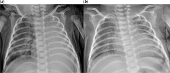
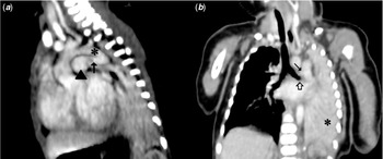
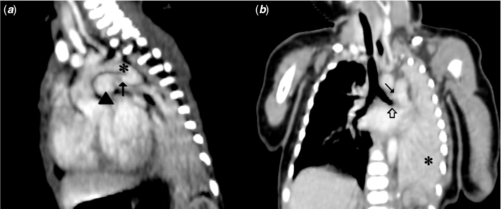
A 2450 g girl was born by caesarean section at the 34th weeks of gestation for placenta previa totalis. The girl was intubated immediately due to respiratory distress, hypoxaemia, and hypotonia. The patient had no other congenital anomaly. A continuous murmur which was heard best at the upper left sternal border and decreased breath sounds on the left side were detected on auscultation. Imaging methods required for diagnosis were performed immediately and consecutively in a few days. During follow-up at the neonatal ICU, a large patent ductus arteriosus was detected. Medical courses of paracetamol and non-steroid antiinflammatory drugs were not able to close the patent ductus arteriosus. Consciousness, tonus, and reflexes were normal, and there were not any signs of perinatal asphyxia. Chest roentgenogram showed cardiomegaly and complete atelectasis of the left lung (Fig 1a). Thereon, transthoracic echocardiography was performed and a large patent ductus arteriosus (4 mm) was detected as the reason of the cardiomegaly. Transthoracic echocardiography also showed dilated left cardiac chambers without any other left-to-right shunt diseases. Thorax CT angiography showed total atelectasis of the left lung due to compression of the left main bronchus by the tortuous patent ductus arteriosus which was 5 mm in diameter (Fig 2). Medical closure of patent ductus arteriosus was not considered because of the large size of the patent ductus arteriosus and the compression at the left main bronchus, and surgical closure of the patent ductus arteriosus was planned immediately with the agreement of all relevant physicians. Because of the patient had patent ductus arteriosus 5 mm in diameter that compressing of the left main bronchus, medical treatment was not performed for closure of patent ductus arteriosus. Our patient weighed less than 5000 g and she had a very large patent ductus arteriosus. Percutaneous patent ductus arteriosus closure procedure has high complication rates in these patients. We also thought that this method might not eliminate the obstruction problem. For all these reasons, we planned performing patent ductus arteriosus closure surgery to our patient immediately.
Chest X-ray posteroanterior view. (a) Pre-operative: Completely collapsed left lung; (b) Post-operative: Reexpanded left lung.

Thorax computed tomography angiography (CTA) with mediastinal window setting. (a) Sagittal maximum intensity projection image shows patent ductus arteriosus as a tubular structure (arrow) between arcus aorta (asterisk) and pulmonary artery (arrow-head); (b) Coronal multi-planar reconstruction image shows patent ductus arteriosus near the pulmonary artery (black arrow) cause external obstruction of left main bronchus (open arrow) and completely collapsed left lung (asterisk).
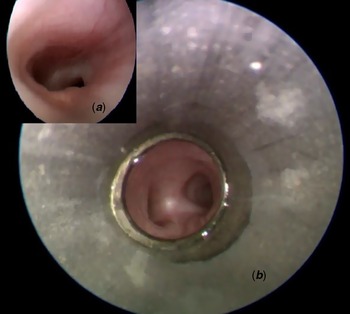
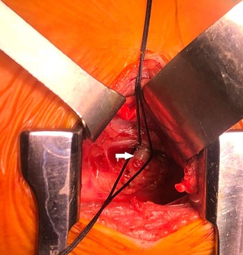
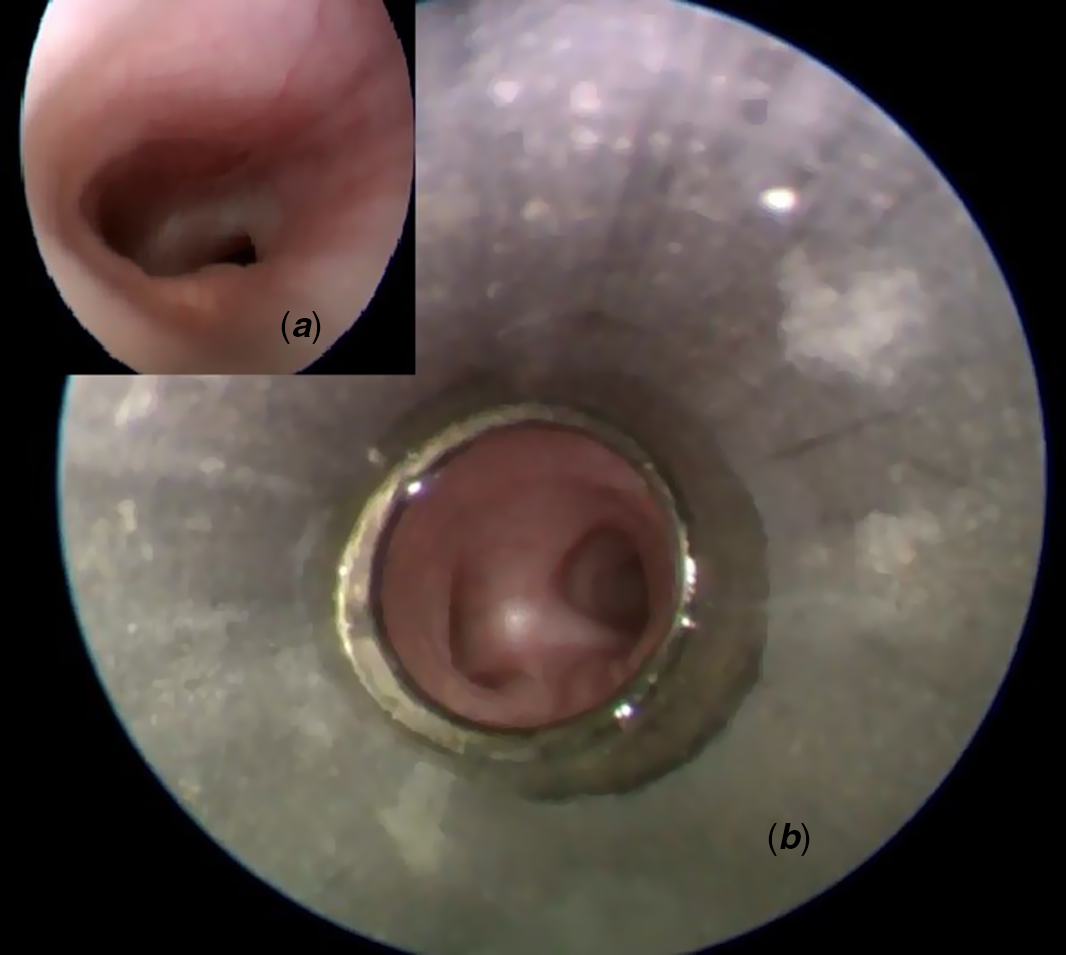
The intubated patient was taken to the operation room. Firstly, rigid bronchoscopy was performed by a paediatric surgeon in the operating room. It was observed that the right bronchus was normal, the left bronchus was lateralised and it was obstructed at the level of the carina. There was no endoluminal pathology. The left main bronchus was constricted due to external compression. There was pulsation in the anterior part of the left main bronchus (Fig 3). In the same session; left posterolateral thoracotomy was performed through the third intercostal space in the right lateral decubitus position. The left lung was atelectasis. The 5 mm diameter PDA was reached using extrapericardial approach and preserving the adjacent neurological structures (nervus vagus, phrenicus, and laryngeus recurrens). The aortic and pulmonary ends of the patent ductus arteriosus that has a large aortic ampulla were clamped and divided. Both ends of the patent ductus arteriosus were closed with 6/0 polypropylene sutures. A chest drain was placed and the operation was completed without any problems (Fig 4). On the first post-operative day, expansion was observed in the left lung on chest X-ray. (Fig 1b). The patient was extubated on the second post-operative day. She was discharged on the 9th post-operative day. Informed consent was obtained from the patient’s parents for publication.
Pre-operative bronchoscopy. (a) Image from obstructed left main bronchus; (b) Image at the level of the carina which demonstrated obstruction secondary to external compression in the left main bronchus.
Intraoperative image of patent ductus arteriosus with large aortic ampulla (white arrow).

Discussion
Extrinsic compression of the trachea and main bronchi by cardiovascular structures may be seen in newborns and infants. Double aortic arch, pulmonary artery sling, right aortic arch, enlarged main pulmonary artery, congestive heart failure, large left to right shunt, and patent ductus arteriosus are pathologies that cause extrinsic compression of the trachea or main bronchi. Some patients may be asymptomatic, but most of the patients have mild or severe symptoms (stridor, wheezing, cyanosis, cough, dyspnoea, recurrent pulmonary infection, growth retardation). The tracheal compression can cause death spells. Reference Rogers, Cunnane and Hartnick3
Extrinsic compression of the main bronchi by the patent ductus arteriosus can be attributed to the large duct diameter or increased shunt flow. Compression due to patent ductus arteriosus commonly occurs at the left and rarely at right main bronchus. The compression effect of patent ductus arteriosus on the left main bronchus may be alone or with enlarged left atrium.
Actual incidence of main bronchial compression caused by the dilated patent ductus arteriosus has not been clearly reported in the literature. Smith et al Reference Smith, Saul, Goldfarb, Biko and OʼByrne4 reported that this incidence should be about 4.6%. Patent ductus arteriosus stenting can be used as an alternative to palliative shunt operation in cyanotic newborns. They routinely evaluated the thoracic cavity and main airways with appropriate imaging methods (X-ray, bronchoscopy, and CT) in all patients before patent ductus arteriosus stenting which was used as an alternative to palliative shunt operation in cyanotic newborns. They determined this incidence (4.6%) from the data they obtained from this algorithm. They also reported that if there is main bronchial compression caused by a large patent ductus arteriosus, there is no clear consensus on whether the obstruction decreases or increases after patent ductus arteriosus stenting. They preferred to perform palliative shunt operations instead of patent ductus arteriosus stenting in patients with main bronchial compression caused by patent ductus arteriosus in their clinics. Because the compression could get worse by patent ductus arteriosus stenting.
Ductus arteriosus aneurysm is a rare pathology, but it is frequently seen in early infancy. It can be asymptomatic or symptomatic (rupture, recurrent pulmonary infection due to obstruction of bronchi, thromboembolism, and compression of the phrenic or recurrent laryngeal nerve). Surgical treatment is indicated for ductal aneurysms that are symptomatic or do not heal spontaneously.
Transcatheter closure of the patent ductus arteriosus is frequently performed. Fitzmaurice et al Reference Fitzmaurice, Coleman, Walsh, Oslizlok, Russell and McGuinness5 reported an 11-month-old girl who was performed closing of patent ductus arteriosus by the Amplatzer muscular ventricular septal defect occluder device. This patient had right aortic arch anomaly. Patent ductus arteriosus had a connection with the right pulmonary artery. They performed bronchoscopy and thorax CT to investigate the causes of prolonged intubation in the post-operative period. They detected external compression of the right main bronchus by a patent ductus arteriosus occluder device. This complication was treated by removal of the device surgically. In conclusion, they recommended surgical closure of the patent ductus arteriosus or using a softer closure device to avoid this complication in infants with additional anomalies. We also decided to close the patent ductus arteriosus surgically in our patient that causes to external compression of the left main bronchus.
External occlusion of the left main bronchus is a rare and life-threatening complication of surgical patent ductus arteriosus closure with clips. Ban et al Reference Ban and Abe6 reported a case of premature girl. They closed patent ductus arteriosus with surgical clips in this patient a day after birth reported. Total left lung atelectasis was detected in this patient on the first post-operative day. They performed bronchoscopy and determined severe external compression of the left main bronchus. They thereupon performed rescue reoperation to the patient. They removed the old clips and placed new clips on the most distal parts of the aortic and pulmonary ends of the patent ductus arteriosus. In this case report, the author emphasises that maximum attention should be paid to the following factors in the premature patients that surgical patent ductus arteriosus closure by using clips was planned: choosing appropriate sizes of clips, placing the clips at a vertical angle and very closely to the aortic end of patent ductus arteriosus. It is very important to perform rescue re-operation immediately (removal of patent ductus arteriosus clips) in patient with suspected main bronchus obstruction to prevent lethal outcome. Bronchoscopy is a very helpful imaging method for early detection of this complication. Late detection of this complication and delayed reoperation carry higher risk of mortality.
In conclusion, enlargement or congenital pathologies of adjacent cardiovascular structures can cause extrinsic compression of the trachea–main bronchi in newborns and infants. A wide range of respiratory problems may be seen in these patients, from wheezing to prolonged intubation. If there is a congenital cardiovascular anomaly/pathology that causes main airway compression, transcatheter or surgical treatment of this pathology should be performed as soon as possible. Compression of the main bronchi by patent ductus arteriosus is rare but it can cause mild to severe respiratory problems. If there are physical findings suggesting major airway obstruction in newborns and infants with patent ductus arteriosus, appropriate radiological imaging methods should be performed to investigate extrinsic compression without delay. Anatomy of trachea–main airways and thorax cavity should be examined with bronchoscopy, thorax CT or magnetic resonance imaging pre-operatively. If there is external compression of the left main bronchus due to patent ductus arteriosus, early surgical closure of patent ductus arteriosus is an effective treatment option. Fast clinical and radiological improvement are expected after surgical closure of patent ductus arteriosus.
Acknowledgements
None.
Financial support
This research received no specific grant from any funding agency, commercial, or not-for-profit sectors.
Conflicts of interest
None.