I. INTRODUCTION
Nacre from the interior of shells is a beautiful biomaterial, both at the macroscopic level, where it exhibits pearlescence colors because of interference effects, and at the microscopic level, where its interesting structure is revealed.
Nacre is a composite material of ∼5 vol% protein/polysaccharide matrix and 95 vol% calcium carbonate in the metastable aragonite (orthorhombic, Pnma) form. Typical dimensions of the tablet-shaped aragonite “bricks” are ∼5–20 μm in length and width, and ∼0.3–1.5 μm in thickness,Reference Bruet, Qi, Boyce, Panas, Lai, Frick and Ortiz1 with organic bridges between the aragonite plates.Reference Rousseau, Lopez, Stempflé, Brendlé, Franke, Guette, Naslain and Bourrat2
Although composites with such high ceramic filler content could be brittle, nacre has a work fracture ∼3000 times greater than that of pure aragonite.Reference Jackson, Vincent and Turner3 Certainly, the structure plays an important role in the mechanical properties of nacre.Reference Lin and Meyers4
Although the “brick-and-mortar” view of nacre is a convenient starting point for discussion of its properties, more recent examination by various techniques has revealed significant structural details.
Consider the aragonite crystals. These can show, on average, long-range preferential orientation, with the c-axes parallel to each other and perpendicular to the sample shell, and the a- and b-axes statistically aligned, over areas as large as 25 mm × 25 mm.2 However, close examination shows oriented domains of 3 to 10 aragonite tabletsReference Bruet, Qi, Boyce, Panas, Lai, Frick and Ortiz1 and differences in orientation at different growth stagesReference Checa and Rodríguez-Navarro5 and different growth rates.Reference Gilbert, Metzler, Zhou, Scholl, Doran, Young, Kunz, Tamura and Coppersmith6
And the aragonite tablets are not monolithic. Each tablet is subdivided by organic matter that can be removed by heating to create pores.Reference Knitter, Odemer and Hausselt7 The intracrystalline organic matrix is in the form of an oriented, crystalline foam with very thin walls and closed cells, dividing each aragonite tablet into nanograins of approximate dimension 40 nm,Reference Rousseau, Lopez, Stempflé, Brendlé, Franke, Guette, Naslain and Bourrat2 and the nanograin structure likely plays a role in nacre’s mechanical properties.Reference Barthelat, Li, Comi and Espinosa8
Furthermore, solid-state nuclear magnetic resonance (NMR spectroscopy) shows that the surface of the aragonite platelets is amorphous and there is no significant (chemical) interaction between this amorphous layer and the protein matrix that surrounds it.Reference Jäger and Cölfen9 However, this amorphous layer surrounds only the plates and is not at the interfaces of the intraplate nanograins.Reference Jäger and Cölfen9
Another subtle factor is that the crystal lattice of aragonite from biogenic sources, such as in nacre, has been found to be anisotropically distorted (up to 0.2% along the c-axis), a distortion that can be relieved by annealing.Reference Pokroy, Fieramosca, Von Dreele, Fitch, Caspi and Zolotoyabko10
Certainly, the hierarchical structure of nacre plays an important role in its mechanical properties.Reference Darder, Aranda and Ruiz-Hitzky11, Reference Launey and Ritchie12
Although most of nacre is calcium carbonate, the proteins are important in the biomineralization process that gives rise to the nacre structure. Recent studies have shown that a single 30 amino acid–protein fragment templates a biomineral rather similar to natural nacre.Reference Metzler, Evans, Killian, Zhou, Churchill, Appathurai, Coppersmith and Gilbert13 Furthermore, the protein layers, which exist between the plates but are virtually absent where plates on the same plane abut,Reference Lin and Myers14 are important for crack deflection, delocalization of compressive damage, and microbuckling mechanisms that contribute to the extraordinary mechanical properties of nacre.Reference Menig, Meyers, Meyers and Vecchio15
With such a unique yet well-characterized structure, nacre offers an important opportunity to investigate the relationship between structure and properties of materials. Here, we have studied the thermal conductivity of nacre as a function of temperature, direction, and chemical modification to advance our understanding of heat conduction in nanostructured materials.
II. EXPERIMENTAL METHODS
An abalone shell (Haliotis fulgens, by appearance), measuring ∼15 cm by 10 cm, was cut using a low-speed diamond-blade cutting saw. Approximately 40 samples of lateral dimension ∼3 to 5 mm were cut from the shell (Fig. 1) in a way that minimized border effects and any natural inhomogeneities found in the shell. The external mantle and periostracum layersReference Lin and Myers14 were removed by polishing, leaving samples 0.5- to 1-mm thick. Thermal conductivity and heat capacity measurements of the (control) nacre samples were carried out without further treatment. Some of the nacre samples were treated with ethylenediaminetetraacetic acid (EDTA, 99%; Sigma-Aldrich, St. Louis, MO) or sodium hydroxide (NaOH, 97%; Sigma-Aldrich, St. Louis, MO), as described later.
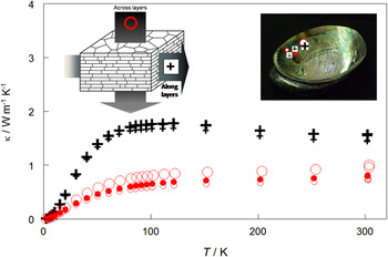
Thermal conductivity of nacre as a function of temperature, showing the results for: three samples across the aragonite layers (〇) and three samples along the aragonite layers (+). The sample locations from the shell are indicated on the inset, with the location symbol corresponding to the data symbol.

Interior structures were exposed for scanning electron microscopic (SEM) studies by immersion in liquid nitrogen followed by freeze fracturing. These samples were then sputter coated with Au–Pd before SEM analysis. The SEM investigations were carried out with a Hitachi S-4700 FEG SEM (Hitachi, Tokyo, Japan); at a 5-kV voltage, the SEM was able to produce images with a resolution of 2 nm.
All NMR experiments were carried out on a Bruker Avance NMR spectrometer (Bruker, Karlsruhe, Germany) with a 9.4T magnet (400.24-MHz 1H Larmor frequency, 100.65-MHz 13C Larmor frequency). Finely ground powder samples [158 mg of the unmodified (control) and 103 mg of the NaOH-treated nacre] were packed into rotors of 4-mm diameter. The samples were spun at 7.0 and 9.0 kHz to determine center bands and to identify spinning sidebands. Proton longitudinal relaxation times were determined by inversion recovery sequences. They were estimated to be on the order of 0.6 s or faster. The recycle delays of 4 s for the 13C cross-polarization (CP)/magic angle spinning (MAS) experiments were chosen because of the probe duty cycle. The other parameters for the 13C CP/MAS experiments with two pulse phase moderation (TPPM) proton decoupling were optimized on glycine, whose carbonyl resonance also served as external, secondary chemical shift standard at 176.06 ppm. Using 1-ms CP contact times, 6,000 and 14,000 scans were accumulated during 6.6 and 15.5 h for the 13C CP/MAS NMR spectra of the control and NaOH-treated samples, respectively.
The heat capacity of nacre from T = 2 K to T = 300 K was determined using a commercial relaxation calorimeter [Physical Property Measurement System (PPMS); Quantum Design, San Diego, CA]. The polished sample had mass of 26.2 mg. The heat capacity was determined by modeling the thermal relaxation response of the sample sitting in vacuum on a sapphire sample stage, based on the methods of Huang et al.Reference Huang, Lin and Tien16 The heat capacity determined with this instrument is accurate to better than 1% over this temperature range.Reference Kennedy, Stancescu, Marriott and White17
The thermal conductivity of nacre was determined from T = 2 K to T = 300 K using the thermal transport option of the PPMS. The samples were attached to gold-plated copper leads using silver-loaded epoxy (Tra-Duct 2902, Bedford, MA) and then mounted on the thermal transport puck. The principle of operation is a one-dimensional heating process in which the heater current is modulated as a square wave and the temperature response is monitored and modeled to give the thermal conductivity, κ.Reference Maldonado18 Typical sample dimensions were 4 × 4 × 1 mm3, with the temperature gradient along the shortest dimension, and measurements were taken in the steady state mode with 2% rise in absolute temperature and temperature stability better than 0.1%. The sample is surrounded by a shield, matched to the temperature of the sample. Correction (Stephan–Boltzmann law) for radiative power losses was 0.09% of the power at T = 2 K, and 1.8% of the power at T = 300 K, lower at the lower temperature because of the much smaller power used to determine the thermal conductivity at lower temperatures.
Both the heat capacity and thermal conductivity measurements were carried out in high vacuum, but, unlike our earlier results on ivory,Reference Jakubinek, Samarasekera and White19 the measurements on nacre were stable over time, showing that no water was removed from the sample in the vacuum chamber.
III. RESULTS AND DISCUSSION
A. Comparisons with other materials and rule-of-mixtures
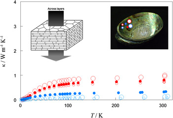
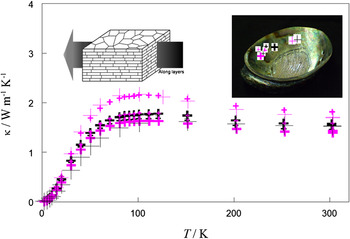
The thermal conductivity, κ, of nacre as a function of temperature is shown in Fig. 1. Given the possible differences in composition and structure from location-to-location within the shell, several samples were measured. To investigate the influence of the anisotropic structure of nacre on its properties, thermal conductivity was determined both along the aragonite layers, and perpendicular to the layers, as indicated in Fig. 1.
There appears to be only one previous investigation of thermal properties of nacre with which we can compare our results. In that case,Reference Gómez, Quintana, Aguilar, Alvarado-Gil, Yánez-Limón, Diaz and Aldana20 the thermal diffusivity (a) of nacre from various sources was determined photothermally, but their room-temperature value for a for aragonite-based nacre was about a factor of six higher than we derived from our results from:

where ρ is the measured density (2.30 ± 0.05 g cm−3) and C s is the specific heat (see below). The difference is outside of the range one might expect for sample-to-sample variation, and we ascribe the discrepancy to the model used in the earlier studyReference Gómez, Quintana, Aguilar, Alvarado-Gil, Yánez-Limón, Diaz and Aldana20 to correct their photothermal results for the overwhelming thermal diffusivity of their aluminum substrate.
The room-temperature thermal conductivity of nacre, κ ∼1 W m−1 K−1, is about twice that of ivory,Reference Jakubinek, Samarasekera and White19 another high-strength bioceramic with a hierarchical structure. The value for nacre is at the low end of typical values for dense, organized solids and similar in value to that of amorphous SiO2.Reference White21 The room-temperature thermal conductivity of nacre is less than that of pure, monolithic aragonite (2.2 W m−1 K−1 along the c-axis and 2.4 W m−1 K−1 perpendicular to the c-axis).Reference Clauser, Huenges and Aherns22 Of course, nacre is not pure aragonite and has ∼5 mass% organic content. A rule-of-mixtures thermal conductivity of 95% aragonite and 5% organic contentReference Tuladhar, Paterson and Wilson23 would be ∼2.2 W m−1 K−1 at room temperature, again about twice that observed. For a simple rigid geometry in which heat is not impeded by, for example, nanostructuring, one could reasonably expect the thermal conductivity to lie between the values expected for thermal resistances in series or in parallel, i.e., the thermal conductivity of nacre, κ N, would be bound by:

where the properties (thermal conductivity and volume) of aragonite are designated by “A” and those of organic by “O.” On the basis of Eq. (2), the room-temperature thermal conductivity of nacre should be between 1.5 and 2.2 W m−1 K−1. It falls below these values, both along the plates and across the plates, indicating that, as we found previously for ivory,Reference Jakubinek, Samarasekera and White19 the structure of nacre plays an important role in impeding heat flow.
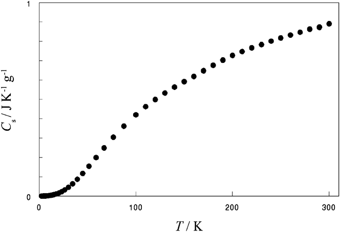
On the other hand, the specific heat is rather accurately described by the rule-of-mixtures value. Using the specific heat of aragoniteReference Staveley and Linford24 and approximating that of the 5% organic component at 1 J K−1 g−1, gives a value within 5% of the observed room-temperature-specific heat of nacre. This is not surprising because, unlike thermal conductivity, heat capacity is not a transport phenomenon and therefore does not depend as strongly on structure.
B. Effective phonon mean free path and thermal anisotropy
Not only is the thermal conductivity of nacre relatively low in comparison with its components but it also is somewhat glass-like. In particular, there is a positive temperature coefficient for the measurement across the layers, as for a glass.Reference White21 Although one would expect a large peak for an ideal simple crystal,Reference White21 only a relatively small peak is observed for the data along the layers (Fig. 1). Clearly the lattice waves are not very efficient at carrying heat through the nacre structure.
Further insight into the relatively low thermal conductivity of nacre can be gained by consideration of the effective phonon mean free path within nacre, where the phonons are the heat carriers. Based on the simple Debye model of heat conduction derived from the kinetic theory of gases, the thermal conductivity, κ, is related to the heat capacity per unit volume, C, the phonon speed, v, and the effective phonon mean free path, λ, asReference Tritt25:

To deduce the effective phonon mean free path, the specific heat of nacre was determined as a function of temperature (Fig. 2). The phonon speed has been assessed in the two principal directions from the elastic modulus, E,Reference Jackson, Vincent and Turner3 and the measured density, ρ, fromReference White21:

Specific heat of (untreated) nacre as a function of temperature.

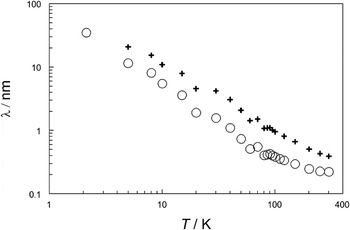
giving phonon speeds of 5400 m s−1 across the plates and 5500 m s−1 along the plates. The resulting effective phonon mean free path as a function of temperature and direction is presented in Fig. 3. We consider this to be an “effective” phonon mean free path because Eq. (3) assumes a uniform composition and it also ignores dispersion, which could underestimate the mean free path by an order of magnitude.Reference Chen26 Furthermore, information derived from use of Eq. (3) presumes that the thermal conductivity in nacre is limited by defects (e.g., boundary resistance or impurities). However, in the absence of better models and further lattice dynamical information, we can draw out some semi-quantitative information. In particular, the effective phonon mean free path is similar to that of ivory,Reference Jakubinek, Samarasekera and White19 another nanostructured biomaterial. Furthermore, as expected, Fig. 3 shows that the effective phonon mean free path in nacre is longer along the layers than across the layers. However, if the main limitation in the mean free path is the size of the aragonite tablets, their aspect ratio would give a ratio of the mean free paths of ∼20, but it is much smaller, about 2–3.
Effective phonon mean free path in nacre: 〇, across the aragonite layers and +, along the aragonite layers.

The similar effective phonon mean free path in both directions in nacre can be understood in terms of its detailed structure. The aragonite crystallites are not monolithic, but are twinned with typically 2–50 sectors subdivided by vertical organic membranes.Reference Bruet, Qi, Boyce, Panas, Lai, Frick and Ortiz1 The domain boundaries within the aragonite tablets could effectively provide a barrier for phononic heat transport and reduce the mean free path by a factor corresponding to the number of sectors, i.e., from ∼20 for a monolithic crystal to ∼2 for a true aragonite tablet of nacre, as observed.
The finding of nearly identical thermal conductivity and effective phonon mean free path in both directions in nacre also fits with the findings that the mechanical properties of nacre, including elastic modulus, toughness, and strength, are nearly isotropic.Reference Jackson, Vincent and Turner3
Of course, from the perspective of the mollusc, it is an advantage to be surrounded by a material that has similar strength and toughness in all directions, to prevent any easy direction for fracture. The nanostructuring that provides the high strength appears to make the thermal conductivity very similar in both principal directions.
C. Chemical modification of nacre
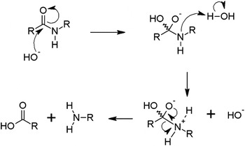
Another interesting aspect of nacre is that the structure can be chemically modified.Reference Checa and Rodríguez-Navarro5 The protein component can be hydrolyzed by the addition of 5% aqueous NaOH as shown in Fig. 4. Alternatively, the calcium can be chelated with 2% aqueous EDTA to demineralize the nacre. Both approaches were used here to explore the influence of structure and chemical composition on thermal conductivity.
NaOH hydrolyzes the proteins to allow their removal from the nacre structure.

1. Demineralized
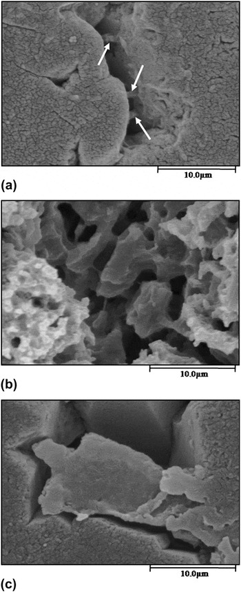
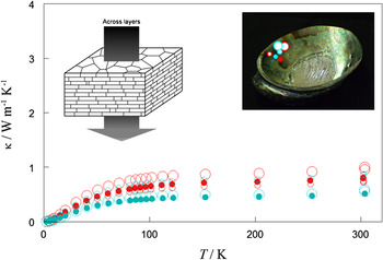
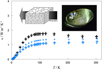
Treatment of nacre with 2% EDTA is known to dissolve the mineral component of nacre’s microstructure.Reference Checa and Rodríguez-Navarro5 The mass of nacre samples of dimensions 4 × 4 × 1 mm3 after such a treatment was followed over time and found to be reduced by ∼30% and still decreasing slowly after about 5 days. The samples were too fragile to handle for property measurement after about 3 days of treatment. However, even 10 min in EDTA led to significant demineralization, as shown in Fig. 5. The thermal conductivity of demineralized nacre (10 min in EDTA; note that the change in density was <5%) was significantly reduced relative to untreated nacre (control), both across the aragonite plates and in the direction along the plates (Figs. 6 and 7). It is not surprising that the thermal conductivity is much lower than the control in both directions, given that the main heat-carrying component in nacre would be expected to be the mineral.
Scanning electron microscope images of nacre: (a) no treatment (control), with bridges highlighted, (b) partially demineralized by treatment for 10 min in ethylenediaminetetraacetic acid (EDTA) as described in the text, and (c) with a portion of the protein matter removed by treatment for 2 h with aqueous NaOH as described in the text.

Thermal conductivity across the aragonite layers of nacre as a function of temperature, showing the results for: red data, three untreated (control) samples; blue data, three samples demineralized for 10 min in EDTAas described in the text. The sample locations from the shell are indicated on the inset.

Thermal conductivity along the aragonite layers of nacre as a function of temperature, showing the results for: black data, three untreated (control) samples; blue data, three samples demineralized for 10 min in EDTA as described in the text. The sample locations from the shell are indicated on the inset.

2. Protein removed
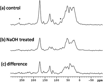
13C NMR spectra were determined for both unmodified nacre (control sample) and nacre treated for 2 h in 5% NaOH (Fig. 8). Both samples show characteristic NMR peaks at: 172.6 ppm, from carbonyl groups in proteins and modified polysaccharides such as chitin; 156, 129, and 117 ppm, from aromatic groups in amino acids, such as tyrosine; 104 ppm, from the acetyl groups of polysaccharides; and highly overlapping signals between 75 and 10 ppm, from substituted and unsubstituted aliphatic groups from both polysaccharides and proteins. Peak predictions for chitin made with the ACD-Spectrum predictor (Chelmsford, MA) give peaks at 23 ppm (CH3), 54 ppm (NCH), 61 ppm (OCH2), 72 ppm, 74 ppm, and 80 ppm (three OCHs), 101 ppm (OCHO), and 175 ppm (NC=O) in agreement with literature spectra.Reference Hu and Schmidt-Rohr27, Reference Gupta, Cody, Tetlie, Briggs and Summons28 The spinning speed comparison for both samples (spectra not shown here) indicated that the carbonyl and the aromatic groups create some spinning sidebands. However, their contribution in the aliphatic region is small and does not dominate the spectral features. The spinning speed of 9.0 kHz was chosen such that the spinning sidebands at 262 ppm and 83 ppm (indicated on Fig. 8) from the strong carbonyl peaks do not overlap with the aliphatic region. The difference between the 13C NMR spectrum of the control sample and the NaOH-treated sample, taking into account the sample mass and number of scans accumulated [Fig. 8(c)], shows that the organic carbon content of the sample is reduced to 60 ± 10% of its original value on NaOH treatment. The difference spectrum also provides information about which material was removed. Most notable is the decrease in signal intensity of the carbonyl groups and the aromatic groups; the latter must originate from the protein because there are no aromatic or unsaturated carbons present in chitin or polysaccharides. Other peaks lost lie in the aliphatic region, which cannot be uniquely distinguished between proteins and chitin. The peak unique to chitin at 104 ppm is weak but present in both the spectra from the control and the NaOH-treated sample and not seen beyond the noise level in the difference spectrum. This indicates, but does not rigorously prove, that the chitin is not affected by the mild basic treatment. Considering the relative intensities of the peak at 104 ppm, compared to, for example, the aromatic peaks, the polysaccharide components are small. Although these spectra were acquired using 13C CP/MAS NMR spectroscopy, and therefore a quantitative comparison between peaks is not rigorously feasible, a qualitative comparison of CH groups, e.g., the 130-ppm aromatic peak and the 104-ppm OCHO peak from polysaccharides is indicative of the relative concentrations given their similar proton-carbon dipole–dipole coupling. The use of the CP-NMR technique also is the reason why we see very little of what could be an overwhelming peak from the carbonate carbon from the aragonite (CaCO3). This peak has been shown to resonate at 170–171 ppm,Reference Jäger and Cölfen9, Reference Papenguth, Kirkpatrick, Montez and Sandberg29, Reference Takahashi, Yamamoto, Onoda, Doi, Inaba, Chiba, Kobayashi, Taguchi, Okumura and Ueyama30 and therefore overlaps with the carbonyl carbons. However, the low concentration of protons close to the carbonate group renders the cross-polarization highly inefficient, especially at the CP conditions chosen here that favor carbons with directly bound protons. In summary, our NMR studies show that the mild NaOH treatment of nacre predominantly removes the proteins and less of the polysaccharides. This removal is not complete under the conditions used. Our finding of this preference is in agreement with other studies on different marine crustaceans.Reference Al Sagheer, Al-Sughayer, Muslim and Elsabee31
13C CP/MAS NMR (cross-polarization/magic angle spinning nuclear magnetic resonance) results for (a) untreated nacre (control) where “*” indicates spinning sidebands, (b) the sample treated for 2 h in NaOH, and (c) the difference. (a, b), taking into account the sample mass and number of scans accumulated, showing which material was removed.

From our SEM studies, at the micron level, the sample with reduced protein content looks much the same as untreated nacre (Fig. 5) and the density was reduced by less than 5%. Across the aragonite layers, the thermal conductivity of nacre after treatment with NaOH is similar to or a little less than that of the control sample (Fig. 9). It is reasonable for the NaOH treatment to increase the thermal resistance across the sample, but not as much as for demineralization. Rather surprisingly, the thermal conductivity in the aragonite plate direction was not much changed, or was even in some cases increased (Fig. 10) after treatment with NaOH. It is known that the protein layer is virtually absent where plates abut on the same plane,Reference Lin and Myers14 so our results show that removal of the protein (presumably from between the plates) maintains the thermal pathway along the plate direction, or, in some cases, allows more plate-to-plate contact, thereby reducing the thermal resistance in this direction.
Thermal conductivity across the aragonite layers of nacre as a function of temperature, showing the results for: red data, three untreated (control) samples; blue data, three samples treated for 2 h with NaOH as described in the text to remove some of the protein content. The sample locations from the shell are indicated on the inset.

Thermal conductivity along the aragonite layers of nacre as a function of temperature, showing the results for: three untreated (control) samples; pink data, three samples treated for 2 h with NaOH as described in the text to remove some of the protein content. The sample locations from the shell are indicated on the inset.

IV. CONCLUSIONS
The thermal conductivity of nacre is rather low compared with other materials with such a high mineral content. This is largely because the effective phonon mean free path is short due to nacre’s nanostructure. The thermal conductivity and effective phonon mean free path show only mild anisotropy. Removal of the mineral component of nacre reduces the thermal conductivity both along the plates and across the plates. Partial removal of the protein reduces the thermal conductivity across the plates, but it is maintained or even slightly increased along the plates.
ACKNOWLEDGMENTS
The authors thank Andrew George, Tolga Goren, Dr. Michael Jakubinek, Dr. Ping Li, Patricia Scallion, and Dr. Catherine Whitman for input. This work was supported by National Sciences and Engineering Research Council of Canada (NSERC) of Canada, NMR-3 at the Department of Chemistry of Dalhousie University, and the Canada Foundation for Innovation, Atlantic Innovation Fund, and other partners, which fund the Facilities for Materials Characterization managed by the Institute for Research in Materials at Dalhousie University.