Chronic thromboembolic pulmonary hypertension is the most worrying complication of acute pulmonary embolism. This condition leads to right ventricular dysfunction and is associated with a poor prognosis.
The rate of acute pulmonary embolism and subsequent chronic thromboembolic pulmonary hypertension is low in children and adolescents. Particularly, it should be in mind in children with hypercoagulable states. A prompt diagnosis requires a high suspicion in combination with a thorough workup. We herein present a child with chronic thromboembolic pulmonary hypertension in the context of Behçet’s disease diagnosed in the outbreak of coronavirus disease 2019.
Case report
The patient was a 13-year-old boy who presented with fever, pleuritic chest pain, and dyspnea on exertion. His past medical and surgical history was unremarkable, and he was under no medication. Nothing of note was found in the boy’s familial history. On physical examination, blood pressure of 100/70 mmHg, pulse rate of 113 beats per minute, respiratory rate of 20 per minute, and temperature of 37.9 ºC were recorded. Fine rales in the lower zones of both lungs and coarse crackles in the upper zone of the left lung were auscultated. Other examinations were unremarkable.
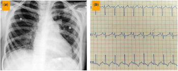
On chest X-ray, there was evidence of the enlargement of the right atrium and the right ventricle, prominent bilateral pulmonary hila, reticulonodular opacities in the right lung, and consolidation in the left lung upper zone. Electrocardiography showed sinus tachycardia, right-axis deviation, and non-specific secondary ST-T segment changes (Fig 1).
(a) Chest X-ray reveals prominent bilateral hila, reticulonodular opacities in the right lung and consolidation in the left lung upper zone. (b) Electrocardiogram (leads I, II, and III) depicts sinus tachycardia, right-axis deviation, and non-specific secondary ST-T segment changes.

Transthoracic echocardiography showed a left ventricular ejection fraction of 50%, mild right ventricular dysfunction, mild right atrium and right ventricle enlargement, moderate eccentric mitral regurgitation, and a peak systolic pulmonary artery pressure of 45 mmHg.
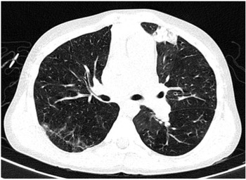
Given the coronavirus disease 2019 pandemic and the possibility of exposure, together with the compatibility of the patient’s suspicious symptoms with coronavirus pneumonia, a chest CT scan was performed (Fig 2). A few patchy bilateral ground-glass opacities and airspace consolidation in the left upper lobe were detected. In addition, reticular densities were detected in the posterior aspect of the right upper lobe. Therefore, the patient was considered a highly suspicious case of coronavirus disease 2019 pneumonia.
Chest CT in the parenchymal window. Patchy bilateral ground-glass opacities and airspace consolidation in the left upper lobe are seen. Reticular densities are noted in the posterior aspect of the right upper lobe.

Laboratory studies showed leukocytosis with the predominance of polymorphonuclear cells, an erythrocyte sedimentation rate of 2 mm/hour, a C-reactive protein level of 4 mg/L, negative cardiac troponin, and creatine kinase–myocardial band. In addition, the polymerase chain reaction test for coronavirus disease 2019 was negative.
Antibiotic therapy with intravenous ceftriaxone (1 g every 12 hours) and oral azithromycin (500 mg daily) was administered with the impression of bacterial pneumonia. The patient responded well, and he was discharged on day 4 of treatment.
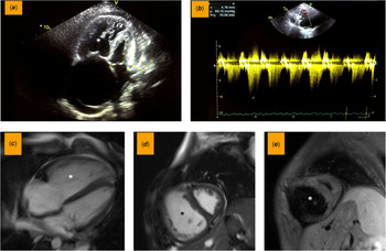
He returned after 2 months with worsened dyspnea. Repeated echocardiography revealed a mildly dilated left ventricle with a left ventricular ejection fraction of 45%, paradoxical interventricular septal motion, moderate right atrium and right ventricular enlargement, moderate right ventricular dysfunction, and a systolic pulmonary artery pressure of 60 mmHg (Fig 3a and b).
Echocardiography and MRI of the patient. ( a ) Echocardiography in semi four-chamber view depicts moderate to severe right atrium and ventricle enlargement. ( b ) A significant increase in maximum tricuspid regurgitation velocity is demonstrated in Doppler mode. ( c and d ) Cine steady-state free precession cardiac magnetic resonance imaging sequences in four-chamber and short-axis mid-ventricular planes reveal notable right-sided chamber enlargement. ( e ) Short-tau inversion recovery sequence depicts no sign of myocardial edema/inflammation. Asterisks: dilated right ventricle.

Laboratory studies showed increased levels of serum B-type natriuretic peptide (759 pg/ml; normal value <87 pg/ml) and proBNP (5950 pg/ml; normal value <125 pg/ml).
The patient was hospitalised, and right heart failure treatment was started immediately with oral spironolactone (25 mg daily), oral propranolol (10 mg twice a day), an intravenous drip of milrinone (50 μg/kg/minute), and an intravenous drip of Lasix (1 mg/hour).
With the suspicion of myocarditis, cardiac magnetic resonance imaging was done, which demonstrated normal left ventricle size with mildly reduced systolic function, severe right ventricle enlargement with an end-diastolic volume index of 144 ml/m2, severe right ventricular dysfunction (right ventricular ejection fraction = 24%), and no sign of myocardial edema or fibrosis (Fig 3c–e).
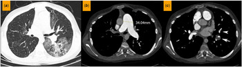
As a part of the workup for right ventricular dysfunction, pulmonary CT angiography was performed. It demonstrated eccentric filling defects and aneurysmal changes in the left upper lobar pulmonary artery with stenosis and obliteration of the segmental branches. Also noted were linear filling defects in the segmental and subsegmental branches of the right lower lobar pulmonary artery. The findings were in favor of chronic pulmonary embolism. In addition, central filling defects were noted in the left upper and right middle lobar and segmental branches, suggestive of acute-on-chronic pulmonary thromboembolism. The parenchymal window revealed multiple patchy bilateral ground-glass opacities with a confluent appearance in the left lower lobe (Fig 4).
( a ) Axial chest CT parenchymal window and ( b and c ) pulmonary CT angiography images. ( a ) Patchy ground-glass densities which are confluent in the superior segment of the left lower lobe are depicted. ( b ) Marked dilatation of the main pulmonary artery is demonstrated. ( c ) Eccentric filling defect and focal aneurysmal dilatation in the distal part of the left lower lobar pulmonary artery (arrow) are noted. Linear filling defect in a segmental branch of the left pulmonary artery (arrowhead) is revealed.

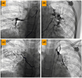
Our overall findings lead us to chronic thromboembolic pulmonary hypertension as the first differential diagnosis. For the confirmation of elevated pulmonary artery pressure, the patient underwent right heart catheterisation and pulmonary angiography. A mean pulmonary artery pressure of 65 mmHg was measured, and multiple narrowing in bilateral lobar and segmental pulmonary arterial subdivisions with multiple collaterals from the aorta and its branches were depicted (Fig 5). Chronic thromboembolic pulmonary hypertension was confirmed as the final diagnosis, and a full workup of underlying diseases was carried out. Appropriate treatment, including anticoagulation therapy, was started.
( a and b ) Pulmonary and ( c and d ) aorta angiography. Multiple narrowing in bilateral lobar and segmental pulmonary arterial subdivisions are depicted in ( a and b ). Collaterals from the aorta and its branches are demonstrated in ( c and d ).

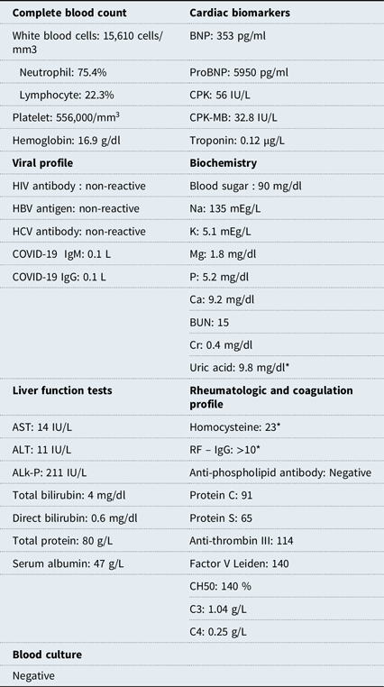
Detailed and concentrated history taking and physical examinations revealed previous suspicious aphthous oral ulcers and oligoarthralgia in the knees and ankles in the preceding 6 months. A comprehensive rheumatologic workup showed elevated homocysteine levels as well as positive HLA-B5 and HLA-B52 (Table 1). Considering the findings including aneurysmal changes in the pulmonary arterial branches, in addition to other clinical and laboratory data, we diagnosed Behçet’s disease as an underlying disease. Treatment with oral prednisolone (5 mg daily), warfarin (2.5 mg daily), and intravenous pulses of cyclophosphamide weekly (total dose of 500 mg/m2 monthly) was commenced. Moreover, the patient was considered a candidate for later pulmonary endarterectomy.
Laboratory findings.

ALK-P = Alkaline phosphatase; ALT = Alanine transaminase; AST = Aspartate aminotransferase; BNP = B-type natriuretic peptide; BUN = blood urea nitrogen; CH50 = C4 hemolytic complement 50; COVID-19 = coronavirus 2019; CPK = creatine phosphokinase; CPK-MB = creatine phosphokinase–myocardial band; HBV = hepatitis B virus; HCV = hepatitis C virus; HIV = human immunodeficiency virus; IgG = immunoglobin G; IgM = immunoglobin M; RF = rheumatoid factor.
* High value.
Discussion
In the present report, we explained an extremely rare case of pediatric chronic thromboembolic pulmonary hypertension with underlying Behçet’s disease. Chronic thromboembolic pulmonary hypertension is the most feared complication of acute pulmonary embolism in which unresolved thrombosis in the pulmonary bed results in chronic inflammation and fibrosis and the subsequent remodeling and obstruction of the pulmonary vasculature. This situation causes a cascade of distal arteriopathy, leading to pulmonary hypertension and the resultant right ventricle failure with a poor prognosis. The incidence of chronic thromboembolic pulmonary hypertension after acute pulmonary embolism is reportedly 0.5–3.8%. Reference Madani, Wittine and Auger1
There is a dearth of data on chronic thromboembolic pulmonary hypertension in children and adolescents owing to the low rates of acute pulmonary embolism in this age group (0.9 in 100,000 children per year); furthermore, chronic thromboembolic pulmonary hypertension happens mostly in patients with underlying hypercoagulable states. Reference Johnson, Driscoll and McGregor2,Reference Verbelen, Cools and Fejzic3
Chronic thromboembolic pulmonary hypertension is underdiagnosed due to its non-specific signs and symptoms. However, it can be an important diagnosis and should be considered in children with hypercoagulable states. It may be a serious situation with significant morbidity and mortality. Chronic thromboembolic pulmonary hypertension is diagnosed with a mean pulmonary artery pressure exceeding 25 mmHg at rest in right heart catheterisation and typical changes in pulmonary CT angiography or ventilation–perfusion mismatch of more than one segment in pulmonary ventilation–perfusion scans. Reference Fallah, Ghodsi and Soleimani4 A timely diagnosis requires a high suspicion of the situation and a full expert workup.
Our case is remarkable from two points of view. First, both chronic thromboembolic pulmonary hypertension and Behçet’s disease are rare conditions in the children. Second, in the outbreak of coronavirus disease 2019, most of the respiratory symptoms are inevitably related to coronavirus disease 2019 pneumonia, causing a diagnostic delay if not a misdiagnosis of the more infrequent, systemic, and complicated diseases like chronic thromboembolic pulmonary hypertension.
Kumbasar et al reported three cases of chronic thromboembolic pulmonary hypertension and suggested that physicians should take note of pulmonary embolism in children who present with dyspnea, chest pain, or syncope. Reference Kumbasar, Aypar, Karagoz, Demircin and Dogan5 Madani et al presented a series of 17 children under 18 years old with chronic thromboembolic pulmonary hypertension, together with their predisposing factors and surgical outcomes. Fifteen patients (88%) had one or more than one risk factor for hypercoagulable states. The lupus anticoagulant was the most common predisposing serum marker (n = 5), followed by the anticardiolipin antibody (n = 4) and protein C deficiency (n = 3). Two patients were positive for both the lupus anticoagulant and the anticardiolipin antibody. They concluded that chronic thromboembolic pulmonary hypertension was a rare condition in children with excellent surgical outcomes and suggested a prompt search for underlying diseases and referral for surgery in pediatric chronic thromboembolic pulmonary hypertension Reference Madani, Wittine and Auger1 . Ma et al described a 13-year-old boy who presented with chronic thromboembolic pulmonary hypertension as the first manifestation of the otherwise-asymptomatic nephrotic syndrome as the predisposing factor. Reference Ma, Zhou and Hua6 These recent reports call for due caution vis-à-vis the incidence of pulmonary embolism and its complications in the children.
Behçet’s disease is a systemic inflammatory disease that often presents with recurrent oral or genital aphthous ulcers, skin lesions, uveitis, arthropathy, and vasculitis. It is more common in the ancient Silk Road, most common in Turkey. Behçet’s disease is associated with an increased risk of endothelial dysfunction, resulting in thrombosis and fibrosis in the arterial and venous vasculature.
In a review of literature by La Regina et al, hyperhomocysteinemia was mentioned to be associated with an increased risk of thrombosis in Behçet’s disease. Reference La Regina, Orlandini, Prisco and Dentali7
Our patient’s first presentation with dyspnea and non-specific symptoms led us to the final diagnosis of chronic thromboembolic pulmonary hypertension. Hyperhomocysteinemia prompted us to perform a more thorough workup for underlying diseases. Furthermore, the history of aphthous ulcers and arthralgia, positive HLA-B5 and HLA-B52, and the presence of pulmonary arterial aneurysmal changes guided us towards the diagnosis and appropriate treatment of Behçet’s disease.
Wang et al reported a 16-year-old boy, a known case of Behçet’s disease, with the complaint of hemoptysis, pulmonary artery aneurysm, established vasculitis, and chronic thromboembolic pulmonary hypertension. The authors introduced him as the first pediatric case of chronic thromboembolic pulmonary hypertension due to Behçet’s disease. To our knowledge, we report the second pediatric case of concomitant chronic thromboembolic pulmonary hypertension and Behçet’s disease. Reference Wang, Rosenzweig and Takeda8
Conclusion
We conclude that a comprehensive examination is mandatory in every child with respiratory symptoms, even during the coronavirus disease 2019 pandemic. Moreover, less frequent conditions, including pulmonary embolism and its complications, should be borne in mind to perform a thorough diagnostic workup for the probable underlying disorder.
Acknowledgements
None.
Financial support
The authors declare that they have no funding support for this case report.
Conflicts of interest
None.
Ethical standards
This article does not contain any studies with human participants or animals performed by any of the authors.