Patients with deficient atrioventricular septation and common atrioventricular junction typically have the common junction shared between the ventricles, although the junction can be guarded by a common valvar orifice, or separate orifices for the two ventricles. Occasionally, nonetheless, such a common atrioventricular junction may be exclusively connected to one or another ventricle. When the junction is exclusively connected to the left ventricle, the right atrium can be blind-ending, as with typical tricuspid atresia, but yet communicating with the left atrium through an ostium primum defect.Reference Van Praagh, Vangi and Sul1 Such cases raise interesting questions concerning the morphology of the patent left atrioventricular valve. We describe two infants having this combination in the setting of a trifoliate left-sided atrioventricular valvar orifice, comparing the situation with the findings reported in mice with trisomy 16Reference Webb, Brown and Anderson2 and a third case with deficient atrioventricular septation and imperforate Ebstein's anomaly.
Case reports
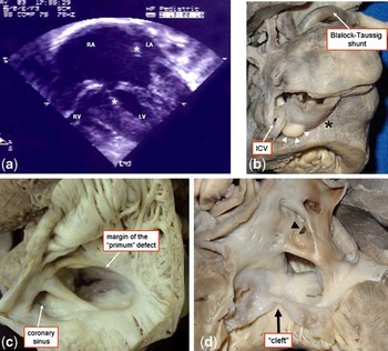
Case I: A male infant aged 2 months was admitted with a history of cyanosis since birth. The echocardiographic diagnosis was a double inlet left ventricle, concordant ventriculo-arterial connections, and a restrictive ventricular septal defect (Fig 1a). The gradient between the ventricles was measured at 76 millimetres of mercury. The infant was referred for construction of a Blalock–Taussig shunt, which produced an immediate improvement in the saturations of oxygen, but he died on the first post-operative day having developed bradycardia followed by cardiorespiratory arrest.
The four-chamber view of our first case (a) shows malalignment between the atrial and ventricular septa (asterisks), with an effective double inlet left ventricle. LA = left atrium; RA = right atrium; RV = right ventricle; LV = left ventricle. The right and inferior view of the heart (b) shows the inferior caval vein (ICV) and the adjacent rhabdomyoma (arrow heads), the dilated right atrium, and the origin of the displaced posterior descending coronary artery (*). Opening the right atrium (c) shows the dilated orifice of the coronary sinus (CS) and mostly the muscular floor. The opened left atrium (d) shows the “primum” defect and patency of the oval foramen (arrow heads). The inlet of the left ventricle shows the “cleft” of the left-sided atrioventricular valve (arrow).

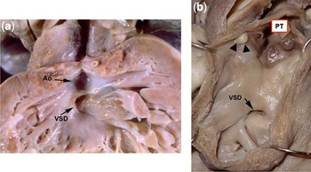
Autopsy examination revealed the usual atrial arrangement and relatively normal venous connections, albeit with a persistent left superior caval vein draining to the right atrium through a dilated coronary sinus. There was an additional separate connection of one hepatic vein to the right atrium. A rhabdomyoma, 0.4 centimetre in diameter, was also seen in the right atrium, protruding on its epicardial surface lateral to the orifice of the inferior caval vein. When examining the inferior aspect of the heart, the inferior interventricular artery was noted to be displaced laterally, marking the boundary of an incomplete right ventricle (Fig 1b). The atrioventricular junction was common, but due to the marked malalignment between the atrial and ventricular septa, the right atrium was connected mainly to the left ventricle. Its floor was mostly muscular, a small anterior area floored by a valvar membrane separating the atrial cavity from the cavity of the left ventricle. (Fig 1c) The atrial septum itself was defective, having a large ostium primum defect in addition to a patent oval foramen. The left-sided atrioventricular valve was made up of three leaflets, with one of its commissures pointing to the top of the inlet component of the muscular ventricular septum (Fig 1d). The inferior leaflet was firmly adhered to the ventricular septum, whereas the superior one was attached to the septal crest at the level of the septal commissure, but floated anteriorly, straddling a restrictive ventricular septal defect, and opening in part in an incomplete right ventricle. The aorta, which arose from the left ventricle, was unwedged relative to the atrioventricular junction. The pulmonary trunk arose from the incomplete right ventricle (Fig 2b). We identified two additional small rhabdomyomas, one bulging in the sub-pulmonary region (Fig 2b), and the other on the epicardial surface of the left ventricular free wall. The arterial duct was patent but restrictive. A patent modified Blalock–Taussig was present between the right subclavian and right pulmonary arteries.
The outflow tract of the left ventricle (a) shows the anterior aorta (Ao) and restrictive ventricular septal defect (VSD). The incomplete right ventricle (b) gives rise to the pulmonary trunk (PT). There is a small rhabdomyoma (arrow heads) on the endocardial surface, in a sub-pulmonary position.

Case II: An antenatal diagnosis of complex congenital cardiac disease had been made in a foetus conceived via an in vitro fertilisation. Delivery was by caesarean section under spinal anaesthesia because of failure to progress and meconium staining of the liquor. The infant was satisfactory at birth, weighing nearly 4 kilograms, and registering Apgar scores of 9 at 1 minute and 5 minutes.
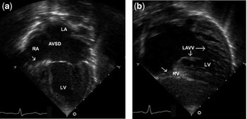
On examination, the infant was found to be well, with normal pulses, but a soft precordial systolic murmur. The electrocardiogram showed sinus rhythm, a frontal axis of +80°, probable biventricular hypertrophy, and widespread inversion of the ST segments and T-waves over the precordial leads. The initial chest X-ray showed a heart of normal size, and with normal pulmonary vasculature. Transthoracic cross-sectional echocardiography confirmed the prenatal diagnosis. An atrioventricular septal defect was observed, with a large primum component (Fig 3a and b), and the atrial septum was malaligned relative to the ventricular septum, being deviated to the left. The right-sided component of the common atrioventricular valve was imperforate, while the left-sided component was trifoliate, a zone of apposition being noted between its bridging leaflets, but with only mild incompetence. The pulmonary trunk arose from the dominant left ventricle and was unobstructed. The aorta arose from the small and incomplete right ventricle. It narrowed significantly when traced to the transverse and distal arches, but the descending aorta was of normal size, being fed through an arterial duct of moderate size, which permitted bidirectional shunting. A left superior caval vein drained to the right atrium through the coronary sinus.
The apical four-chamber view in our second case (a) shows “normal” left-sided component of the common AV valve, with the arrow pointing to the imperforate right-sided component of the common AV valve. LA = left atrium; RA = right atrium; RV = right ventricle; LV = left ventricle; AVSD = atrioventricular septal defect; LAVV = left-sided component of common AV valve. The subcostal view (b) confirms the findings.

The infant was transferred to a tertiary surgical centre on the second day of life, having been maintained on an infusion of Prostaglandin. On her third day of life, the proximal pulmonary trunk was connected to the aorta and the aortic arch repaired. The atrial septum was resected and a modified right-sided Blalock–Taussig shunt constructed. After the initial surgery to the aorta, a significant gradient persisted, and therefore the connections were reconstructed a second time. Post-operatively, the infant was troubled by excessive pulmonary flow, and required a prolonged period of continuous positive airway pressure, also requiring ongoing diuretics and an inhibitor of angiotensin converting enzyme. She developed paralysis of the left vocal cord, and initially was fed via gavage. She eventually settled, and was able to be discharged home, gaining weight satisfactorily. On review, increasing cardiomegaly led to the discovery of increasing atrioventricular valvar regurgitation. Further surgery, performed in two stages, consisted of initial repair of the incompetent atrioventricular valve and further revision of the aortic arch. After a further 2 months, she underwent a successful bidirectional cavopulmonary connection.
Case III: A 3-year-old female patient previously palliated by construction of a modified Blalock–Taussig shunt in the neonatal period was referred to hospital with fever and bronchospasm. Despite therapy with antibiotics, she developed septicaemia and died three days after admission. A previous echocardiographic study had revealed isomerism of the right atrial appendages with mixed biventricular atrioventricular connections, right-handed ventricular topology, atrioventricular septal defect with Ebstein's anomaly of the right-sided valvar component, and double outlet right ventricle.
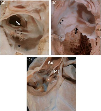
At necropsy, we confirmed the diagnosis of right isomerism of the atrial appendages, with the right pulmonary veins connected to the right-sided atrium. The atrial septum was deficient, showing a huge primum defect (Fig 4a). There was an atrioventricular septal defect with separate valves and complete plastering of the right-sided component of the inferior bridging leaflet against the right ventricular inlet. The right-sided component of the superior bridging leaflet lacked tendinous cords, and was fused with the inferior bridging leaflet to produce an imperforate Ebstein's malformation (Fig 4b). The left atrioventricular valve was trifoliate, albeit with dual orifices (Fig 4b). A small interventricular communication was found below the superior bridging leaflet. Both arterial trunks emerged exclusively from the right ventricle (Fig 4c), the aortic valve being in fibrous continuity with the superior bridging leaflet, and the pulmonary valve supported by a muscular infundibulum. The atrial and ventricular septa were aligned.
The right-sided cardiac chambers in our third case (a) show a huge atrial primum defect and an imperforate Ebstein's malformation of the right-sided component of the atrioventricular septal defect (asterisk). There is complete fusion of the bridging leaflets on the crest of the ventricular septum (open rectangle). The left atrioventricular valve is trifoliate, with the zone of apposition between the left ventricular components of the bridging leaflets pointing towards the outlet (“cleft”- white arrow). Further examination of the left-sided cardiac chambers (b) reveals that the left atrioventricular valve has two orifices. The arrow shows the zone of apposition between the superior and inferior bridging leaflets, whereas the arrowheads show the opening of the second orifice, located between the superior bridging leaflet and the mural leaflet. Opening the right ventricle (c) shows a double outlet with a right-sided aorta (Ao). The pulmonary trunk (PT) is supported by a complete muscular infundibulum. The asterisk shows the outlet septum.

Discussion
The morphology of atrioventricular septal defects with a common atrioventricular junction has been extensively studied in humans. The phenotypic feature is now recognised as the commonality of the atrioventricular junction, with variation depending on the presence of a common or separate valvar orifices, variation in the level of shunting across the atrioventricular septal defect, and the balance between the atrial and ventricular chambers. The presence of malalignment between the atrial and ventricular septa can also provide the potential for a spectrum from biventricular to univentricular atrioventricular connections.
In infants born with Down's syndrome, the most frequent presentation is that of a common valve with interatrial and interventricular communications and balanced ventricles.
Owing to the homology between the trisomic chromosomes, the mouse with trisomy 16 has long been recognised as a potential genetic model for humans with trisomy 21. The cardiac malformations typically seen in the mice, however, are markedly different from this typical morphology seen in man. A frequent arrangement in the mice, nonetheless, with a common atrioventricular junction exclusively connected to the left ventricle, is comparable to the anatomy observed in two of our patients.
This anatomy, although unexpected in the global sense, was observed in a relatively large series of 13 human cases reported by Van Praagh et al.Reference Van Praagh, Vangi and Sul1 In nine of their cases, the arrangement of the atrioventricular junction was as described in our first two patients, and in the mouse hearts. Of the remaining cases, three had an imperforate Ebstein's malformation of the right component of the common atrioventricular valve, as in our third case. The final case showed severe stenosis of the right atrioventricular valvar orifice. At least one of their cases could be seen to exhibit malalignment of the atrial septum, thus resulting in a double inlet left ventricle. Other series of hearts exhibiting tricuspid atresia are reported to show coexistence of “ostium primum” atrial septal defects. In one such series,Reference Anderson, Wilkinson, Gerlis, Smith and Becker3 all six cases were reported to show a “cleft” in the left atrioventricular valve. Nonetheless, in one of the cases reported by Williams et al,Reference Williams, Tandon and Edwards4 and in one from the series reported by Van Praagh and colleagues,Reference Van Praagh, Vangi and Sul1 the mitral valve was described as being “normal”. An examination of the photographic documentation in the article by Anderson et al Reference Anderson, Wilkinson, Gerlis, Smith and Becker3 demonstrates in at least one case the presence of a double inlet left ventricle, as in our first case.
It is the use of “tricuspid atresia” in the description of these anomalies that creates potential problems. The common atrioventricular junction lacks a definitive mitral component, although the right half of the common valve is more akin to the normal tricuspid valve. As is shown in one of our cases, and in those reported by Van Praagh et al,Reference Van Praagh, Vangi and Sul1 nonetheless, the right side of the common valve can exhibit Ebstein's malformation. The haemodynamic effect of the imperforate valve, furthermore, produces the clinical picture of tricuspid atresia. It is the term “absence of the right atrioventricular connection” that is inapplicable. The atrioventricular connection is certainly univentricular, but with a double inlet via a patent left valve and an imperforate right valvar component.