Introduction
There has been considerable interest to systematically tune the electronic properties of nanocrystals, quantum dots, and semiconductors in general.[ Reference Brown, Kim, Lunt, Zhao, Bawendi, Grossman and Bulović 1 − Reference Zhang, Kong, Xu, Su, Gao and Cheng 8 ] Recently, Brown et al. analyzed how the energy levels of the valence and conduction bands of lead sulfide nanocrystals can be tune using different ligands.[ Reference Brown, Kim, Lunt, Zhao, Bawendi, Grossman and Bulović 1 ] Ligand exchange methods have demonstrated that depending on the identity of the ligand, it can become either an electronic trap site or affect the electronic carrier inside semiconductor core.[ Reference Brown, Kim, Lunt, Zhao, Bawendi, Grossman and Bulović 1 , Reference Voznyy, Zhitomirsky, Stadler, Ning, Hoogland and Sargent 9 ] This is very important in the moment to design and systematically construct devices that include from solar energy harvesting applications to amperometric sensors that includes the inclusion of functionalized semiconductors. However, we wanted to analyze how the crystal structure is affected due to functionalization. In this study, we use a functionalized zinc oxide material to address this fundamental understanding of how the crystal structure of the material is affected upon functionalization.
Zinc oxide (ZnO) materials have attracted attention due to their facile synthesis and their use in different applications such as medicine, sensors, and solar energy harvesting applications, among others.[ Reference Öztürk, Tasaltin, Kilinç and Öztürk 10 ] The semiconductor has a band gap of 3.37 eV, which makes it an attractive material for the construction of solar cells, pH sensors, biosensors, etc.[ Reference Fulati, Usman Ali, Riaz, Amin, Nur and Willander 5 , Reference Dong, Licheng, Weilin, Guangzhong, Zhiping, Yingsan, Jie and Chuanxi 11 – Reference Petrella, Cosma, Lucia Curri, Rochira and Agostiano 13 ] Therefore, since inclusion of ligands do have a profound effect in the electronic properties of the semiconductor, we present using x-ray techniques to demonstrate how the surface of the crystal can be affected. We chose 3-mercaptopropionic acid (3-MPA) as our ligand of interest because: (1) is a relatively short ligand that either can bind through the carboxylate or the thiol group; and (2) similar and larger ligands have been used for further functionalization for amperometric biosensors.[ Reference Lokesh, Balakrishnan, Jeganathan, Layek, Verma and Gopalakrishnan 14 , Reference Roberts, Ford and Sparks 15 ]
Results and discussion
Scanning electron microscopy (SEM)
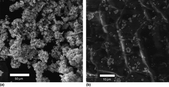
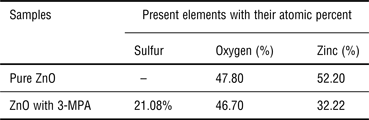
We prepared two types of samples, pure ZnO and ZnO functionalized with 3-MPA. Upon functionalization, we observed that the surface of the crystal was dramatically changed. Figure 1 shows the SEM micrographs of these samples of ZnO and ZnO with 3-MPA. Pure ZnO shows that the material is polycrystalline, whereas ZnO with 3-MPA has a film-like surface. Energy-dispersive spectroscopy (EDS) analyses were also performed with these materials. As can be observed in Table I, the amount of Zn atoms and oxygen atoms of pure ZnO and ZnO with 3-MPA are comparable, suggesting that these samples have a stoichiometric amount of Zn and oxygen atoms.
SEM micrographs of unmodified ZnO (a) and (b) ZnO-treated 3-MPA.

EDS quantitative results.

The EDS data show that practically the formation of the semiconductor remained unaffected, and the inclusion of the ligand was successful. However, the difference in morphology observed in Fig. 1, clearly states that the surface of the crystal dramatically changed. To further analyze the crystal structure of the semiconductor, we performed x-ray powder diffraction (XRPD). Fourier transform infrared spectroscopy data (see Supplementary Data) demonstrate that upon functionalization, the –SH vibration band about 3234 cm−1 is still present; however, the C = O vibration band about 1569/cm is drastically diminished due to functionalization on the Zn2+ active sites. Therefore, the 3-MPA is attached through the carboxylate group rather through the thiol group.
X-ray absorption and diffraction techniques
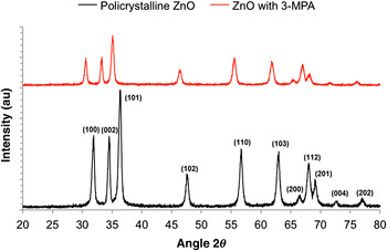
ZnO has a wurtzite crystal structure and the XRPD data are well documented. Figure 2 shows the characteristic x-ray powder diffraction data of pure ZnO and ZnO functionalized with 3-MPA. Upon functionalization of the material during the synthesis, the wurtzite structure is retained; however, all the diffraction peaks seem to shift to lower angles. Previous reports suggest that the ligands are located in the defects of the crystal.[ Reference Niepelt, Schröder, Sommerfeld, Slowik, Rudolph, Möller, Seise, Csaki, Fritzsche and Ronning 7 , Reference Öztürk, Tasaltin, Kilinç and Öztürk 10 , Reference Roberts, Ford and Sparks 15 ] Therefore, during functionalization strains in the crystal structure might have present the shift in the diffraction peaks as observed in the XRPD.
XRPD diffractogram of ZnO and of ZnO functionalized with MPA. The diffraction peaks correspond to the wurtzite structure of zincite database (PDF #36–1451).

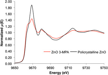
To obtain more information of the shifts to lower angles of the diffraction peaks due to the surface functionalization of ZnO, we performed x-ray absorption near-edge structure (XANES) and extended x-ray absorption fine structure (EXAFS) analysis of the materials. We performed Zn K-edge XANES and EXAFS using the Cornell high-energy synchrotron source (CHESS) to have an insight how the crystal structure and the bond lengths of Zn–O are affected during the formation. XANES and EXAFS studies of ZnO materials are explained elsewhere.[ Reference Roberts, Ford and Sparks 15 – Reference Jeong, Yu, Kim, Yi, Choi and Han 17 ] Figure 3 shows the normalized absorption in the XANES region near the Zn K-edge from pure ZnO and of ZnO capped with 3-MPA. The XANES region, although gives a qualitative information data, shows how the chemical properties of the Zn atom and the semiconductor band gap are affected upon functionalization. Both the graphs in Fig. 3 show differences, which indicates that the number of coordination of the Zn atom is affected by the presence of the ligand.[ Reference Roberts, Ford and Sparks 15 ] The K-edge peak of Zn in the ZnO with 3-MPA sample is less intense suggesting that the Zn ion is reduced. This is not entirely surprising since the conjugated base of 3-MPA can behave as a reducing agent. ZnO, in the other hand the K-edge peak is intense demonstrating that Zn is oxidized with presence of oxygen.
XANES spectra from polycrystalline ZnO (black line) and ZnO capped with 3-MPA (red line) near the Zn K-edge.

To determine the local structural properties around zinc atom we have to look into the EXAFS region. EXAFS can describe the bonding lengths, bonding length disorders, and atomic species of neighboring atoms around a probe atom.[ Reference Roberts, Ford and Sparks 15 , Reference Jeong, Yu, Kim, Yi, Choi and Han 17 , Reference ES, HJ, Han, An, Yoo, Kim and Yi 18 ]
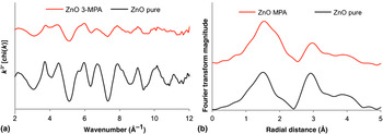
The k 3—weighted Zn EXAFS spectra (chi) and corresponding Fourier transforms magnitudes appear in Fig. 4. The R space (Fig. 4, right) of pure ZnO is similar from those that appear in the literature,[ Reference Roberts, Ford and Sparks 15 – Reference Jeong, Yu, Kim, Yi, Choi and Han 17 ] that corresponds to a wurtzite structure. This result also confirms the results obtained in Fig. 2, where the XRPD of both samples are very similar. In Fig. 4 (right spectra), the ZnO modified with 3-MPA spectrum shows that it has a wurtzite structure since it follows the same patterns as the spectrum of ZnO. The first peak (pure ZnO sample r = 1.4673 ± 0.02917 Å; ZnO with 3-MPA, r = 1.5747 ± 0.014884 Å) corresponds to the Zn–O distances. The second peak (pure ZnO sample r = 2.9532 ± 0.01432 Å; ZnO with 3-MPA, r = 2.9759 ± 0.044837 Å) corresponds to the Zn–O–Zn distances. However, as reported by Roberts et al.,[ Reference Roberts, Ford and Sparks 15 ] due to the complexation of Zn atoms at the crystal surface, an increase of bond lengths is observed. As can be observed in the k 3—weighted spectra (Fig. 4, left) of ZnO with 3-MPA shows the same pattern of pure ZnO, but the peaks are broadened.
(a) Zn-EXAFS chi-spectra weighted by k 3 and (b) corresponding Fourier transforms for ZnO and ZnO with 3-MPA. The results were generated using the ATHENA software.[ Reference Ravel and Newville 19 ]

Diffuse reflectance
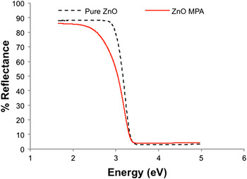
To determine whether the presence of the ligand affects the band gap of the semiconductor, we performed diffuse reflectance spectroscopy of the solids. Figure 5 shows the diffuse reflectance spectra of these materials. The band gap of pure ZnO appears to be on 3.45 eV similar to what appears in the literature.[ Reference Aneesh, Vanaja and Jayaraj 20 ] ZnO with 3-MPA shows a similar band gap at 3.42 eV as a result that crystal structure of the material remains unchanged. Therefore, if the band gap of the materials remained unchanged, the energy levels of the valence and conduction bands may have shifted during functionalization. To test this theory, we performed cyclic voltammetry of the materials.
Diffuse reflectance spectra of pure ZnO and of ZnO with 3-MPA.

Electrochemistry
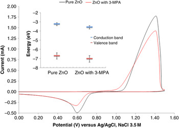
We performed electrochemical studies, using cyclic voltammetry of the materials demonstrated that although the band gap remains unaffected, the position of the conduction band and valence band might have shifted. Figure 6 shows the cyclic voltammogram of pure ZnO and of ZnO with 3-MPA. As can be observed from the cyclic voltammetry the onset of the oxidation process and the onset of the reduction reaction are shifted toward more reductive potentials.
Cyclic voltammogram of ZnO and of ZnO with 3-MPA-modified gold electrodes in a 0.1 M NaCl aqueous solution. Scan rate was 100 mV/s and an Ag/AgCl, NaCl 3.5 M reference electrode was used. (Inset) Energy levels of pure ZnO and of ZnO functionalized with 3-MPA as determined by cyclic voltammetry and diffuse reflectance spectroscopy. The blue line corresponds to the conduction band and the magenta line corresponds to the valence band. The error bars correspond to 5% of standard deviation of four samples.

The electrochemical reaction that occurred at the electrode surface corresponds to[ Reference Pourbaix 21 , Reference Zhang 22 ]:
 $${\rm Zn}{\rm O}_{{\rm surface}} + {\rm H}^ + _{{\rm solution}} \to {\rm ZnO} \cdot {\rm H}^{\rm +} \to {\rm Z}{\rm n}^{{\rm 2 +}} + {\rm O}{\rm H}^ -.$$
This electrochemical reaction is pH dependent and also depends on the adsorption of protons at the electrode surface. In our experiments, the pH of the solution was 7.0, using NaCl as the electrolyte. At the potentials that we worked in this study, the stability of ZnO-modified electrode is maintained up to 1.5 V versus Ag/AgCl reference electrode.
However, due to the presence of the ligand a shift to more reductive potentials is observed in the cyclic voltammogram of ZnO with 3-MPA. This shift could be an effect of different factors. First, the presence of the ligand affects the inner Helmhotz plane of ZnO. However, if the ligand is preventing charge transfer, it will require more energy to oxidize the electroactive species. This is not the case in Fig. 6. The presence of the ligand shifts the oxidative potential to more reductive potentials, suggesting that the electrode surface is passivated (or reduced). With these data, we calculated and determine the HOMO-LUMO levels (Highest Occupied Molecular Orbital–Lowest Unoccupied Molecular Orbital) of the material as a result of the shift of the onset potential. The separation between the first reduction and first oxidation of a material corresponds to the approximate HOMO–LUMO gap. Nevertheless, from the electrochemical data we can estimate the HOMO and LUMO energy levels by using the following empirical equation[ Reference Liu, Liu and Jen 23 ]:
 $$E_{1/2}^{{\rm HOMO/LUMO}} = [ - e(E_{{\rm onset(versus}\,\,{\rm reference}\,\,{\rm electrode)}})] - 4.8\,{\rm eV}.$$
As can be observed in Fig. 6, the energy levels have shifted to lower energy levels. De la Cueva et al. observed a similar response using Chloride ligands in cadmium selenide nanocrystals.[ Reference de la Cueva, Lauwaet, Otero, Gallego, Alonso and Juarez 24 ] The authors also observed that upon functionalization of the surface of the crystal there is a profound effect of the chemical composition of the electrode surface affecting the electrochemical response. More recently, Brown et al. observed similar behavior with different ligands using lead sulfide nanocrystals.[ Reference Brown, Kim, Lunt, Zhao, Bawendi, Grossman and Bulović 1 ] These results can help us to get an understanding of how to improve electrochemical response of these materials in the construction of sensors, photovoltaic devices, etc. These results are going to be presented in a near future, along with photoelectrochemical data.
Conclusions
In summary, we have studied the complexation of ZnO with 3-MPA. Upon functionalization the crystal structure is affected and the XANES data suggested that the crystal surface is passivated. XRPD, EXAFS, and diffuse reflectance data confirmed that the crystal structure and the electronic properties of the materials remained unaffected. However, the electrochemical response of modified gold electrodes with ZnO or with ZnO with 3-MPA the oxidative process is shifted to more reductive potentials. Future photoelectrochemical experiments are necessary to test these materials in operando conditions.
Experimental section
Current functionalization and synthesis of ZnO crystals and nanocrystals are mostly based in hydrothermal growth,[ Reference Aneesh, Vanaja and Jayaraj 20 ] which is a Lewis acid–base reaction of zinc nitrate (or zinc acetate) with sodium hydroxide (NaOH) at 55 °C. However, most of the functionalization in the literature is a separate step, just after the nanoparticle or nanowire is formed.[ Reference Niepelt, Schröder, Sommerfeld, Slowik, Rudolph, Möller, Seise, Csaki, Fritzsche and Ronning 7 , Reference Zhang, Kong, Xu, Su, Gao and Cheng 8 , Reference Lokesh, Balakrishnan, Jeganathan, Layek, Verma and Gopalakrishnan 14 , Reference Liu, Wu, Qiu, Yang, Xiao, Wang, Ding and Wang 25 ] In this paper, we present a one-step synthesis of functionalized ZnO with 3-MPA.
All materials were obtained from Sigma Aldrich. The zinc oxide particles were synthesized by the hydrothermal method by titration of NaOH, 0.9 M with zinc nitrate, [Zn(NO3)2], 0.45 M as precursor. The temperature was maintained constant at 55 °C. The solution of NaOH was maintained at constant stirring. With addition of a minimum amount of Zn(NO3)2 in the NaOH solution zinc oxide begins to precipitate. After the solution was titrated with Zn(NO3)2, we sealed the flask with paraffin and let stand for 2 h at room temperature. The white precipitate was vacuum filtered and washed with nanopure water and last with ethanol. Finally, it was then dried under vacuum. To functionalize ZnO with 3-MPA we performed the same reaction synthesis of pure ZnO. However, 500 µL of 3-MPA were added to the NaOH solution. The rest of the synthesis was done as explained before.
SEM micrographs and EDS were taken using a fully Integrated Tescan/Oxford variable pressure SEM system with an EDS Microanalysis System with X-Max 20 large area Analytical Silicon Drift Detector (127 eV, 20 mm2 active area, Peltiercooled). X-ray powder diffraction was done using a Benchtop Miniflex from Rigaku and the data were analyzed using the software Jade. XANES and EXAFS were done using the F3 beamline at the CHESS measuring the K-edge of the Zn atom. The data were analyzed using the Athena software, which is part of the Demeter software.[ Reference Ravel and Newville 19 ]
Supplementary material
The supplementary material for this article can be found at http://dx.doi.org/10.1557/mrc.2016.14.
Acknowledgments
The authors want to thank Dr. Victor M. Rodríguez from Universidad Metropolitana, Kenneth David Finkelstein, and Matthew James Ward from CHESS and Dr. Carlos R. Cabrera from the University of Puerto Rico for constructive discussion during the preparation of this manuscript. Financial support was possible due to PR REU: Research training in cross-disciplinary chemical sciences. NSF CHE 1262826.