Introduction
Microbial mats comprise an assemblage of microorganisms involving a functionally integrated system (Paerl et al. Reference Paerl, Pinckney and Steppe2000). They are amongst the most productive and successful of known microbial communities, and constitute the largest non-marine biomass present in polar regions (Quesada et al. Reference Quesada, Fernández-Valiente, Hawes and Howard-Williams2008). The production of these benthic communities might represent a significant input of allochthonous carbon for the unproductive pelagic food webs of nearby lakes (Reference Rochera, Toro, Rico, Fernández-Valiente, Villaescusa, Picazo, Quesada and CamachoRochera et al. this issue). For this very reason, they must be considered crucial to high latitude ecology. As described for cyanobacterial mats from the McMurdo Ice Shelf (De los Ríos et al. Reference De Los Ríos, Ascaso, Wierzchos, Fernández-Valiente and Quesada2004), a regular feature in these microbial communities is the occurrence of differently coloured layers in which different species are distributed. Steep light gradients occur inside the mat due to the strong absorption of the incident radiation (Quesada & Vincent Reference Quesada and Vincent1997, Quesada et al. Reference Quesada, Vincent and Lean1999). The pigment composition, which covers a wide spectrum from chlorophylls to carotenoids, can vary notably along this vertical gradient of light.
Microbial mats are conspicuous on Byers Peninsula (Toro et al. Reference Toro, Camacho, Rochera, Rico, Bañón, Fernández-Valiente, Marco, Justel, Avendaño, Ariosa, Vincent and Quesada2007, Fernández-Valiente et al. Reference Fernández-Valiente, Camacho, Rochera, Rico, Vincent and Quesada2007, Velázquez et al. Reference Velázquez, Rochera, Camacho and Quesada2011). They are widespread in the region and cover extensive surfaces up to several hundred square metres in some locations. These mats can vary in thickness, pigmentation and degree of vertical stratification (Fernandez-Valiente et al. 2007). The more laminated mats on Byers Peninsula are located over moist soils and at the bottom of ponds. They are dominated by cyanobacteria throughout their vertical profiles, yet they show evident and distinct layers.
Extreme habitats are characterized by the conjunction of several environmental stressors. For instance, Antarctic microbiota experience a high exposure to UV radiation, which can be partially offset by the synthesis of screening pigments (Quesada & Vincent Reference Quesada and Vincent1997). More particularly, some environmental factors such as water or nutrients availability may vary on Byers Peninsula depending on mat location (Fernandez-Valiente et al. 2007). The purpose of this paper is to report on the vertical structure of these microbial mats, paying attention to those traits that might represent a response or strategy for coping with all these stressors.
Material and methods
Study site and sampling
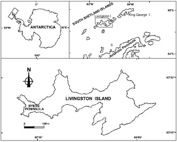
The study was conducted during January and February of 2003–04 on the central plateau of Byers Peninsula (Fig. 1; 62°34′35″–62°40′35″S, 60°54′14″–61°13′07″W), a 60.6 km2 ice-free area, located at the western end of Livingston Island (South Shetland Islands, Antarctica). Six mat samples were obtained from different locations within or near the catchment area of Lake Limnopolar. For all analytical determinations, samples were cored randomly using a metal core-taker with an internal diameter of 13 mm. Samples were then sectioned with a blade into the two distinctive layers that compose the mat and either examined fresh or stored frozen until analysis.
Map showing the location of Byers Peninsula.

Analytical determinations
Fresh mat samples were inspected by fluorescence microscopy within a few hours of sampling and the taxonomic affiliation of dominants phototrophs was determined based on morphology (Krammer & Lange-Bertalot Reference Krammer and Lange-Bertalot1986, Reference Krammer and Lange-Bertalot1988, Reference Krammer and Lange-Bertalot1991a, Reference Krammer and Lange-Bertalot1991b, Broady & Kibblewhite Reference Broady and Kibblewhite1991, Komarek & Anagnostidis Reference Komarek and Anagnostidis1999, Reference Komarek and Anagnostidis2005). On return to Spain, up to three months later, the vertical distribution in the mat of photosynthetic organisms was inferred from a high-performance liquid chromatography (HPLC) analysis of the chlorophyll and carotenoid content from frozen samples. Samples were extracted with pure acetone. Subsequently they were prefiltered through 0.2 μm nylon filters and injected into a Waters® C18 Spherisorb S5 ODS2 column connected in tandem with a NovaPack C18 column. The chromatographic runs were performed following the procedure described by Pinckney et al. (Reference Pinckney, Millie, Howe, Paerl and Hurley1996). Peak identities were determined by comparing retention times and spectra with those of pure standards purchased from DHI (Denmark) and quantified against the curves obtained with standards by integration of the area under the cross-section at 440 nm. The UV-screening scytonemin was quantified from chromatograms with an extinction coefficient of 112.6 l (g cm) at 384 nm as described by Garcia-Pichel et al. (Reference Garcia-Pichel, Sherry and Castenholz1992). Myxoxantophyll and fucoxantin were used as taxa-specific biomarkers to assess the relative abundances of cyanobacteria and diatoms respectively.
Dry weight of samples was determined after drying at 105°C for 6 h. To assess the stoichiometric composition of mats, total carbon and nitrogen contents were quantified using an elemental analyser (Perkin-Elmer 2400CHN) with a thermal conductivity detector in homogenized dry samples from each mat layer. The total phosphorus was determined from the same samples after a persulphatic-acid hydrolysis at 135°C for 2 h. Subsequently, orthophosphate was measured using the phosphomolybdic acid-ascorbic method after the pH-neutralization of samples. The exopolymeric substances (EPS) were extracted from each mat layer with 2% EDTA from known-weight fractions of previously lyophilized samples. The carbohydrate content of these extracts was measured using the phenol-sulphuric acid method (Herbert et al. Reference Herbert, Phipps and Strange1971) with glucose as standard. The protein amount in the EPS extracted was estimated according to Bradford (Reference Bradford1976) taking bovine serum albumin (BSA) as standard. ANOVA tests were used to compare differences between mat strata. To observe the predominant patterns in the data, a principal component analysis (PCA) was conducted with the descriptive variables obtained. The correlation matrix between variables obtained in the PCA was used to explore the relationships between them. The SPSS 15.0 software was used for all statistical procedures.
Results
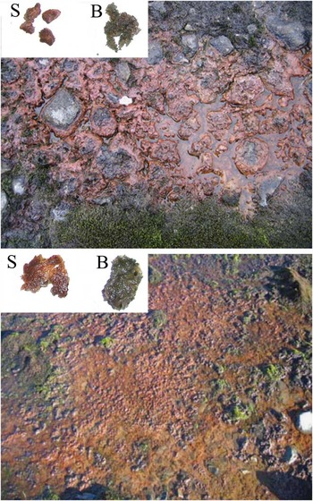
Microbial mats were observed frequently over moist soils and shallow, ephemeral ponds that were inundated for weeks or months. Surface colour of mats varied from purple to orange-brown and had a smooth surface and a cohesive texture (Fig. 2). They also displayed, within total thickness of about 3–5 mm, a deep green basal layer in which microorganisms alternated with fine-grained sand and mineral deposits. Mats harboured a variety of recognizable cyanobacterial morphotypes and both non-heterocystous and heterocystous taxa were observed, the former being dominant in biomass. Filamentous species of cyanobacteria, such as Oscillatoriales of the genera Phormidium, Leptolyngbya and Porphyrosiphon were usually the major constituents. Among heterocystous genera, a morphotype assigned to Nostoc was also observed, mainly in the mats occurring in the dryer sites. Other less abundant cyanobacterial morphotypes found in mats were assigned to the genera Gloeocapsa or Schizothrix. When examined using an epifluorescence microscope, trichomes of cyanobacteria, mainly those unsheathed taxa that occurred in the bottom layer, fluoresced red under green excitation, indicating the presence of phycoerythrin. Commonly occurring diatom genera were Navicula, Fragilaria, Stauroneis, Nitzschia, Gomphonema and Pinnularia, among other genera. They were observed in both layers but, damaged diatoms recognized as empty frustules, were mainly observed in the top layer.
Pictures showing common microbial mat types from Byers Peninsula. The pictures on the left show the surface (S) and basal (B) layers of each mat.

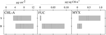
The chlorophyll a (chl a) concentration in the mats tended to be higher in deep rather than surface layers (Fig. 3), although differences were not statistically significant. Values ranged from 0.67–16.15 μg cm-2 in the surface layer and from 3.08–14.81 μg cm-2 in the deep layer. The HPLC analysis revealed the major presence of taxon-specific carotenoids of cyanobacteria (myxoxantophyll) and diatoms (fucoxanthin), although pigments of green algae were also detected in some chromatograms (i.e. chlorophyll b and lutein). Carotenoids occurred throughout the mat, but their relative amounts (μg (μg chl a)-1) and variation between mats differed greatly between layers (Fig. 3). Concentrations of fucoxanthin were high and variable in the lower layer, showing higher concentrations of 0.15 (μg (μgchl a)-1). However, this pigment was almost absent from the upper layer while myxoxanthophyll displayed the opposite pattern being low in the lower layer and high at the surface, where relative concentrations ranged up to one order of magnitude from 0.09–0.98 (μg (μg chl a)-1). On the other hand, the photoprotective pigment scytonemin, when present, occurred in the upper layers, and its concentration varied broadly from 9.4–301 μg cm-2. Scytonemin was mainly observed in mats with a higher occurrence of Nostoc sp. but not specifically. Also, amounts of red-scytonemin, a reduced derivative of scytonemin, were detected in some of the surface layers.
Box plots representing statistical values of the chlorophyll a (chl a) and the major taxa-specific carotenoids (FUC = fucoxanthin, MYX = myxoxantophyll) concentrations in the two layers of microbial mats. S = surface layer and B = bottom layer. The boundaries of the box indicate the 25th and 75th percentiles. Solid and dashed lines indicate median and mean respectively.

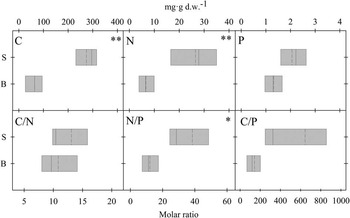
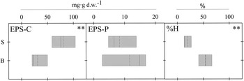
The stoichiometric composition of mats showed that, the carbon, nitrogen and phosphorus content per dry weight in the upper layer were greater than in the bottom layers of the mats, but differences were only statistically significant in the case of carbon and phosphorus (Fig. 4). Molar ratios of carbon with respect to nitrogen and particularly phosphorus were greater in the surface compared to the bottom layer although not significantly due to the high scattering of replicates. By contrast, N/P ratios were significantly higher in the upper stratum. At the surface, the C/N and N/P molar ratios ranged from 6.4–25.4 and from 3.9–94.9, respectively. Moisture in mats also varied between layers. The water content at the bottom was significantly higher, with values ranging 47–83%, whereas water content in the surface layer averaged 20% (Fig. 5). All the mats studied had EPS varying from 1–11% of the total dry weight. The distribution with depth of the carbohydrate and protein fractions of these EPS is shown in Fig. 5. The EPS-carbohydrate fraction was significantly higher in the surface layer, while the protein fraction tended to be more abundant in the lower layer. With all, the protein fraction was always considerably lower than the carbohydrate fraction.
Box plots representing statistical values of the elemental composition (C = carbon, N = nitrogen, and P = phosphorus) and molar ratios at layers S = surface, and B = bottom. The boundaries of the box indicate the 25th and 75th percentiles. Solid and dashed lines indicate median and mean respectively. Symbols * and ** indicate significant differences between the mat layers at P > 0.05 and P > 0.01 respectively.

Box plots representing statistical values of the exopolymeric substances (EPS) content (EPS-C = carbohydrates, and EPS-P = proteins) and water content (%H) at layers S = surface, and B = bottom of the mats. The boundaries of the box indicate the 25th and 75th percentiles. Solid and dashed lines indicate median and mean respectively. Symbols * and ** denote significant differences between the mat layers at P > 0.05 and P > 0.001 respectively.

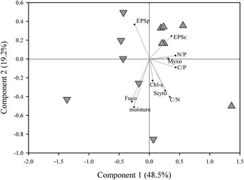
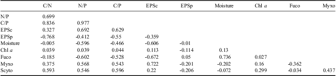
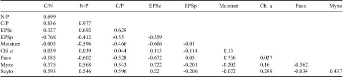
The PCA performed to describe the variance observed in data recognized both layers of the mats (Fig. 6). The first axis explained 48.5% of variance and was mainly related with these layers. Thus, variables with higher positive loadings on this axis were the molar ratios of carbon and nitrogen with the phosphorus, and the carbohydrate fraction of EPS. The relative content of myxoxantophyll also contributed notably to the positive end of this axis. On the other hand, the relative content of fucoxanthin, moisture and the protein fraction of EPS were at the negative end of this axis. The correlation matrix obtained in the analyses (Table I) showed higher positive correlations between C/P and the other molar ratios (N/P and C/N), the carbohydrate fraction of EPS and the relative content of myxoxantophyll, and the moisture and the fucoxanthin. On the other hand, the higher negative correlations were observed between the C/N ratio and the protein fraction of EPS, and between fucoxanthin and the carbohydrate fraction of EPS.
Principal component analysis performed with data obtained in the study of bi-layered microbial mats. Triangles top and down represent samples from surface and bottom mat layers respectively. The labels of variables are: N/P (nitrogen/phosphorus molar ratio), C/P (carbon/phosphorus molar ratio), C/N (carbon/nitrogen molar ratio), EPSc (carbohydrate fraction of the exopolymeric substances), EPSp (protein fraction of the exopolymeric substances), chl a (areal chlorophyll a concentration), fuco (fucoxanthin concentration relative to chl a), myxo (myxoxanthophyll concentration relative to chl a), scyto (scytonemin concentration relative to chl a), moisture (% of water content).

Correlation matrix of variables obtained in the principal component analysis (PCA). EPSc = exopolymeric substances - carbohydrate fraction, EPSp = exopolymeric substances - protein fraction, chl a = chlorophyll a, fuco = fucoxanthin, myxo = myxoxanthophyll, scyto = scytonemin.
Discussion
The microbial mats studied are distributed across the central plateau of Byers Peninsula, where water retention in land depressions has produced freshwater wetlands with small watersheds. These mats proliferate mainly in moist soils of depressed areas, in the bottom of shallow ponds and on the lakeshores. These microbial mats typically show a bi-layer structure which can be a habitat-linked trait. They have to cope with some environmental stresses such as low nutrients availability and periods of desiccation. In this sense, mats show a top layer comprised of an unstructured stratum, largely composed of empty or half-empty cyanobacterial sheaths and diatom frustules, and a lower layer assembled from more functional photosynthetic biomass. Also, the surface layers show a higher content of the sheath pigment scytonemin, higher EPS and some stoichiometric unbalance. These traits indicate that surface layers are particularly inactive and ones in which senescing biomass accumulates, but they are probably improving the functional capability of the whole community. The synthesis of scytonemin has been suggested to be induced by exposure to UV under conditions of restricted metabolism (Castenholz & Garcia-Pichel Reference Castenholz and Garcia-Pichel2000) such as low water and/or nutrient availability. Some of the surface layers analysed show red-scytonemin, a derivative of scytonemin, which has also been observed in other cold environments (Mueller et al. Reference Mueller, Vincent, Bonilla and Laurion2005, Bonilla et al. Reference Bonilla, Rautio and Vincent2009, Jungblut et al. Reference Jungblut, Lovejoy and Vincent2010). These authors propose that this is a result of a slow accumulation over time, which demonstrates the perennial nature of these slow-growing cyanobacterial mats. The accumulation of these non-functional pigments in the upper layers is probably exacerbated by their low degradation rates under cold environmental conditions (Vincent & Howard-Williams Reference Vincent and Howard-Williams1989). In this sense, a higher thickness of the upper layer relative to the bottom layer might be an indication of the age of the mat. Nevertheless, this cannot exceed certain limits because it would reduce by too much the light available to the deeper active photosynthetic community. Scytonemin did not occur in all the studied mats, indicating that this could be a species-dependent trait (Garcia-Pichel & Castenholz Reference Garcia-Pichel and Castenholz1991). Alternatively, these differences might also respond to differences in the environmental conditions because the synthesis of scytonemin can be enhanced under conditions of restricted metabolism, which probably exacerbate the damage produced by the UV radiation. In this sense, our multivariate analysis indicates some relationship between scytonemin concentrations and C/N molar ratios. This agrees with experimental observations showing Nostoc synthesizing higher scytonemin amounts while fixing nitrogen when the combined nitrogen (i.e. ammonium and nitrate) was scarce (Fleming & Castenholz Reference Fleming and Castenholz2008). Indeed, previous studies performed on Byers Peninsula microbial mats have already demonstrated the co-occurrence of high scytonemin amounts and the potential for diazotrophic metabolism (Fernández-Valiente et al. Reference Fernández-Valiente, Camacho, Rochera, Rico, Vincent and Quesada2007).
Carotenoid pigments are useful biomarkers in assessing the relative dominance of the photosynthetic taxa present in the microbial mats. In our case, they suggest a segregation of cyanobacteria and diatoms in the upper and bottom layers respectively. However, carotenoids may also vary depending on their functional role. In this sense, carotenoids as well as scytonemin can perform a photo-protective function (Quesada & Vincent Reference Quesada and Vincent1997). Nevertheless, the role of myxoxantophyll, which was higher in the upper layer of mats has been supposed to act as a first protection barrier against an excess of UV light intensity but also has a photosynthetic role stabilizing thylakoid membranes (Mohamed et al. Reference Mohamed, van de Meene, Roberson and Vermaas2005). Temperature may also regulate the concentration of carotenoids as demonstrated by Kleinteich et al. (Reference Kleinteich, Wood, Küpper, Camacho, Quesada, Frickey and Dietrich2012), indicating that low temperatures could trigger a higher content of these photoprotective pigments. Pigment amount may also vary because of the physiological state of the mat. In other respects, Quesada et al. (Reference Quesada, Vincent and Lean1999) demonstrated that the surface layers of Arctic microbial mats very efficiently absorbed the UV radiation and some photosynthetically relevant wavelengths were also absorbed in the mat profile. This partial absorption was considered by these authors as an ecological strategy of the community providing the appropriate radiation conditions for each component of the community. The vertical profiles of photosynthetic activity previously performed on these microbial mats have shown maximum oxygen evolution occurs at non-superficial layers (Fernández-Valiente et al. Reference Fernández-Valiente, Camacho, Rochera, Rico, Vincent and Quesada2007), which also suggested that the uppermost stratum was essentially non-active and had a mainly protective role. Other than the pigment composition, there are further features that might also regulate the light regime inside the mat. Thus, it has been suggested that the EPS matrix might indirectly explain the deepening of the photosynthetic maximum by increasing the penetration of the active radiation (Decho et al. Reference Decho, Kawaguchi, Allison, Louchard, Reid, Stephens, Voss, Wheatcroft and Taylor2003).
Our findings on the elemental composition of mats systematically show a higher content of nutrients per dry weight in the upper layer, which might be due to a higher mineral content in the lower layer. However, C/N and C/P molar ratios indicate the higher nutrient imbalance of the upper layer. This uppermost layer also has a higher EPS content and lower moisture. The total amount of EDTA-extractable EPS includes both colloidal and capsular fractions, the latter being constituents of the cyanobacterial sheaths. The EPS determine the cohesive properties of mats, but they also increase the capability to retain water because of the existence of large amounts of hydrogen bonding in their structure (Sutherland Reference Sutherland2001). However, EPS may also be related to the nutrient imbalance observed in the upper layers. Cell growth is limited when nutrients are scarce, but the photosynthetic inorganic carbon uptake continues. Accordingly, photosynthetic cells can maintain a relatively balanced nutrient composition by driving out secondary metabolites with high carbon content such as EPS, since neither N or P are incorporated in significant quantities into carbohydrates. Otero & Vincenzini (Reference Otero and Vincenzini2004) showed experimentally how EPS production in a cyanobacterial strain of Nostoc worked as a sink of carbon when the stoichiometry in the culture media was unbalanced. On the other hand, a decrease of EPS amounts with depth could also be due to their microbial utilization. In this sense, the constituent macromolecules of these EPS can be degraded to mono and dimeric unities by the bacterial exoenzymatic activity, and end products can be then reabsorbed and used again as an organic carbon source. Exudates produced by cyanobacteria can comprise both high molecular weight (> 1000Da) substances, probably aromatic compounds and proteins, and low molecular weight compounds (< 1000Da) such as simple sugars (Kirkwood et al. Reference Kirkwood, Nalewajko and Fulthorpe2006), which are all part of the EPS pool. The microbial mats that we have studied diverge from seasonal phototrophic biofilms that conspicuously occur in Byers during some periods (Velazquez et al. 2011), which demonstrates the occurrence of diverse ecological strategies in the same geographic location. Differently to our mats, the biofilms are dominated by green algae and rapidly grow on seasonal ice during short time periods at the beginning of summer. By contrast, the net balance between photosynthesis and respiration probably comes close to zero in the mats studied here during the summer climax, as observed for instance in microbial mats from the McMurdo Dry Valleys (Hawes & Howard-Williams Reference Hawes and Howard-Williams1998). Consequently, there should be no accretion of the active parts of these mat but they will be able to maintain important populations because of the paucity of grazers and the slow decomposition rates.
In summary, our approach combining different analyses on the structure of bi-layered microbial mat from Byers Peninsula leads us to think that environmental stressors, such as UV damage, dryness or nutrient scarcity, which are regular characteristics of these maritime Antarctic ecosystems, might potentially have implications for their vertical structure. This would demonstrate the existence of a specific ordered structure within the photic layer of mats which varies depending of the surrounding environmental conditions.
Acknowledgements
Fieldwork was supported by grant REN2000-0435-ANT from the Science and Technology Ministry (Spain) to AQ. Sample processing was supported by grant CGL2005-06549-C02-02/ANT from the Spanish Ministry of Education and Science to AC, which was co-financed by European FEDER funds. We are in debt to the UTM (Maritime Technology Unit, CSIC) and the Las Palmas crew (Spanish Navy) who provided us with the logistics to make this expedition possible. We also acknowledge the constructive comments of the reviewers.