Introduction
In recent years, nanotechnology is becoming the most active research area in the field of materials science as nanoparticles shows novel and enhanced properties based on their characteristics such as particle size, morphology, and distribution.[ Reference Saxena, Tripathi, Zafar and Singh 1 ] Among the different nanoparticles used in various fields, silver nanoparticles (AgNPs) have its own top place. AgNPs open up a path for fascinated and demandable research due to its diverse properties such as catalytic activity, good conductivity,[ Reference Hussain, Kumar, Hashmi and Khan 2 ] and its application in sensor,[ Reference Dar, Khare, Cole, Karnac and Srivastava 3 ] biofilms,[ Reference Percival, Bowler and Dolman 4 ] coating medical tools,[ Reference Wijnhoven, Peijnenburg, Herberts, Hagens, Oomen, Heugens, Roszek, Bisschops, Gosens, van de Meent, Dekkers, de Jong, van Zijverden, Sips and Geertsma 5 ] and antimicrobial activity.[ Reference Prabhu and Poulose 6 ] Thus, an increase in the usage of AgNPs led to the development of different methodologies for its synthesis. Currently, chemical, physical, and biological methods are employed for the synthesis of AgNPs.[ Reference Ahmad, Sharma, Alam, Singh, Shamsi, Mehta and Fatma 7 , Reference Xu and Hu 8 ] Even though the chemical and physical synthesis methods are good, they have certain drawbacks such as time consuming, difficulty in purification, expensive, and the release of hazardous byproducts.[ Reference Nagajyoti, Prasad, Sreekanth and Lee 9 ]
Hence, the only way to overcome these drawbacks could be the biosynthesis approach, in which different biological samples such as bacteria, algae, fungi, polysaccharide, enzymes, and plant extracts are being used for the synthesis.[ Reference Gopinath, Saha, John, Khanum, Ganesh and Patil 10 ] Modern research is working on the biosynthesis of AgNPs using different plant parts such as leaf,[ Reference Logeswari, Silambarasan and Abraham 11 ] fruits,[ Reference Gopinath, Mubarak Ali, Priyadarshini, Priyadharsshini, Thajuddin and Velusamy 12 ] and seed extracts[ Reference Bar, Bhui, Sahoo, Sarkar, Pyne and Misra 13 ] because they contain different phytochemicals which plays a key role in the reduction of silver nitrate (AgNO3) to AgNPs.[ Reference Logeswari, Silambarasan and Abraham 11 ] However, biosynthesis of AgNPs using flower extract is a novel approach as well as an emerging area of research as very less work is being carried out in this aspect. Hence, in this study we mainly focused on green synthesis of AgNPs using flower extract of Allamanda cathartica and investigated its antibacterial and antioxidant properties.
Experimental
Materials used and Biosynthesis of AgNPs
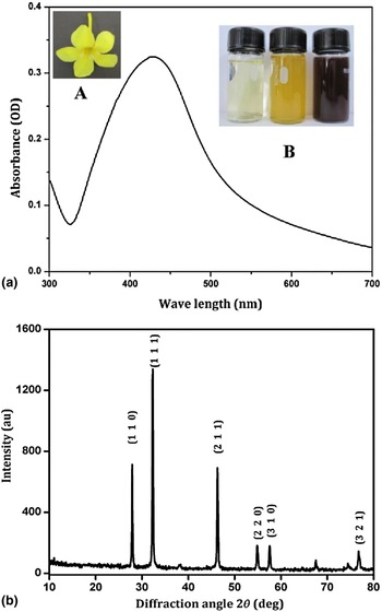
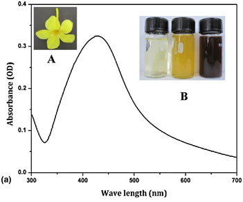
AgNO3 was procured from Merck, India, and it was used without any further purification. The flower of A. cathartica plant [Fig. 1(a.A)] was collected from the garden of K. S. Rangasamy College of Arts and Science, Tiruchengode, Tamil Nadu, India. Fresh flowers of A. cathartica were washed with sterile de-ionized water and air dried for few hours. Aqueous extract was prepared using 10 g of flowers with the help of a mortar and pestle. The extract was filtered repeatedly through Whatman no.1 filter paper using a separating funnel until a clear solution was obtained. Subsequently 1 mM concentration of AgNO3 was prepared using de-ionized distilled water. The clear yellow extract and AgNO3 solution were mixed in an equal ratio and incubated in dark condition for 24 h followed by centrifugation at 6000 rpm for 30 min. The settled particles in the centrifuge tubes were collected and dried in hot air oven at 100 °C for 2 h.
(a) UV–vis absorbance of AgNPs—(A) Flower of A. cathartica, (B) Color change due to AgNPs formation; (b) XRD pattern of AgNPs.

Characterization of synthesized AgNPs
The obtained powder was characterized using different techniques. The formation of AgNPs was confirmed by measuring the wave length of the reaction mixture in UV–visible (UV–vis) spectrum using spectrophotometer (LAMBDA 35, PerkinElmer, USA). The scanning range for the sample was between 200 and 800 nm at a speed of 480 nm/min. The nature and the crystalline phase of the reduced AgNPs were identified by X-ray powder diffraction (XRD) patterns (X'Pert PRO; PANalytical, the Netherlands) using CuK α as a radiation (l51.54060 Å) source. The diffractometer was scanned in the 2θ range from 10° to 80° at the scanning rate of 5°/min. The observed peak positions and the relative intensities of the powder pattern were identified in comparison with the reference powder diffraction data.
The presence of functional groups and chemical bonds on the surface of AgNPs was investigated using Fourier transform infrared (FTIR) spectrophotometer (Spectrum 100; PerkinElmer, USA). The spectrum was recorded between the ranges of 4000 and 400 cm−1 at a resolution of 4 cm−1. The presence of silver (Ag) was confirmed through energy-dispersive spectrum (EDS) (EDX-720; Shimadzu, Japan). The sample was focused directly at 10/5 mm on a thin film of Mylar. Particle size distribution (PSD) was used to determine the particle size using a particle size analyzer (Nanophox; Sympatec, Germany) using the well-known dynamic light-scattering technique. Transmission electron microscopy (TEM) was used to figure out the dimension and the form of AgNPs. The sample was analyzed using high-resolution TEM (TEMCM200; Philips, USA). For analysis, the well-dispersed samples were loaded on the copper grids and scanned at 120 kV. Particle size was then manually calculated by taking into account the maximum and the minimum particle size as marked in the observed image.
Antibacterial activity
Antibacterial activity of the synthesized AgNPs was determined against the selected clinical bacterial strains (collected from NCIM, India) such as Salmonella typhimurium (NCIM 2501), Staphylococcus aureus (NCIM 2127), Escherichia coli (NCIM 2065), and Klebsiella pneumoniae (NCIM 2883) using the Kirby–Bauer disc diffusion method[ Reference Bauer, Kirby, Sherris and Turck 14 ] in the Muller & Hinton Agar medium (Hi-Media Pvt. Ltd). Roughly 10 mL of sterile media was poured in Petri dishes. Once the medium got solidified, 100 µL of freshly prepared overnight inoculums was spread on to it using sterile swabs. Sterile filter discs were kept over the medium and the biosynthesized nanoparticles were loaded onto the disc under aseptic condition. The plates were subsequently incubated at 37 °C for 24–48 h after which the zone of inhibition (in mm diameter) was measured and tabulated.
Antioxidant activity
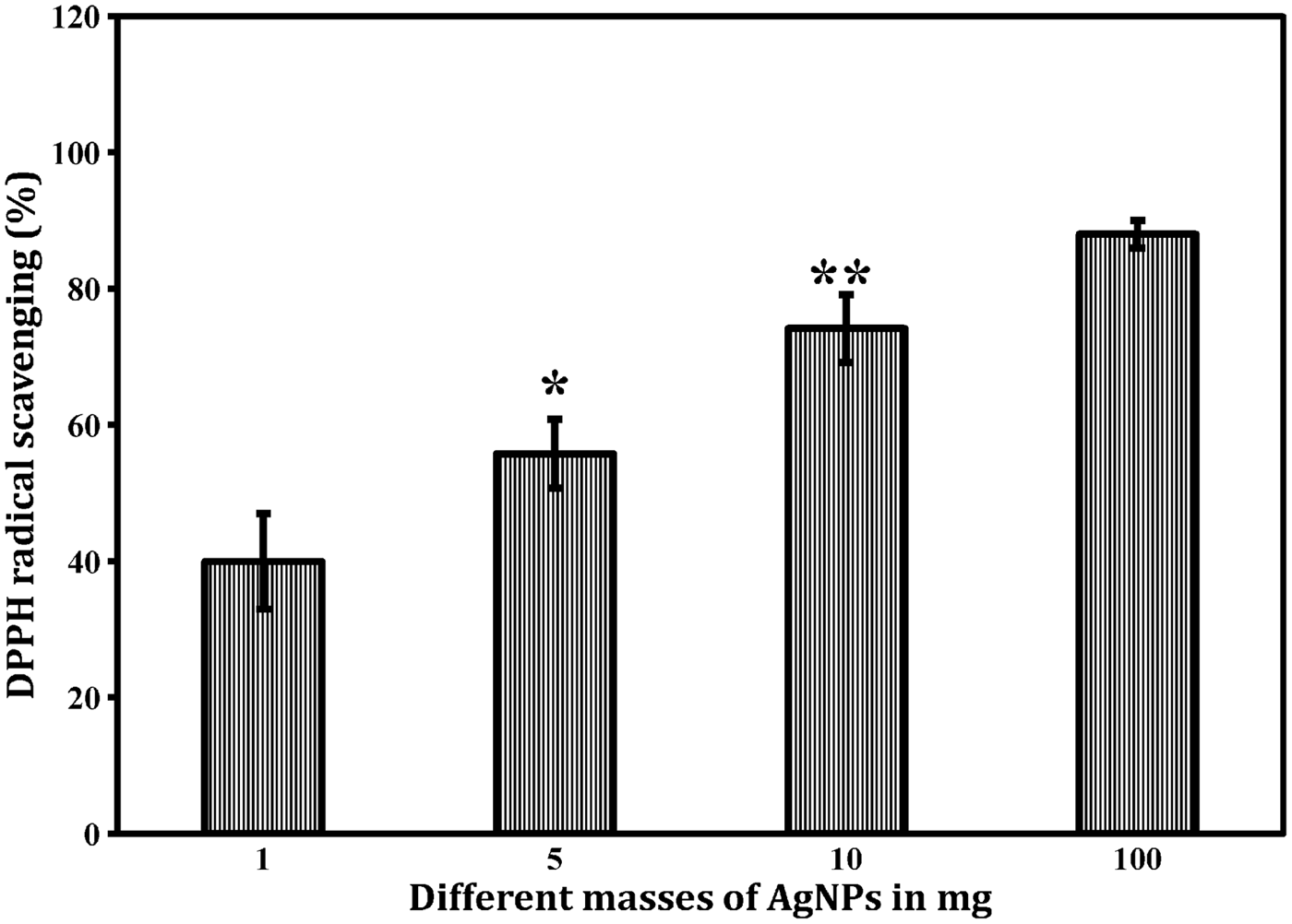
Antioxidant activity was examined by the 1′–1′ diphenylpicryl-hydrazyle (DPPH) method.[ Reference Serpen, Capuano, Fogliano and Gokmen 15 ] To determine the antioxidant property 1, 5, 10, and 100 mg of AgNPs were used. Roughly, 1.7 mL of DPPH reagent was added to AgNPs in Eppendorf tubes and vortexed for about 3 min. Control (C) tube contained only the DPPH reagent, whereas AgNPs added tubes served as the test (T). The mixture was then incubated for 30 min at room temperature followed by the centrifugation at 1000 rpm for 2 min. The optical density of the supernatant was measured using UV–vis spectrophotometer (U-2900/2910, Japan) at 517 nm. The radical scavenging was calculated by applying the formula as below:
 $$\hbox{DPPH scavenging activity}\;(\% ) = \displaystyle{{{C_{\rm A}} - {T_{\rm A}}} \over {{C_{\rm A}}}} \times 100,$$
whereas T A and C A are absorbances of the test and the control.
Results and Discussion
The flower of A. cathartica is well known for possessing broad varieties of phytocompounds such as saponins, flavonoids, terpenes, anthraquinones, and tannins.[ Reference Essiett and Udo 16 ] These compounds play a key role in the conversion of AgNO3 to AgNPs.[ Reference Logeswari, Silambarasan and Abraham 11 ] On incubation of the aqueous extract of the flower with AgNO3 in dark condition for 24 h, a change in color from yellow to brown [Fig. 1(a.B)] was observed. This color change indicated the conversion of Ag ions to AgNPs. The similar result was observed for bioreduction mediated by the mango peel extract for the formation of AgNPs.[ Reference Yanga and Li 17 ]
UV–vis spectroscopy showed [Fig. 1(a)] that the maximum absorbance peak of the particles was in the range of 350–470 nm. Similarly, a study on the synthesis of AgNPs from tissue culture derived callus and leaf extracts of saltmarsh plant had reported a peak at 420 nm indicating AgNPs production.[ Reference Nabikhan, Kandasamy, Raj and Alikunhi 18 ] The XRD pattern was used to confirm the crystalline nature of the AgNPs. Fig. 1(b) showed the XRD pattern of the synthesized nanoparticles corresponded to Bragg's reflection of Ag nanocrystals (Joint Committee on Powder Diffraction Standard-JCPDS). The observed XRD pattern was compared and matched with the JCPDS data file no. 43–0997. The pattern observed after matching with the JCPDS file reveals that AgNPs crystallized in cubic phase, with sharp bands of Bragg's peaks at approximately 27° (110), 32° (111), 46° (211), 54° (220), 57° (310), and 76° (321) respectively in the spectrum of 2θ value in the range between 10 and 80. Our result was in well agreement with a similar study of AgNPs synthesis using the leaf extract of Caesalpinia coriaria.[ Reference Jeeva, Thiyagarajan, Elangovan, Geetha and Venkatachalam 19 ]
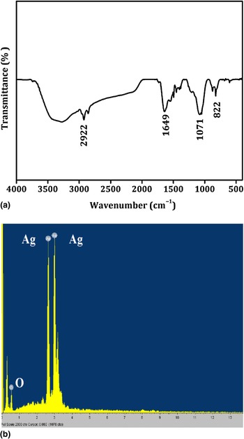
FTIR gives the information about the functional groups present in the synthesized AgNPs for understanding their transformation from simple inorganic AgNO3 to elemental Ag by the action of different phytochemicals present in the flower extracts such as (E,E)-geranyl linalool, n-pentacosane, 1,8-cineole and n-tricosane.[ Reference Baez, Pino and Morales 20 ] FTIR spectrum obtained clearly illustrated [Fig. 2(a)] the biofabrication of AgNPs mediated by the extract. The spectrum exhibited absorption peaks at 2922, 1649, 1071, and 822 cm−1, respectively. The strong peak at 2922 was due to O–H stretching vibration corresponding to phenolic and carboxylic acid groups capped on nanoparticles. The prominent bands at 1649, 1071, and 822 cm−1 indicated C–H, C = O stretching vibration, and C–H bending vibration. Phytochemicals in the flower extract might have acted as reducing, stabilizing, and capping agents during conversion of AgNO3 to AgNPs as evidenced from the previous studies.[ Reference Dipankar and Murugan 21 , Reference Mallikarjuna, John Sushma, Narasimha, Manoj and Deva Prasad Raju 22 ]
(a) FTIR spectrum and (b) elemental analysis (EDS) of AgNPs.

The EDS analysis was helpful in identifying the elemental composition of the synthesized nanoparticles. The obtained spectrum confirmed the presence of AgNPs [Fig. 2(b)] in which the vertical axis displayed the x-ray counts and the horizontal axis displayed the keV. The identified lines for the energy emission for Ag were shown and these peaks were matched with the spectrum of Ag and correctly identified. This analysis revealed the presence of Ag more than 60.75 percentages by weight. PSD [Fig. 3(a)] showed that the synthesized AgNPs ranged between 28 and 55 nm in size. However, the average particles size was found to be 39 nm. TEM was used to study the morphology and size of the nanoparticles. TEM image [Fig. 3(b)] indicated almost spherical morphology and the size of the nanoparticles correlated with the particle size distribution results. This result was in accordance with previous reports on AgNPs synthesis using pepper leaf[ Reference Mallikarjuna, John Sushma, Narasimha, Manoj and Deva Prasad Raju 22 ] and Eclipta alba leaf extracts.[ Reference Subbaiah, Ramanjaneyulu and Savithramma 23 ]
(a) PSD and (b) TEM image of AgNPs.

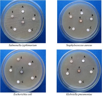
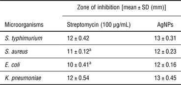
Antibacterial activity of the synthesized AgNPs from the flower extract was tested against four important clinical bacterial strains: S. typhimurium, S. aureus, E. coli, and K. pneumoniae. These particles exhibited promising antibacterial activity (Fig. 4) when compared with the standard antibiotic streptomycin (Table I). The results of antibacterial activity varied with respect to the bacterium used. Biosynthesized AgNPs showed higher zone of inhibition against S. typhimurium (13 ± 0.31 mm) and K. pneumoniae (13 + 0.45 mm) respectively compared with streptomycin; whereas for E. coli and S. aureus lesser order of inhibition was noticed as 12 ± 0.16 and 12 ± 0.23 mm. The antibacterial activity was attributed to the penetration of AgNPs across the bacterial cell membrane and causing cellular destruction.[ Reference Mallikarjuna, John Sushma, Narasimha, Manoj and Deva Prasad Raju 22 , Reference Subbaiah, Ramanjaneyulu and Savithramma 23 ] The synthesized AgNPs found to render more antibacterial activity in respect of S. aureus and E. coli compared with a similar study with E. alba leaves.[ Reference Subbaiah, Ramanjaneyulu and Savithramma 23 ]
Antibacterial activity of AgNPs [(A) antibiotic, (B) flower extract, (C, D) only disc, (E) distilled water, (F) AgNPs].

Antibacterial activity of AgNPs synthesized from aqueous extract of A. cathartica flower.

a Represents the level of significant at P < 0.05.
The analysis of free radical scavenging activity of AgNPs (Fig. 5) showed mass dependency. As the dosage of AgNPs was increased, its scavenging potential was enhanced from 40% (1 mg) to 55% (5 mg), 74% (10 mg), and 88% (100 mg), respectively. Similar antioxidant activity was reported for iron and nickel oxide nanoparticles.[ Reference Paul, Saikia, Samdarshi and Konwar 24 , Reference Saikia, Paul, Konwar and Samdarshi 25 ] This excellent antioxidant property of AgNPs will be very helpful to scavenge the free radicals generated inside the living system.[ Reference Watal, Watal, Rai, Rai, Sharma and Sharma 26 ] Thus, these particles can be taken as an additive for different biomedical applications.
Antioxidant activity of AgNPs (*, ** represents the level of significant at P < 0.05).

Conclusion
A. cathartica flower extract showed great capability to synthesize AgNPs at room temperature. UV absorption peak from 350 to 450 nm clearly indicated the formation of AgNPs. XRD patterns confirmed the phase composition and nature of the synthesized nanoparticles. FTIR studies confirmed the biofabrication of the AgNPs by the action of different phytochemicals with its different functional groups present in the extract solution. EDS confirmed the presence of elemental signal of AgNPs as well as the significant presence of Ag without any impurities. TEM and PSD studies were helpful at deciphering the spherical morphology and average size distribution. By comparing the different characteristic data, we concluded that, AgNPs synthesized from A. cathartica was more stable. Synthesized AgNPs possessed significant antibacterial activity against S. typhimurium, S. aureus, E. coli, and K. pneumoniae compared with the standard antibiotic. In addition, it also exhibited excellent free radical scavenging property. Thus, green synthesis of AgNPs using A. cathartica flower extract is a cost-effective, safe, non-toxic, and an eco-friendly approach for mass production as well as biomedical applications.
Acknowledgments
The authors gratefully acknowledge the financial support of the Ministry of Education and Science of the Russian Federation in the framework of Increase Competitiveness Program of NUST “MISiS” (No. K4-2015-017).