





Introduction
Accurate and reproducible patient positioning is essential in radiotherapy, as setup uncertainties directly influence target coverage and normal tissue sparing. Reference Verellen, De Ridder and Storme1,Reference van Herk2 Standardised supine or prone positions are widely adopted to ensure geometric stability and workflow efficiency. However, in routine clinical practice, enforcing these positions can exacerbate discomfort and distress in patients with dyspnoea, post-operative pain, musculoskeletal limitations or advanced disease. Reduced tolerance to treatment positioning may threaten treatment compliance and continuity, highlighting the need for practical positioning strategies that prioritise patient comfort. Reference Hsu, Lee and Huang10,Reference Stroom and Heijmen11
The semi-Fowler’s position, characterised by elevation of the upper body at approximately 30–45°, is commonly used in clinical care to improve respiratory mechanics and patient comfort. Despite its recognised role in supportive care, concerns regarding gravitational effects, longitudinal slippage and vertical instability have limited its adoption in radiotherapy. Reference van de Water, Bijl and Schilstra6,Reference Li, Qi, Pitterle, Kalakota and Mueller7 From a radiotherapy practice perspective, validating such alternative positioning strategies is essential to ensure that improvements in patient comfort do not compromise daily setup accuracy or workflow efficiency.
Previous studies evaluating setup reproducibility in radiotherapy have primarily focused on conventional supine or prone positioning, reporting setup uncertainties typically within ±0.3–0.4 cm under image guidance. Reference Verellen, De Ridder and Storme1–Reference van Elmpt, McDermott, Nijsten, Wendling, Lambin and Mijnheer3 Evidence regarding the reproducibility of semi-inclined or non-standard positions remains limited, although recent studies have begun to explore the feasibility of image-guided reproducibility in alternative treatment postures. Reference Li, Citrin and Camphausen9,Reference Hsu, Lee and Huang10,Reference Feldman, Pryanichnikov, Shwartz, Hillman, Wygoda, Blumenfeld, Marash and Popovtzer12
Therefore, this study aims to evaluate the setup reproducibility of the semi-Fowler’s position as a practical positioning strategy applicable within routine radiotherapy workflows, using existing immobilisation systems and standard image-guided verification.
From a practical radiotherapy perspective, alternative positioning strategies must be assessed not only for patient comfort but also for their impact on daily setup reproducibility and treatment workflow. Radiotherapy practitioners are frequently required to adapt positioning approaches for patients with limited tolerance, yet objective data supporting such adaptations remain scarce. This study addresses this gap by providing reproducibility data for the semi-Fowler’s position under routine clinical conditions.
Materials and Methods
Patients
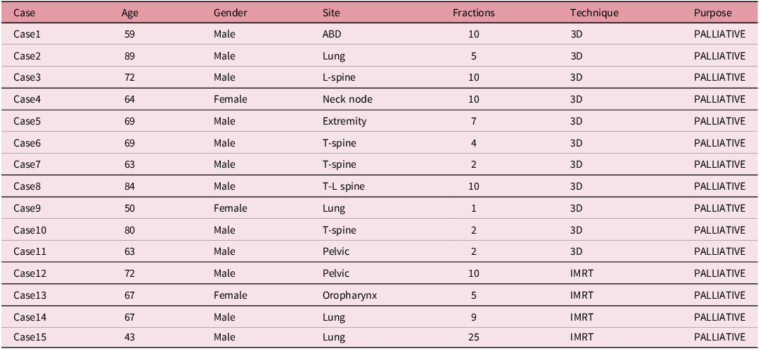
Between February 2020 and March 2025, 15 patients treated at our institution who were unable to tolerate standard supine or prone positioning were retrospectively included. This study was approved by the Institutional Review Board (IRB No. 2025-03-014). The cohort predominantly consisted of palliative cases with diverse treatment sites, reflecting real-world clinical scenarios in which conventional positioning was difficult to tolerate (Table 1).
Clinical characteristics of the patients included in this study

Table 1. Long description
The table presents clinical characteristics of 15 patients treated at an institution between February 2020 and March 2025. It includes details such as case number, age, gender, treatment site, number of fractions, technique used, and purpose of treatment. The table has 15 rows and 6 columns, with headers labeled Case, Age, Gender, Site, Fractions, Technique, and Purpose. The data shows a diverse range of treatment sites including ABD, Lung, L-spine, Neck node, Extremity, T-spine, T-L spine, and Pelvic. Techniques used include 3D and IMRT, with all cases being palliative. Notable trends include varying ages from 43 to 89 years, a predominance of male patients, and different numbers of fractions ranging from 1 to 25.
Simulation and positioning
Patients were positioned in the semi-Fowler’s position with an upper-body inclination of approximately 30°–45° using routinely available immobilisation components, including a vacuum-lock cushion, a Kneefix system and elevation blocks. The treatment couch itself remained at 0°, and the semi-inclined posture was achieved by combining these support devices rather than by changing the couch rotation angle. All equipment was compatible with a standard CT bore (GE Discovery CT590 RT16, 80 cm), and the arrangement was selected to maintain adequate bore clearance during simulation.
Simulation CT images were acquired according to institutional protocols. For treatment reproducibility, the same immobilisation arrangement was reproduced at each fraction using the same combination and placement of support devices established at simulation. The inclination was visually checked at each treatment session and maintained within a clinically consistent range throughout the treatment course.
A representative image of the semi-Fowler’s setup configuration is shown in Figure 1.
Representative setup of the semi-Fowler’s position used for radiotherapy simulation and treatment. The semi-inclined posture was achieved using standard immobilisation components, including a vacuum-lock cushion, a Kneefix system and Kneefix elevation blocks.

Figure 1. Long description
A person lies on a medical table in a semi-Fowler’s position, wearing a white mask and a green surgical gown. The person’s head rests on a vacuum-lock cushion, and their knees are supported by a knee fix system with elevation blocks. The setup includes labels pointing to the vacuum-lock cushion, knee fix, and knee fix elevation blocks.
Image guidance and setup error assessment
Orthogonal kilovoltage (kV) images were acquired prior to treatment using on-board imaging systems integrated into Varian linear accelerators (TrueBeam and Clinac iX; Varian Medical Systems, Palo Alto, CA, USA). Two-dimensional image matching was performed against digitally reconstructed radiographs generated from the planning CT. Translational displacements were recorded along the lateral (X), longitudinal (Y) and vertical (Z) axes in accordance with IEC 61217 conventions. Planning target volume margins were determined according to site-specific institutional protocols and were not expanded solely because of the use of the semi-Fowler’s position.
Statistical analysis
Setup reproducibility was evaluated using the intraclass correlation coefficient, Lin’s concordance correlation coefficient and Bland–Altman analysis. Patient-averaged displacement values were used for primary analysis. All statistical analyses were performed using Python version 3.10.
Results
Setup error distribution
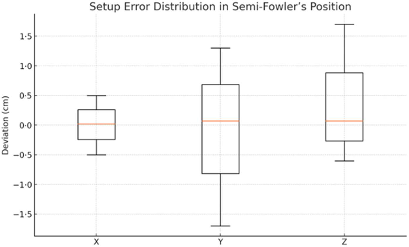
Across all analysed fractions, median setup errors in all translational directions were close to zero, indicating the absence of systematic directional bias (Figure 2). Variability was relatively limited in the lateral and vertical directions, while slightly greater dispersion was observed along the longitudinal axis.
Distribution of setup displacements along the lateral (X), longitudinal (Y) and vertical (Z) axes for patients treated in the semi-Fowler’s position. Boxplots represent median values, interquartile ranges and overall variability across analysed fractions.

Figure 2. Long description
A box-and-whisker plot displays the distribution of setup displacements along the lateral (X), longitudinal (Y), and vertical (Z) axes for patients treated in the semi-Fowler’s position. The plot includes three vertical box plots, each representing median values, interquartile ranges, and overall variability across analyzed fractions. The X-axis labels the axes (X, Y, Z), and the Y-axis measures deviation in centimeters, ranging from -1.5 to 1.5 centimeters. The box plots show the median as a horizontal line within each box, with the boxes representing the interquartile range (Q1 to Q3). The whiskers extend to the minimum and maximum values, indicating overall variability. The X-axis box plot has a smaller interquartile range and shorter whiskers compared to the Y and Z axes, suggesting less variability. The Y and Z axes show wider interquartile ranges and longer whiskers, indicating greater variability in setup errors along these axes. All values are approximated.
The median setup errors were 0.02 cm (X), 0.07 cm (Y) and 0.07 cm (Z), with interquartile ranges remaining within ±0.5 cm across all directions. These values are consistent with clinically acceptable limits commonly reported for image-guided radiotherapy. Reference Verellen, De Ridder and Storme1–Reference van Elmpt, McDermott, Nijsten, Wendling, Lambin and Mijnheer3
This directional pattern reflects predictable gravitational influences associated with semi-inclined positioning rather than random setup instability, as longitudinal variations remained within accepted clinical tolerance ranges and did not demonstrate systematic drift over the treatment course.
Agreement analysis
Bland–Altman analysis demonstrated acceptable agreement between repeated setup measurements across all translational directions (Figures 3–5). Increased variability in the longitudinal direction was consistent with gravitational effects associated with semi-inclined positioning; however, the magnitude of this variability remained within clinically acceptable limits. Vertical reproducibility was maintained despite the semi-inclined support geometry used to achieve the semi-Fowler’s position.
Bland–Altman plot for lateral (X-axis) setup displacements comparing repeated fractions in the semi-Fowler’s position. The solid line represents the mean difference, and dashed lines indicate the 95% limits of agreement.

Figure 3. Long description
A Bland-Altman plot for lateral X-axis setup displacements comparing repeated fractions in the semi-Fowler’s position. The x-axis represents the mean of setup displacements in centimeters, ranging from negative two to two centimeters. The y-axis represents the difference between repeated setups in centimeters, ranging from negative two to two centimeters. The plot includes a blue dashed line indicating the mean difference, which is close to zero. Red dashed lines represent the 95% limits of agreement, positioned approximately at 0.5 centimeters above and below the mean difference line. Several data points are clustered around the mean difference line, indicating minimal variation in setup displacements. The data points are marked with black crosses. The plot suggests that the setup displacements are generally consistent, with most differences falling within the limits of agreement. All values are approximated.
Bland–Altman plot for longitudinal (Y-axis) setup displacements. Slightly increased variability is observed compared with the lateral direction, reflecting gravitational influence associated with the semi-inclined posture.

Figure 4. Long description
A Bland-Altman plot titled ‘Bland-Altman Plot (Y-axis)’ displays the difference between repeated setups on the Y-axis in centimeters versus the mean of setup displacements on the X-axis in centimeters. The plot includes several data points marked with black crosses. A blue dashed line represents the mean difference, which is close to zero. Two red dashed lines indicate the upper and lower limits of agreement (LOA). The data points are clustered around the mean difference line, with slight variability observed. The plot reflects slightly increased variability compared to the lateral direction, likely due to gravitational influence associated with a semi-inclined posture. The X-axis ranges from negative two to two centimeters, and the Y-axis ranges from negative two to two centimeters. The legend in the top right corner explains the lines: blue for mean difference and red for upper and lower LOA.
Bland–Altman plot for vertical (Z-axis) setup displacements. Most data points fall within the 95% limits of agreement, indicating acceptable reproducibility under routine image-guided radiotherapy.

Figure 5. Long description
A Bland-Altman plot showing the difference between repeated setups on the Z-axis against the mean of setup displacements. The x-axis represents the mean of setup displacements in centimeters, ranging from negative two to two centimeters. The y-axis represents the difference between repeated setups in centimeters, ranging from negative two to two centimeters. The plot includes a blue dashed line indicating the mean difference, which is close to zero. Two red dashed lines represent the upper and lower limits of agreement. Most data points cluster around the mean difference line, with a few outliers. The data points are marked with black crosses. The plot demonstrates that most data points fall within the 95% limits of agreement, indicating acceptable reproducibility under routine image-guided radiotherapy.
Discussion
This study demonstrates that the semi-Fowler’s position can achieve setup reproducibility comparable to that reported for conventional supine positioning under routine image guidance. Reference Verellen, De Ridder and Storme1–Reference van Elmpt, McDermott, Nijsten, Wendling, Lambin and Mijnheer3 Although non-standard patient positioning is often avoided due to concerns regarding geometric uncertainty, the present findings indicate that acceptable reproducibility can be maintained using standard immobilisation systems and established verification workflows.
The slightly increased variability observed in the longitudinal direction is consistent with previous reports describing gravitational influences on patient stability in inclined positions. Reference van de Water, Bijl and Schilstra6,Reference Li, Qi, Pitterle, Kalakota and Mueller7 Importantly, these variations did not translate into clinically meaningful setup errors that suggested the need for additional margin expansion beyond site-specific institutional practice. The combined use of intraclass correlation coefficients, Lin’s concordance correlation coefficients and Bland–Altman analysis provides a robust and complementary assessment of setup reproducibility.
In the semi-Fowler’s position, gravitational forces may contribute to increased longitudinal and vertical displacement due to a tendency for inferior sliding. The use of knee fixation systems, such as the Kneefix, can counteract this effect by stabilising the lower extremities and facilitating weight redistribution. In the present study, these routinely available immobilisation components likely helped limit gravitational drift, resulting in longitudinal and vertical variations that remained within clinically acceptable ranges.
Beyond geometric reproducibility, the semi-Fowler’s position may provide a meaningful clinical benefit for patients who would otherwise struggle to complete radiotherapy due to positioning intolerance. In this cohort, semi-inclined positioning was used to improve tolerability and support treatment continuity when standard setups were not feasible. This underscores the value of adaptable positioning strategies that enable treatment completion without compromising geometric accuracy in daily radiotherapy practice.
In routine radiotherapy practice, maintaining treatment continuity for patients with limited positioning tolerance is a frequent challenge. Our findings suggest that the semi-Fowler’s position can be implemented using standard immobilisation equipment without disrupting established workflows. For radiotherapy practitioners, this approach offers a pragmatic balance between patient comfort and geometric accuracy, supporting selective use in daily clinical settings.
This study has several practical limitations. First, although the semi-Fowler’s position was feasible within our routine workflow, its use may be constrained by CT bore clearance and by limitations in beam angle selection, particularly when steeper beam arrangements are required. Second, this study focused on translational setup reproducibility derived from two-dimensional image guidance and did not separately quantify rotational errors such as pitch, roll or yaw. In clinical environments where a 6-degree-of-freedom couch is not available, correction of rotational misalignment may require additional repositioning attempts and could increase setup time. These workflow-related factors were not systematically measured in the present study and should be evaluated in future investigations.
Conclusions
The semi-Fowler’s position represents a practical and reproducible positioning option for selected radiotherapy patients who are unable to tolerate standard supine or prone setups. When implemented using existing immobilisation systems and routine image-guided verification, this approach supports daily clinical practice without compromising setup accuracy.



 Open access

