Introduction
Fellhaneropsis Sérus. & Coppins is a small genus originally segregated from Fellhanera Vězda (Sérusiaux Reference Sérusiaux1996), characterized by a dispersed thallus, emarginate apothecia with a prosoplectenchymatous excipulum, and filiform-sigmoid conidia. It occurs on various substrata, such as living leaves, bark, soil and rock. Although the genus is distributed worldwide, it is uncommon and only sporadically reported. Indeed, among a large number of foliicolous lichen specimens we recently collected in tropical China, only a small number of samples representing this genus were found.
Currently, 11 species of Fellhaneropsis are known worldwide: three foliicolous species, F. australiana Lücking, F. kurokawana G. Thor et al. and F. rhododendri Aptroot; three corticolous species, F. macrocarpa P. M. McCarthy et al., F. pallidonigrans (Müll. Arg.) Kantvilas & Lücking and F. tasmanica P. M. McCarthy et al.; two terricolous species, F. humicola P. M. McCarthy and F. subantarctica Øvstedal; one saxicolous species, F. almquistiorum S. Ekman; and two species known from various substrata, F. myrtillicola (Erichsen) Sérus. & Coppins and F. vezdae (Coppins & P. James) Sérus. & Coppins.
So far, species of Fellhaneropsis have been mainly reported from Europe, as well as Australia and neighbouring islands. Only F. kurokawana has been reported from Asia (Japan), and this is also the only species of Fellhaneropsis so far known to occur in China (Aptroot & Sparrius Reference Aptroot and Sparrius2003). The new taxon described in this study is the second species of Fellhaneropsis reported from Asia. It is a rare and typically foliicolous species, collected in tropical forest in Guangxi Province, China, in 2019.
Materials and Methods
The material is deposited in the herbarium of the Institute of Microbiology of the Chinese Academy of Sciences (HMAS-L). Morphological, chemical and molecular analyses (DNA extraction and sequencing) were performed as described in Wang et al. (Reference Wang, Sangvichien, T-Z and Wei2020).
Sequence alignment and phylogenetic analysis
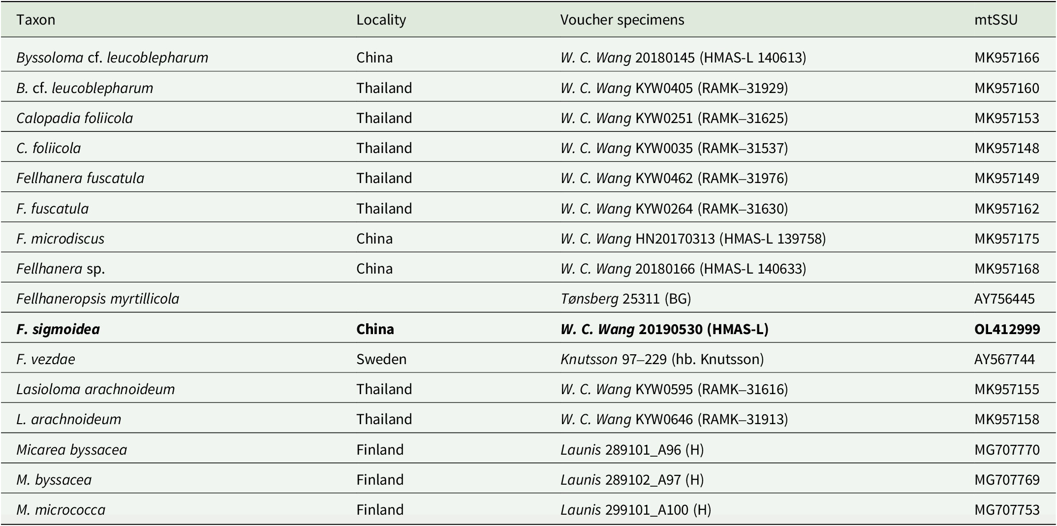
Geneious v. 6.1.2 (Biomatters Ltd, Auckland, New Zealand) was used to assemble and edit the newly generated mtSSU sequence reads. The assembled mtSSU sequence of the new taxon was aligned with 15 mtSSU sequences of Byssolomataceae retrieved from GenBank (Table 1). Two species of Micarea Fr. were chosen as outgroup based on previous phylogenetic analyses (Andersen & Ekman Reference Andersen and Ekman2005). The assembled sequences were aligned using MAFFT v. 7 online (Katoh et al. Reference Katoh, Asimenos and Toh2009). Gblocks v. 0.91b (Castresana Reference Castresana2000) was used to delimit ambiguous regions, implementing all the options for a less stringent selection. A matrix consisting of 769 unambiguously aligned mtSSU positions was produced.
Specimens used in the phylogenetic analyses (Fig. 1) with voucher information and GenBank Accession numbers. The new sequence and new species are in bold.
Maximum likelihood (ML) analysis was used to infer phylogenetic trees based on the mtSSU dataset, using RaxML-HPC v. 8.2.6 (Stamatakis Reference Stamatakis2014) on the Cipres Science Gateway (http://www.phylo.org). Bootstrap support values were based on 1000 non-parametric pseudoreplicates. The phylogenetic tree was visualized with the program FigTree v. 1.4.3. and edited in Adobe Illustrator CC 2019.
Results and Discussion
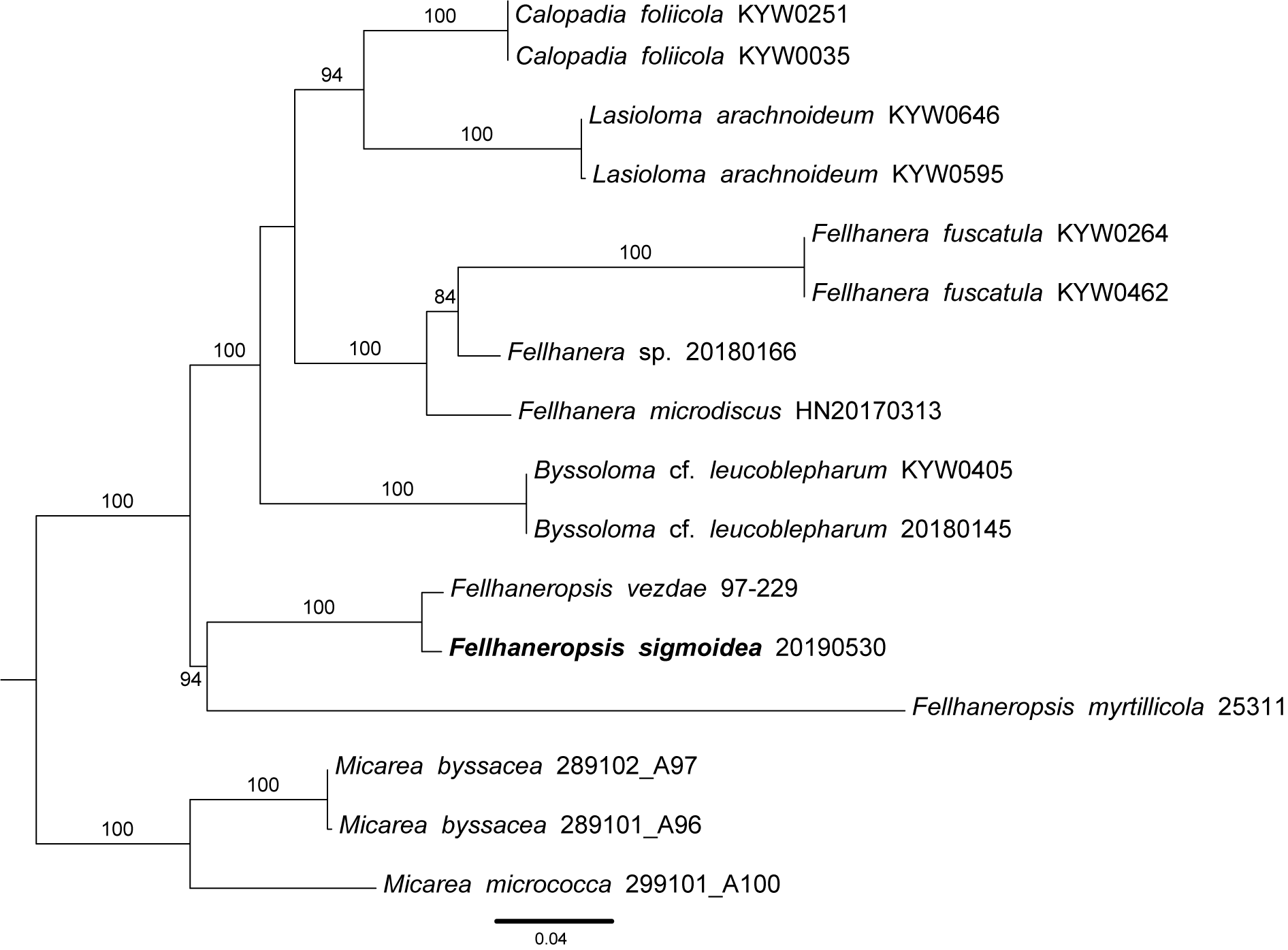
Most known species of Fellhaneropsis are based on morphological assessments, with molecular data being available for only two species: F. myrtillicola and F. vezdae. We compared these with the new sample (20190530) and constructed a phylogenetic tree together with sequences of related genera, including Byssoloma Trevis., Calopadia Vězda, Fellhanera, Lasioloma R. Sant. and Micarea, to determine the taxonomic position of the new sample (Fig. 1).
Phylogram using maximum likelihood (ML) inferred from the mtSSU dataset to show the phylogenetic position of Fellhaneropsis sigmoidea. Bootstrap values ≥ 75 are indicated at the branches. The tree was rooted using species of Micarea. Scale = 0.04 substitutions per site.

The new taxon forms a well-supported clade, together with F. myrtillicola and F. vezdae (BS = 94); it is supported sister to F. vezdae (BS = 100) but the two species have obvious differences in morphology (see notes below), whereas F. myrtillicola forms a long branch sister to the other two species. Other genera all form strongly supported (BS = 100) clades which in turn form a large, highly supported (BS = 100) sister to Fellhaneropsis.
Although the genus was originally segregated from Fellhanera, it is not closely related to that genus but appears to form an early diverging lineage closer to Micarea. Morphologically, Micarea resembles Fellhaneropsis in the seemingly emarginate apothecia, the transversely septate, fusiform ascospores, and the filiform-sigmoid macroconidia, but differs by having a granulose thallus and an exciple composed of branched, radiating hyphae (Smith et al. Reference Smith, Aptroot, Coppins, Fletcher, Gilbert, James and Wolseley2009). In contrast, Fellhanera has a paraplectenchymatous excipulum and ellipsoid to oblong or pyriform conidia (Lücking Reference Lücking2008).
Taxonomy
Fellhaneropsis sigmoidea W. C. Wang & J. C. Wei sp. nov.
Fungal Names No.: FN 571246
Differing from F. kurokawana and F. vezdae in having brown apothecia and pycnidia, 3-septate, smaller ascospores (14–20 × 3.5–5 μm) and longer conidia (38–42 × c. 1 μm).
Type: China, Guangxi Province, Shi’wan Big Mountain National Forest Park, 21°53′N, 107°54′E, 400 m alt., on living leaves, 21 March 2019, W. C. Wang 20190530 (HMAS-L—holotype!).
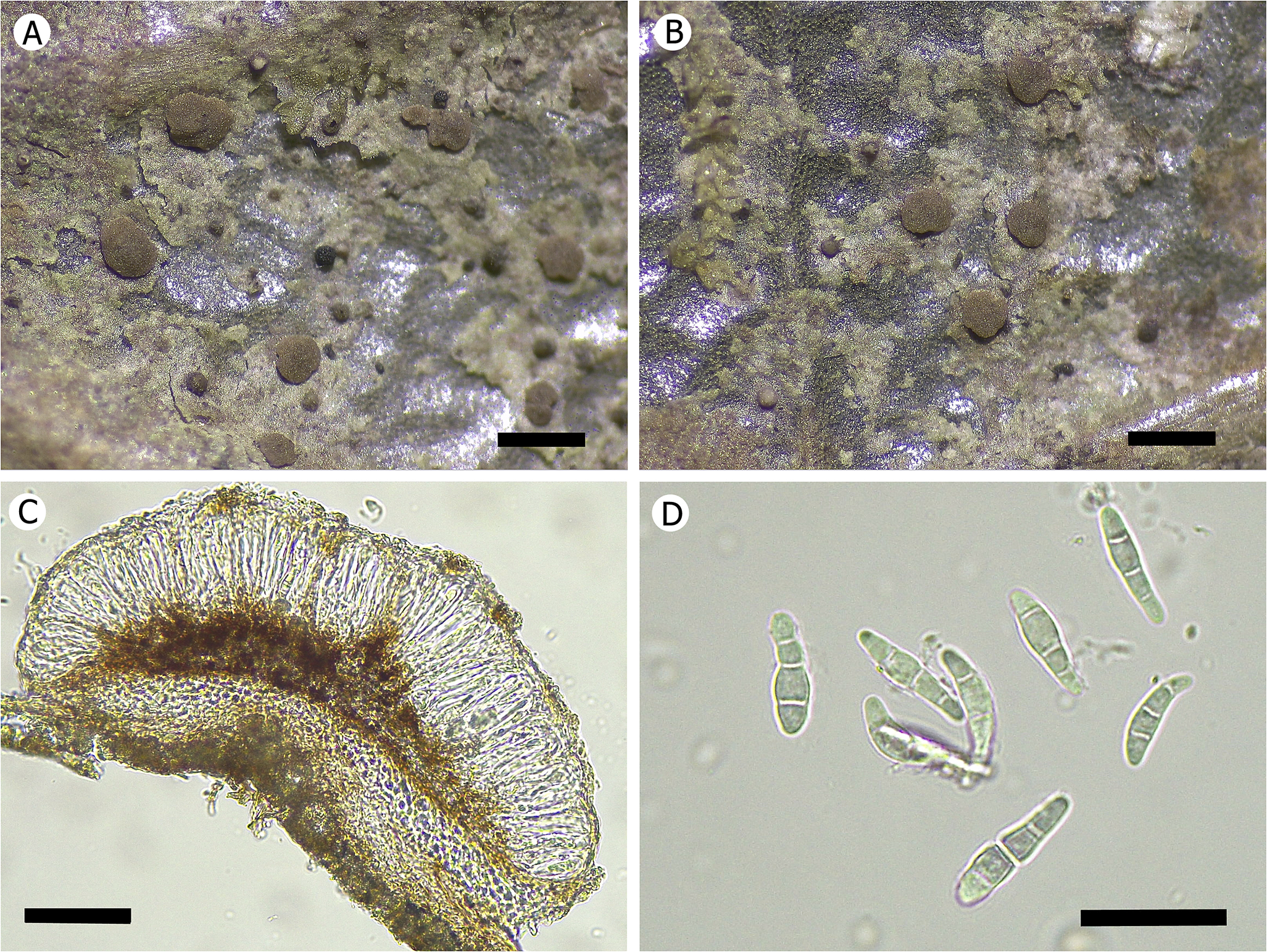
Fellhaneropsis sigmoidea (W. C. Wang 20190530, HMAS-L). A & B, dispersed thallus with emarginate apothecia and pycnidia. C, section of apothecium showing pigmentation. D, 3-septate ascospores. Scales: A & B = 500 μm; C = 50 μm; D = 20 μm. In colour online.
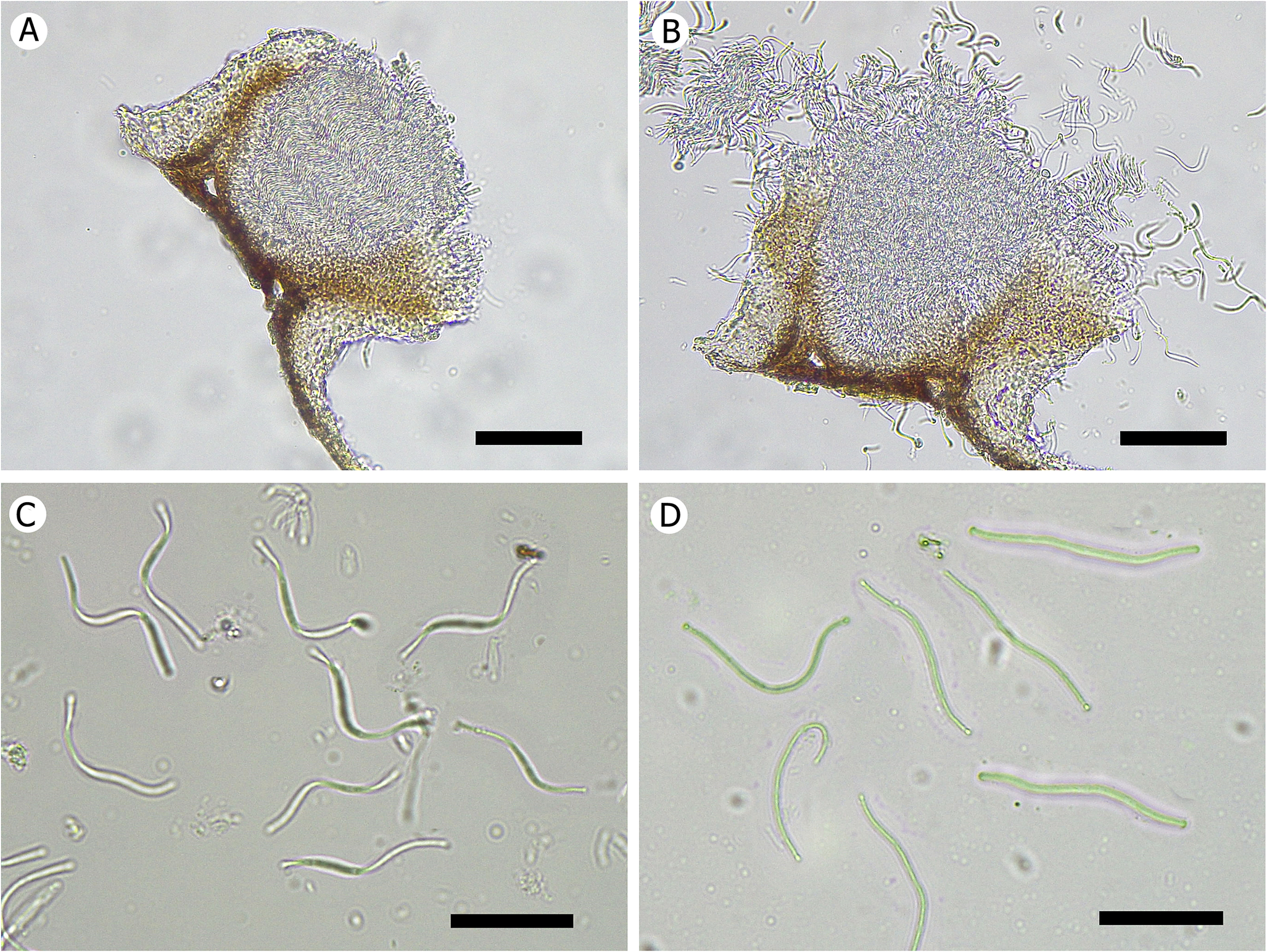
Fellhaneropsis sigmoidea (W. C. Wang 20190530, HMAS-L). A & B, section of pycnidium. C, filiform-sigmoid conidia (in water). D, conidia (after drying). Scales: A & B = 50 μm; C & D = 20 μm. In colour online.
Thallus epiphyllous, crustose, thin, 20–30 μm thick, 1–2 cm diam., continuous but in marginal parts made of dispersed patches, patches irregular in outline, surface smooth, greyish green, easily detaching from the leaves after drying. Prothallus not observed. Photobiont a species of Chlorococcaceae; algal cells green, globose, 7–10 μm diam.
Apothecia sessile, rounded to slightly irregular when mature, flat when young and becoming slightly convex when mature, epruinose, 0.1–0.3 mm diam. and 120–150 μm high; disc greyish brown; margin thin, soon disappearing, of the same colour. Excipulum poorly developed, colourless, prosoplectenchymatous; hypothecium distinctly brown, 45–55 μm, K−; epithecium pale brown, K−; hymenium colourless, 45–55 μm, K−; apothecial base pale brown, 30–35 μm, K−; paraphyses thin, branched and anastomosing. Asci clavate, tholus with I+ darker tubular structure. Ascospores 8 per ascus, fusiform, 3-septate, without constrictions at septa, some slightly curved, without halo, upper median cell somewhat enlarged, hyaline, 14–20 × 3.5–5 μm.
Pycnidia sessile, subglobose with rather wide ostiole, 140–160 μm wide and 90–110 μm high, brown, top white due to the conidia mass, only black ostiole left after the conidia have been released. Wall indistinctly paraplectenchymatous, outer part colourless, inner part and base brown, upper part forming densely arranged, palisadic hyphae surrounding ostiole, hyphae multiseptate, unbranched, colourless, 60–80 × 2.5 μm. Conidia numerous, densely and orderly arranged inside pycnidium, filiform-sigmoid, non-septate, colourless, 38–42 × 1 μm.
Secondary chemistry
Not tested (scanty material).
Etymology
The epithet sigmoidea refers to the filiform-sigmoid conidia.
Habitat and distribution
Fellhaneropsis sigmoidea is so far known from Guangxi Province, China, found in montane rainforest in tropical areas.
Notes
Fellhaneropsis sigmoidea is readily characterized by the foliicolous habitat, dispersed thallus, brown apothecia with indistinct margin, and 3-septate ascospores, 14–20 μm in length. Fellhaneropsis vezdae is phylogenetically close to F. sigmoidea; both are foliicolous but F. vezdae has 5–7-septate ascospores (30–35(–42) × 3–4.5 μm) and is known from Europe and South America (Sérusiaux Reference Sérusiaux1996; Lücking Reference Lücking2008).
Fellhaneropsis australiana also resembles the new species in its foliicolous habitat, but differs by its 7-septate ascospores, 20–30 μm in length, and 3–7-septate conidia, 40–50 μm in length, while the conidia of the new species are non-septate and shorter (Lücking et al. Reference Lücking, Streimann and Elix2001).
Fellhaneropsis kurokawana is a foliicolous species reported from Japan (Thor et al. Reference Thor, Lücking and Matsumoto2000) which agrees with F. sigmoidea in the 3-septate ascospores, but differs in having pale ochraceous yellow apothecia, pale flesh-coloured pycnidia and shorter conidia. Fellhaneropsis rhododendri Aptroot (Aptroot Reference Aptroot2012), reported from the Netherlands, is also foliicolous but differs by the dull, continuous thallus, pinkish brown apothecia, and an excipulum with black-walled hyphae.
Morphologically, the new species is also close to Fellhaneropsis pallidonigrans (Müll. Arg.) Kantvilas & Lücking (Reference Kantvilas and Lücking2009), since both have brown, seemingly emarginate apothecia and 3-septate ascospores of comparable size. However, F. pallidonigrans is corticolous and has a colourless to pale yellow hypothecium and a green epihymenium, while the new species is foliicolous and has a dark brown hypothecium and a colourless epihymenium (sometimes with a brown tinge but never green).
Additional specimen examined
China: Guangxi Province: Shi’wan Big Mountain National Forest Park, 21°53′N, 107°54′E, 400 m alt., on living leaves, 2019, W. C. Wang 20190531 (HMAS-L).
World key to species of the genus Fellhaneropsis

Acknowledgements
This research was funded by the National Natural Science Foundation of China (32400009), the Natural Science Foundation of Fujian Province, China (2023J05184), and the Project for Fundamental Research on Science and Technology, Ministry of Science and Technology of China (2013FY110400). I thank Dr Robert Lücking for very careful modifications and valuable suggestions on the manuscript.
Author ORCID
Wei-Cheng Wang, 0000-0002-8391-5120.