Introduction
Since the 1960s epidemiological studies of psychosis have shown increased rates of schizophrenia in many migrant and ethnic minority groups (Cantor-Graae & Selten, Reference Cantor-Graae and Selten2005). Compared with the white UK population the incidence rate of psychosis in people from the African-Caribbean community living in the UK has been reported to range from between two and 18 times higher (Sharpley et al. Reference Sharpley, Hutchinson, McKenzie and Murray2001; Fearon et al. Reference Fearon, Kirkbride, Morgan, Dazzan, Morgan, Lloyd, Hutchinson, Tarrant, Fung, Holloway, Mallett, Harrison, Leff, Jones and Murray2006). The incidence rates for schizophrenia in the English-speaking Caribbean are, on the other hand, thought to be similar to the incidence rates for schizophrenia in the white UK population (Hickling, Reference Hickling2005). The reasons underlying the increased rate of diagnosed psychosis in African-Caribbeans living in the UK are poorly understood although some have suggested that black patients are frequently misdiagnosed due to a failure of UK psychiatrists to take cultural factors into consideration (Littlewood & Lipsedge, Reference Littlewood and Lipsedge1981; Littlewood, Reference Littlewood1992).
Brain structural abnormalities represent one of the most consistently reported findings amongst white patients diagnosed as having psychosis. Neuroimaging studies have shown a range of neuro-anatomical abnormalities in first-episode schizophrenia and other forms of psychosis, including ventricular enlargement (Cahn et al. Reference Cahn, Pol, Lems, van Haren, Schnack, van der Linden, Schothorst, van Engeland and Kahn2002) and subtle grey-matter deficits in the whole brain (Fannon et al. Reference Fannon, Chitnis, Doku, Tennakoon, O'Ceallaigh, Soni, Sumich, Lowe, Santamaria and Sharma2000a), frontal and temporal lobes (Job et al. Reference Job, Whalley, McConnell, Glabus, Johnstone and Lawrie2002). If black patients are frequently misdiagnosed as having psychosis in the UK, one might expect that they would be less likely to have such abnormalities than their white counterparts.
Using high resolution magnetic resonance imaging (MRI) and voxel-based morphometry methods of image analysis, we therefore set out to examine whether, in comparison with their ethnically matched healthy controls, there would be more extensive structural brain abnormalities in first-episode psychosis patients who are white British than in those who are African-Caribbean or black African.
Methods
Sample
Patients
Subjects were inner city South London residents. Inclusion criteria for MRI analysis were: (a) age 16–65 years; (b) resident in defined area; (c) belonging to one of the following ethnic groups: (i) white British; (ii) African-Caribbean; (iii) black African; (d) presenting for the first time to local psychiatric services (in-patient and out-patient) with symptoms meeting functional psychosis criteria [ICD10: F20 (schizophrenia) and F30–39 (affective disorders – psychotic codings)] (WHO, 1992) Exclusion criteria were: (a) head trauma history with >1 h unconsciousness; (b) central nervous system disease; (c) poor English fluency; (d) transient psychotic symptoms resulting from acute intoxication (ICD-10), following administration of a psychoactive substance. A total of 161 patients meeting the above criteria were invited to participate in the MRI study and 97 consented to MRI scanning. Nine of these patients did not complete the full scanning procedure and therefore were not included in the analysis. A total of 13 further scans were excluded due to: (a) subject motion=11; (b) congenital hydrocephalus=1; (c) sub-arachnoid cyst=1. Therefore, 75 patients were included in the analysis; 34 of these were white British patients [male (n=20), mean age 28 years, ICD-10 schizophrenia (n=16), other psychosis (n=18)] and 41 either African-Caribbean (n=23) or black African (n=18) patients [male (n=27), mean age 27 years, ICD-10 schizophrenia (n=23), other psychosis (n=18)]. These 75 patients were younger [mean age 27.2 (s.d.=8.0) years v. 33.6 (s.d.= 11.7) years, t=3.9, p<0.001] compared with the 86 patients not included in the MRI analysis. In terms of ethnicity there were proportionately more white British patients included in the MRI analysis [34/75 (62%) v. 21/86 (38%)] than African-Caribbean/black African patients [41/75 (39%) v. 65/86 (61%), χ2=7.8, p=0.005]. There were no significant differences between the patients included and those not included in the analysis in terms of the proportion of male patients (χ2=0.78, p=0.38) and those diagnosed with schizophrenia (χ2=0.48, p=0.49).
Controls
In total, 68 controls were recruited from the same community as the patients; 33 were white British and 35 either African-Caribbean (n=22) or black African (n=13). Exclusion criteria were the same as those for the patients. Evidence of past or present psychosis, screened with the Psychosis Screening Questionnaire (Bebbington & Nayani, Reference Bebbington and Nayani1995), was an additional exclusion criterion. In addition to these 68 controls included, a further eight control subjects who had MRI scans were excluded from the analysis due to subject motion (n=7) and suspected hydrocephalus (n=1).
Classification of ethnicity
The coding of participants to ethnic groups was based on 2001 census categories according to self-ascribed ethnicity collected with the Medical Research Council Sociodemographic Schedule. If self-ascribed ethnicity was not recorded, other sources of information were used including other informants and case notes.
Study design
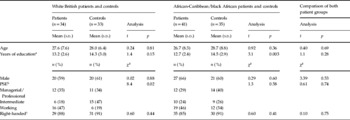
We conducted two separate between-group analyses. In the first analysis, the MRI scans of the white British patients were compared with those of the white British controls. In the second analysis, the African-Caribbean/black African patient scans were compared with African-Caribbean/black African control scans. Thus, 34 white British patients were compared with 33 white British controls, and 41 African-Caribbean/black African patients were compared with 35 African-Caribbean/black African controls (Table 1). A direct comparison of the two patient groups was not performed as the principal aim of the study was to evaluate patterns of structural brain abnormalities in white British and African-Caribbean/black African patients when compared with their ethnically matched healthy controls. Ethical approval was granted by the South London and Maudsley Trust Research Ethical Committee. All participants gave written, informed consent.
Sociodemographic characteristics of the sample

PSE, Parental socio-economic status.
a Data missing for two controls.
b data missing for one control.
c Data missing for two patients and three controls.
Clinical assessments
Patients were all interviewed using the WHO Schedules for Clinical Assessment in Neuropsychiatry (WHO-SCAN) (WHO, 1994). We made ICD-10 diagnoses in consensus meetings with senior clinicians (R.M. or J.L.), using WHO-SCAN information and clinical notes. Using the WHO-SCAN data, we encoded (in weeks) duration of illness (DOI) as the onset date of psychotic symptoms to MRI date and lifetime duration of antipsychotic exposure (DAE) (to MRI date). Total symptomology was scored by summing the WHO-SCAN's individual item scores using the algorithm of Wing & Sturt (Reference Wing and Sturt1978). Data on cannabis use in the patients and controls (lifetime use) were collected from patient and control interviews, medical histories and informant interviews.
Structural MRI acquisition
Scans were acquired with a GE Signa 1.5-T system (GE Healthcare, USA), at the Maudsley Hospital, London. Contiguous, interleaved proton-density- and T2-weighted 3-mm-thick coronal plane dual-echo images were acquired, providing whole brain coverage. A repetition time of 4000 ms and effective echo times of 20 ms and 85 ms were used with eight-echo train length. Matrix size was 256×192, collected from a rectangular field-of-view of 22 cm×16.5 cm, giving an in-plane resolution of 0.859 mm. Total acquisition time was 10 min, 12 s.
Structural MRI processing
The methods used for segmentation and registration of each fast spin echo dataset are described in detail elsewhere (Bullmore et al. Reference Bullmore, Suckling, Overmeyer, Rabe-Hesketh, Taylor and Brammer1999; Suckling et al. Reference Suckling, Sigmundsson, Greenwood and Bullmore1999). Briefly, subject masks were generated to identify neural tissue. Extra-cerebral tissues were removed initially, using an automated algorithm. Manually editing the skull-stripped images was necessary only to remove brainstem and cerebellum from the cerebral hemispheres and diencephalon. The probability of each intracerebral voxel belonging to each of four possible tissue classes (grey matter, white matter, cerebrospinal fluid (CSF) or dura/vasculature) was estimated with a modified fuzzy clustering algorithm (Suckling et al. Reference Suckling, Sigmundsson, Greenwood and Bullmore1999). This type of segmentation assigns for each voxel, a value in the range 0–1 indicating the fraction of the voxel comprised by each tissue type (e.g. a grey-matter value of 0.7 means 70% of tissue represented by that voxel is grey matter; therefore, the value indicates the proportion of the voxel occupied by grey matter). Total grey tissue volume was calculated at this stage of the analysis. The construction of the sample's template image is described elsewhere (Dazzan et al. Reference Dazzan, Morgan, Orr, Hutchinson, Chitnis, Suckling, Fearon, Salvo, McGuire, Mallett, Jones, Leff and Murray2004). In summary, a template image was constructed using the Analysis of Functional Neuroimages program from six proton-density images acquired from six healthy controls and then averaging these images. Tissue distribution maps were registered on to the template by first registering each subject's proton density image using a nine-parameter affine registration, minimizing between image grey-level difference between images. This registration aligns all the images together and scales them to the same gross dimensions. The derived mapping was then applied to the corresponding tissue maps.
Ventricular volume
Additional masks were generated per subject by tracing around the lateral and third ventricles in native space, in every slice in which they were visible. Ventricles were traced by one rater, blind to age, gender, ethnicity and patient/control status. Within the masked area, CSF volume was calculated using the data generated from the previously described modified fuzzy clustering algorithm.
Statistical analysis
Between-group regional differences in grey-matter volume were estimated by fitting an analysis of covariance (ANCOVA) model at each intracerebral voxel in standard space covarying for age and total grey-matter volume. Permutation testing was used to assess statistical significance and regional relationships were tested at voxel cluster level (Bullmore et al. Reference Bullmore, Suckling, Overmeyer, Rabe-Hesketh, Taylor and Brammer1999; Sigmundsson et al. Reference Sigmundsson, Suckling, Maier, Williams, Bullmore, Greenwood, Fukuda, Ron and Toone2001). Given that structural brain changes are likely to extend over a number of contiguous voxels, test statistics incorporating spatial information, such as 3D cluster mass (the sum of suprathreshold voxel statistics), are generally more powerful than other possible test statistics informed only by single voxel data. We set the statistical threshold for cluster significance in all analyses so that the expected number of false positive clusters (p value times number of tests) was <1 false positive. ANCOVA (controlling for age) were used to analyse between-group differences in ventricle:brain volume ratio and total grey-matter volume.
Results
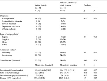
t Tests and χ2 analyses showed there were no significant differences between the white British and African-Caribbean/black African patients in terms of age, gender, years of education, parental socio-economic status, DOI, type of antipsychotic taken (typical or atypical), lifetime DAE, lifetime use of cannabis, total symptom scores, handedness and the proportion of patients with schizophrenia. Current dosage of antipsychotic medication (chlorpromazine equivalents) was significantly higher in the African-Caribbean/black African patients [219.5 mg (s.d.=240.3) v. 139.4 (s.d.=133.2) t=1.9, p=0.05]. Rates of involuntary admission were significantly higher in the African-Caribbean/black African patients (n=24, 60%) than in the white British patients (n=10, 30%) (χ2=6.41, p=0.01) (Tables 1 and 2).
Comparison of clinical characteristics of the white British and African-Caribbean/black African patients

CPZ, Chlorpromazine.
a Data missing for three cases.
b Data missing for three cases.
c Data missing for six cases.
d Data missing for two cases.
Analysis 1: White British patients compared with white British controls
t Tests and χ2 analyses showed there were no significant differences between the white British patients and control groups in terms of age, gender, years of education and handedness. White British patients did differ significantly from white British controls in terms of parental socio-economic status, with a higher proportion of patients rated as ‘working class’ than the controls (χ2=8.40, p=0.02) (Table 1).
Total tissue and ventricular volumes
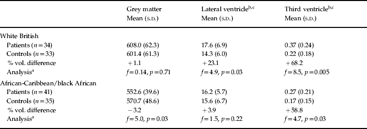
There were no between-group differences in total grey matter (Table 3). However, compared with the white British controls, the white British patients had a significantly larger lateral ventricular:brain volume ratio [17.6 ml (s.d.=6.9) v. 14.3 ml (s.d.=6.0), f=4.9, p=0.03] and significantly larger third ventricle:brain volume ratio [0.37 ml (s.d.=0.24) v. 0.22 ml (s.d.=0.18), f=8.5, p<0.01]. In a repeated analysis, the significant differences in ventricular:brain volume ratio remained after controlling for parental socio-economic status and years of education.
Mean total tissue and ventricular volumes (ml) in patients and healthy controls

a Analysis of covariance controlling for age performed. (Significant differences remain when gender of participant added as control factor.)
b Volume (ml) shown, analysis calculated according to ratio of ventricle:total grey-matter volume.
c Lateral and third ventricle data missing for two patients and one control.
Regional proportional tissue volume differences
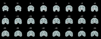
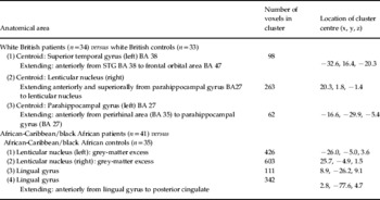
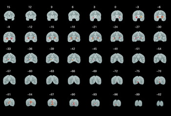
In comparison with the white British controls, the white British patients had three clusters of grey-matter excess centred at: (1) the left superior temporal gyrus; (2) the left parahippocampal gyrus; (3) the right lenticular nucleus (Table 4 and Fig. 1). There were no clusters of grey-matter deficit.
Regional differences in grey matter in white British patients (n=34) versus white British controls (n=33). Red/yellow regions denote areas of grey-matter excess in the patients relative to the control subjects. The results are displayed on averaged grey-matter maps. The left side of the image corresponds to the right side of the brain. Numbers refer to the approximate y coordinates in the standard space of Talairach and Tournoux.

Regional differences in grey-matter volume: patients versus controls

BA, Brodmann area.
Analysis 2: African-Caribbean/black African patients compared with African-Caribbean/black African controls
t Tests and χ2 analyses showed there were no significant differences between the African-Caribbean/black African patient and control groups in terms of age, gender and handedness. The patients had, on average, <1.8 years of education [mean 12.7 (s.d.=2.4) years v. 14.5 (s.d.=2.9) years, t=3.1, p=0.003] (Table 1).
Total tissue and ventricular volumes
Compared with the African-Caribbean/black African controls, the African-Caribbean/black African patients had a significantly smaller total grey-matter volume [551.4 ml (s.d.=40.8) v. 570.8 ml (s.d.=48.5), f=5.0, p=0.028] and significantly larger third ventricle:brain volume ratio [0.27 ml (s.d.=0.21) v. 0.17 ml (s.d.=0.15), f=4.75, p<0.03]. There were no between-group differences in total lateral ventricular:brain volume ratio (Table 3). In a repeated analysis, the significant differences in total grey-matter volume and third ventricle:brain volume ratio remained after controlling for parental socio-economic status and years of education. The significant difference in total grey-matter volume remained after controlling for total body height.
Regional proportional grey-matter volume differences
Compared with the African-Caribbean/black African controls, the African-Caribbean/black African patients had two clusters of significant grey-matter excess located in the left and right lenticular nucleus and two clusters of significant grey-matter excess located bilaterally in the lingual gyrus (Table 4 and Fig. 2).
Regional differences in grey matter in African Caribbean/black African patients (n=41) versus African Caribbean/black African controls (n=35). Red/yellow regions denote areas of grey-matter excess in the patients relative to the control subjects. The results are displayed on averaged grey-matter maps. The left side of the image corresponds to the right side of the brain. Numbers refer to the approximate y coordinates in the standard space of Talairach and Tournoux.

Post hoc analysis: antipsychotic dosage and admission status
There was no significant correlation between antipsychotic dosage and total grey-matter volume and lateral or third ventricle:brain volume ratio in either the African-Caribbean/black African or white British patients. ANCOVA showed no significant difference in total grey-matter volume, lateral or third ventricle:brain volume ratio between voluntary and involuntary patients in either the African-Caribbean/black African or the white British groups.
Discussion
To our knowledge, this is the first study to report on ethnicity and structural brain abnormalities in first-episode psychosis. We found structural brain abnormalities in both white British and African-Caribbean/black African patients that are similar to those that have been reported in other first-episode samples. In the regional analysis, ventricular enlargement and increased lenticular nucleus volume were shown in the two patient groups compared with their ethnic controls. Increased ventricular volume is one of the most frequently found structural brain abnormalities in studies of schizophrenia and other forms of psychosis, including samples of first-episode patients (Fannon et al. Reference Fannon, Tennakoon, Sumich, O'Ceallaigh, Doku, Chitnis, Lowe, Soni and Sharma2000b; Cahn et al. Reference Cahn, Pol, Lems, van Haren, Schnack, van der Linden, Schothorst, van Engeland and Kahn2002). The lenticular nucleus is part of the striatum, and striatal enlargement has been reported in the early stages of psychosis, but only following antipsychotic exposure (Lawrie & Abukmeil, Reference Lawrie and Abukmeil1998). It is thought that typical rather than atypical antipsychotics are linked to increased striatal volume as typicals have a strong affinity to sub-cortical D2 dopamine receptors and receptor blockade may induce cellular growth and increase blood flow (Corson et al. Reference Corson, O'Leary, Miller and Andreasen2002). In the present study, 89% of the patient sample were receiving antipsychotic medication, with the majority of them (59%) taking some form of typical antipsychotic.
The finding of reduced total grey-matter volume in the African-Caribbean/black African patients is also consistent with other reports (Fannon et al. Reference Fannon, Chitnis, Doku, Tennakoon, O'Ceallaigh, Soni, Sumich, Lowe, Santamaria and Sharma2000a) concerning first-episode psychosis. However, the absence of both global and regional grey-matter deficits (and the observation of two regions of increased temporal lobe grey matter) in the white British patients was not expected and ran contrary to our hypothesis of finding more extensive structural abnormalities in that patient group. It is unlikely the findings can be accounted for by anomalies in image acquisition or processing as such effects would not occur systematically in one group only.
The validity of comparing the findings from the white British patient–control analysis with those of the African-Caribbean/black African patient–control analysis needs to take into account any potential bias arising from the sociodemographic and clinical composition of the different patient and control groups. The controls were recruited from the same community as the patients. The African-Caribbean/black African patients and controls did not differ significantly on any sociodemographic variable, with the exception of the number of years spent in education, which was longer in the controls (14.5 years v. 12.7 years). The finding of reduced total grey-matter volume in the African-Caribbean/black African patients remained after a repeated ANCOVA that controlled for years of education and there was no significant correlation between years of education and grey-matter volume in either those patients or their ethnically matched controls, making it unlikely that the patient versus control difference in this variable contributed to the patient versus control differences in brain tissue volume. In the white British sample, the mean age of the patient and control groups was closely comparable, as was the distribution of males and females. Parental socio-economic status was the only sociodemographic variable in which there was a difference between patients and controls. If patient versus control differences in this variable could account for the absence of grey-matter deficits and the presence of grey excesses in the white British patients, one would expect a higher parental socio-economic status in the patients. The analysis of the white British patients and controls revealed, however, that a higher proportion of patients was classified at the lowest level (47% patients v. 18% controls) and a smaller proportion of patients at the intermediate level (19% patients v. 47% controls). In summary, the small differences in sociodemographic status observed between patients and controls in both sets of analysis does not explain the finding of grey-matter reductions in the African-Caribbean/black African patients and grey-matter increases in the white British patients.
The criteria applied to the recruitment of patients were the same for both the white British and African-Caribbean/black African groups. A comparison of the two patient groups showed no significant differences on the socio-economic measures. In terms of the patients' clinical characteristics, both groups were similar with regard to diagnostic classification, DOI, duration of exposure to antipsychotics, type of antipsychotic (typical or atypical) and total symptom ratings. The African-Caribbean/black African patients were, however, receiving a significantly higher dosage of antipsychotic medication and were significantly more likely to be admitted to hospital on a compulsory basis. While dosage of antipsychotics was not statistically correlated with grey-matter volume, it is notoriously difficult to establish such relationships. We cannot therefore exclude the possibility that this is at least in part a drug effect. Higher rates of involuntary admission in the African-Caribbean/black African patients raise the possibility of a more severe form of illness, which might explain the prescription of a higher antipsychotic dosage in that group. However, the similarities in total symptom ratings between the two patient groups suggest that, in terms of illness severity, any differences between white British and African-Caribbean/black African patients was not substantial.
A further issue that may be of relevance is the rate of substance use amongst the patient and control groups. A significantly higher proportion of African-Caribbean/black African patients had used cannabis (65%) compared with the African-Caribbean/black African controls (36%). In the white British group the proportion of patients using cannabis (72%) was also higher than in the controls (59%) but this difference was not statistically significant. A possible interpretation from this is that less difference in cannabis use between white British patients and controls may have contributed to the finding of less structural brain differences when compared with the analysis of the African-Caribbean/black African group. However, any such interpretation should be treated with caution as data on cannabis use were available only on 80% of the control sample. Furthermore, there was insufficient information available on the use of other recreational psychoactive substances (including alcohol) on the patients and controls taking part in this MRI study. Whilst the unavailability of further data on substance use is a limitation, data collected on drug use and ethnicity in the British Crime Survey (BCS) (Aust & Smith, Reference Aust and Smith2003) are of relevance here. The BSC data were collected at the same time as the current study and showed that, amongst the general UK population, levels of lifetime use of illicit drugs (including class A drugs) in 16- to 59-year-olds were similar amongst white (35%) and black (39%) people. The closely comparable levels of illicit substance use in white and black people living in the UK indicate, therefore, that between-ethnic group differences in structural brain changes observed at the first onset of psychosis are unlikely to be attributable to variations in substance use.
We hypothesized that white British patients would have more structural brain abnormalities when compared with the African-Caribbean/black Africans patients. This was posited as a basis upon which to investigate the notion that misdiagnosis of psychosis may be more likely to occur in black patients. If this was the case, one might expect that in the black patients there would be less evidence of structural brain abnormalities similar to those observed in a series of psychosis neuroimaging studies conducted over the last 15 years (when such abnormalities are not observed in healthy controls). The findings did not support this view.
The absence of more widespread differences in these patients, in particular the white British patients, might be accounted for by the method of recruitment. The patient sample consisted of people presenting to psychiatric services for the first time in their lives with a functional psychotic illness, irrespective of admission status, symptom severity or family history. This is in contrast, for example, to some other reported studies of first-episode psychosis (Job et al. Reference Job, Whalley, McConnell, Glabus, Johnstone and Lawrie2002; Pantelis et al. Reference Pantelis, Velakoulis, McGorry, Wood, Suckling, Phillips, Yung, Bullmore, Brewer, Soulsby, Desmond and McGuire2003), where patients are enrolled from university clinics, referral centres and in-patient samples, which attract subjects not necessarily representative of first-episode psychosis in general. Thus, it is possible that our findings do not reflect the findings reported in patients with more severe illnesses. Another potential explanation for the less marked brain differences found could be the diagnostic heterogeneity of the sample, which comprised a range of psychotic disorders. Additional ANCOVA including only those patients with a diagnosis of schizophrenia showed that the African-Caribbean/black African patients (n=23) had significantly smaller grey-matter volume than their ethnically matched controls (n=35), while in the white British group there was no significant difference between patients and controls in total grey-matter volume. Thus, the pattern of findings in relation to total grey-matter volume is similar to those of the main analyses, indicating that diagnostic heterogeneity is unlikely to account for the absence of more widespread patient–control brain differences.
Without replicating the study design, explaining the finding of more structural abnormalities in the African-Caribbean/black African patients will at best be speculative. It has been suggested, for example, that stress arising from social adversity and racial discrimination may play an important role in the development and onset of psychosis in individuals with an existing biological or genetic vulnerability. A national cohort study in Sweden found social adversity to be a risk factor for the onset of psychosis amongst first and second generation migrants (Hjern et al. Reference Hjern, Wicks and Dalman2004) and in the UK higher rates of psychosis have been reported in members of ethnic minority groups who are subject to either verbal or physical racist abuse (Karlsen and Nazroo, Reference Karlsen and Nazroo2004). Social stress may increase the likelihood of structural brain changes through a stress-induced neuro-toxic pathway. There have been suggestions that in schizophrenia such a pathway exists. A recent study of schizophrenia has shown an association between a dysregulated metabolic stress response and lower grey-matter volume (Marcelis et al. Reference Marcelis, Suckling, Hofman, Woodruff, Bullmore and van Os2006). Increased rates of self-reported life stress have also been associated with ventricular enlargement and white-matter loss in psychosis patients (Marcelis et al. Reference Marcelis, Myin-Germeys, Suckling, Woodruff, Hofman, Bullmore, Delespaul and van Os2003) and hippocampal grey-matter loss in a non-clinical sample (Gianaros et al. Reference Gianaros, Jennings, Sheu, Greer, Kuller and Matthews2007).
In conclusion, the analysis of structural MRI brain scans in this study provided no evidence to support the notion that increased rates of psychosis in black people living in the UK is attributable to misdiagnosis. However, there are no similar studies to compare our findings with, and it would have been desirable to have analysed a larger sample of patients more homogeneous in terms of diagnostic classification. Explaining the increased rates of psychosis amongst people from ethnic minorities remains a priority for researchers and clinicians.
Acknowledgements
The AESOP study was funded by the Medical Research Council (UK) and the Stanley Medical Research Institute. We thank the NIHR Biomedical Research Centre for Mental Health for their support. The research was undertaken at the Biomedical Research Centre, Department of Psychological Medicine, Institute of Psychiatry, London, UK.
Declaration of interest
None.