INTRODUCTION
Piroplasms, including Babesia, Theileria and Cytauxzoon species, are one of the most common blood parasites of mammals. These tick-transmitted protozoans have been reported in many domestic animals (Ristic, Reference Ristic1988) where they can cause asymptomatic infection or severe disease, including fever, anaemia, depression, diarrhoea, lethargy, anorexia and abortion (Homer et al. Reference Homer, Aguilar-Delfin, Telford, Krause and Persing2000; Zintl et al. Reference Zintl, Mulcahy, Skerrett, Taylor and Gray2003). Piroplasms are thus of considerable economic, medical and veterinary importance. Because most zoonotic Babesia are maintained in wildlife reservoir, surveillance of wild mammals living in area where undescribed Babesia are emerging has been suggested (Yabsley and Shock, Reference Yabsley and Shock2013).
In a wide range of wildlife, Babesia have been molecularly described (Schnittger et al. Reference Schnittger, Rodriguez, Florin-Christensen and Morrison2012; Yabsley and Shock, Reference Yabsley and Shock2013). In mongooses (Herpestidae), although several piroplasms have been detected, they all have been described based entirely on morphology (Grewal, Reference Grewal1957; Bandyopadhyay and Ray, Reference Bandyopadhyay and Ray1985; Penzhorn and Chaparro, Reference Penzhorn and Chaparro1994). The meerkat (Suricata suricatta), one of South Africa's most endearing wildlife celebrities, is a small burrow-living mongoose (van Staaden, Reference van Staaden1994) that can be infested by several ixodid tick species, including Haemaphysalis zumpti and Rhipicephalus theileri (Hoogstraal and El Kammah, Reference Hoogstraal and El Kammah1974; Horak et al. Reference Horak, Chaparro, Beaucournu and Louw1999, Reference Horak, Braack, Fourie and Walker2000). Meerkats can live on ranchlands, in close proximity to humans, pets or livestock, which increases the potential for the movement of pathogens, such as Babesia, between species. The presence of piroplasms in meerkats has never been studied. Here we investigated the occurrence and molecular phylogeny of piroplasms infecting wild meerkats using 18S rRNA and internal transcribed spacer 1 (ITS1) rRNA assay.
METHODS
Animal sources
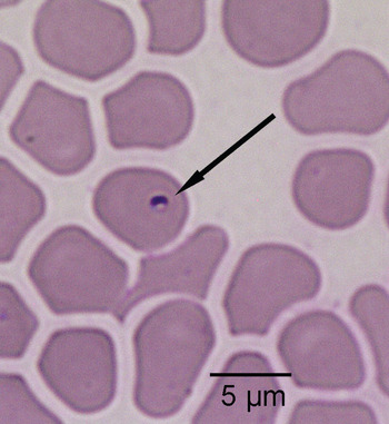
This study was conducted on a wild population of meerkats at the Kalahari Meerkat Project (Kuruman River Reserve, Northern Cape; 26°58′S, 21°49′E), on ranchland in the South African Kalahari. Thirty two meerkats from 10 social groups were sampled in March 2011, while 18 meerkats from 8 social groups were sampled in November 2011. All meerkats in the study population were individually recognizable (Jordan et al. Reference Jordan, Cherry and Manser2007) and habituated to close observation. Meerkats were blood sampled in the morning (between 6:00 AM and 9:00 AM) while they were sunning themselves at a burrow entrance. Meerkats were caught by gently but firmly lifting them by the tail base, and they were immediately placed in a dark-coloured cotton bag. The meerkat was supported by placing an arm under the bag while it was carried away from the rest of the group to a sampling area approximately 100 m away. Anaesthesia induction and maintenance was by isoflurane inhalation in air administered by face mask. Blood (2 mL) was withdrawn from the jugular vein using a 25-gauge needle and a 2-mL syringe and placed in a heparinized tube. Just after collection, a drop of blood was either transferred on FTA® Elute Cards (Whatman) with a glass capillary or smeared on glass slide (Fig. 1).
Photomicrograph of a Giemsa-stained thin blood film from a meerkat showing an intra-erythrocytic piroplasm (Babesia or Cytauxzoon sp.). Original magnification: ×1000.

European captive-born meerkats have presumably never been in contact with the parasites, given their several generations in captivity and no documentation of contact with African ticks during this time. They were, therefore, used as negative controls. We used blood stored in EDTA-tubes from 3 captive-born meerkats housed at Mulhouse Zoological Garden (France), and 2 captive-born meerkats housed at Thoiry Zoological Garden (France).
DNA extraction and PCR
DNA from filter papers of wild meerkats was extracted using Whatman DNA extraction procedure. DNA from blood smears and from blood of captive meerkats was extracted using DNA extraction kits (DNeasy Blood and Tissue kit, QIAGEN, Southern Cross Biotechnologies, SA), following the manufacturer's protocol.
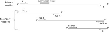
Primary outside amplification for 18S rRNA gene was conducted with 5·1 (Yabsley et al. Reference Yabsley, Davidson, Stallknecht, Varela, Swift, Devos and Dubay2005) and B primers (Fig. 2; Medlin et al. Reference Medlin, Elwood, Stickel and Sogin1988). One μL of DNA was added to 9 μL of a master mix containing 1X Green GoTaq reaction Buffer (Promega), 200 μ m of each dNTP (Promega), 0·4 μ m of each primer and 0·05 unit of GoTaq DNA Polymerase (Promega). Cycling conditions were 95°C for 3 min followed by 40 cycles of 95°C for 30 s, 48°C for 30 s and 72°C for 2 min, and a final extension at 72°C for 5 min.
Schematic illustration of the directions and combinations of the different primary and secondary primers used to obtain the near full-length 18S rRNA gene sequences.

To obtain near full-length 18S rRNA gene sequence, overlapping sequences were amplified using 3 secondary reactions. We used 5·1 (Yabsley et al. Reference Yabsley, Davidson, Stallknecht, Varela, Swift, Devos and Dubay2005) and RLB-R (Gubbels et al. Reference Gubbels, De Vos, Van der Weide, Viseras, Schouls, De Vries and Jongejan1999) primers, or RLB-F (Gubbels et al. Reference Gubbels, De Vos, Van der Weide, Viseras, Schouls, De Vries and Jongejan1999) and BabRev (Blaschitz et al. Reference Blaschitz, Narodoslavsky-Gfoeller, Kanzler, Stanek and Walochnik2008) primers, or BabFor (Blaschitz et al. Reference Blaschitz, Narodoslavsky-Gfoeller, Kanzler, Stanek and Walochnik2008) and B (Medlin et al. Reference Medlin, Elwood, Stickel and Sogin1988) primers (Fig. 2). To obtain sequence of the hypervariable region of the 18S rRNA gene, secondary reaction was realized using RLB-F and BabRev primers. All secondary reactions were realized using 1 μL of primary product as a template in a 20 μL reaction. PCR master mix composition and cycling conditions were the same as primary reaction except the annealing temperature was 44°C, and cyclic extensions were for 1 min.
Primary outside amplification for ITS1 gene was conducted using ITS-15C and ITS-13B primers (Bostrom et al. Reference Bostrom, Wolf, Greene and Peterson2008). PCR master mix composition was the same as for primary outside amplification for 18S rRNA gene. Cycling conditions were 95°C for 3 min followed by 40 cycles of 95°C for 30 s, 55 °C for 30 s and 72°C for 1 min and a final extension at 72°C for 10 min. Secondary amplification was performed using ITS-15D and ITS-13C primers (Bostrom et al. Reference Bostrom, Wolf, Greene and Peterson2008) with the same condition as primary reaction except the annealing temperature was 49°C.
All amplifications included a negative control consisting of sterile molecular-grade water in the primary reaction and the negative-control primary PCR product for the secondary reaction. The PCR products were purified and sequenced by Eurofins MWG Operon (Ebersberg, Germany) or MilleGen (Labège, France) sequencing services.
Phylogenetic analyses
Phylogenetic analyses were conducted on the near full-length 18S rRNA sequences obtained during the present study and additional sequences retrieved from GenBank. Sequences were aligned using Bioedit Sequence Alignment Editor. Before generating the phylogenetic trees, a series of likelihood ratio tests were completed using MEGA 6 (Tamura et al. Reference Tamura, Stecher, Peterson, Filipski and Kumar2013) to determine the best nucleotide substitution model to use for phylogeny analyses. The best model predicted using the Bayesian Information Criterion (BIC) was a Tamura-Nei model (TN93) with a proportion of invariant sites (I) and among site heterogeneity (G). A maximum-likelihood phylogenetic tree based on the Tamura-Nei model with evolutionary rate differences among sites modelled using a discrete Gamma distribution (number of categories = 5), a neighbour joining tree and a maximum parsimony tree model were then inferred using MEGA 6 (Tamura et al. Reference Tamura, Stecher, Peterson, Filipski and Kumar2013).
RESULTS
Molecular characterization
For 1 meerkat, DNA was extracted both from filter paper and from blood smear. ITS1 sequences were identical suggesting that sequences collected from the 2 DNA extraction procedures can be reliably compared. Blood from 4 meerkats was collected both in March 2011 and November 2011. For 2 meerkats, species and strain detection based on ITS1 sequences gave similar results across the 2 seasons. For the 2 remaining meerkats, infection was different between the 2 seasons, with meerkats being infected by the 2 piroplasm species in 1 season, but infected by a single species in the other season. However, for each of these 2 meerkats, species-specific sequences were identical between the 2 seasons.
All the 5 captive-born meerkats were PCR negative, while only 9% (n = 4) of wild meerkats were. The 4 PCR-negative wild meerkats belonged to 4 different social groups.
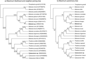
We amplified a hypervariable region of the 18S rRNA gene (~780 pb) in 13 wild meerkats, the near full-length 18S rRNA gene (~1700 pb) in 6 of these 13 wild meerkats, and the ITS1 region (~540 pb) in all wild meerkats (n = 51 samples). The 18S rRNA and ITS1 sequences of wild meerkats separated into 2 groups, suggesting infection by 2 piroplasm species (Fig. 3). Based on the ITS1 sequences, co-infection with the 2 species was detected in 46% of individuals (n = 21). Prevalence of co-infection may, however, be underestimated because primers designed to amplify all piroplasms may preferentially amplify the more common template in the exponential phase of amplification.
(a) Maximum-likelihood, neighbour-joining and (b) maximum parsimony bootstrap consensus trees showing the phylogenetic relationships of the new Babesia sp. and Cytauxzoon sp. with other piroplasms based on full-length 18S rRNA sequences. The numbers represent the percentage of 1000 replicates (bootstrap support) for which the same branching patterns were obtained for (a) maximum likelihood and neighbour joining analyses, respectively and (b) maximum-parsimony analysis. Toxoplasma gondii was used as an out-group. Sequences obtained during the present study are shown in bold. The number after the species designation refers to the GenBank number (indicated in the first tree only for clarity).

Four wild meerkats had a 100% similar amplicon of the 18S rRNA hypervariable region. The near full-length 18S rRNA gene was sequenced for 2 of these individuals and was shown to be 100% identical to one another (GenBank accession number: KM025200). By BLAST analysis (Altschul et al. Reference Altschul, Gish, Miller, Myers and Lipman1990), they shared the highest homology with Cytauxzoon felis (identity score: 95%; query cover: 100%; Fig. 3) and Cytauxzoon manul (identity score: 95%, query cover: 96%; Fig. 3). The ITS1 region of this novel Cytauxzoon sp. was detected in 57% of individuals (n = 26; Table 1). However, because many meerkats were co-infected, ITS1 sequences of Cytauxzoon sp. were reliably sequenced for 7 individuals only. Within the 485-pb ITS1 region, we found 1 single nucleotide polymorphism (SNP) (GenBank accession number: KM025207 and KM025208). Out of the 7 individuals, 5 individuals had a single ITS1 sequence, while the remaining 2 individuals had the 2 ITS1 sequences. All meerkat social groups were infected by the Cytauxzoon species, except 1 group for which only 1 meerkat was sampled (Table 1).
Percentage of wild meerkats infected by the Babesia species, the Cytauxzoon species and being co-infected within each social group

Nine wild meerkats had similar sequence of the 18S rRNA hypervariable region. The near full-length 18S rRNA gene was sequenced for 4 of these individuals and was shown to be 100% identical to each other (GenBank accession number: KM025199). They shared the highest homology with Babesia lengau (identity score: 99%; query cover: 93%; Fig. 3). Based on the ITS1 sequence, infection by this Babesia sp. was detected in 80% of samples (n = 40), and all meerkat social groups were infected (Table 1). Eighty-three percent of meerkats had more than one Babesia ITS1 sequence. Based on the electropherograms containing Babesia sequences only, 13 SNPs, one 1-nucleotide insertion and one 2-nucleotide deletions were detected in the 480-pb ITS1 sequence of Babesia sp. The 2-nucleotide deletion sequence was detected in 51% of Babesia-infected meerkats, while the 1-nucleotide insertion sequence was detected in 50% of Babesia-infected meerkats. From the unambiguous electropherograms, we reliably characterized 6 distinct ITS1 sequences (GenBank accession number: KM025201 to KM025206).
Microscopy
In all blood smears where piroplasms were observed, the organisms were single intraerythrocytic inclusions, small in size (measuring ca. 1 μ m in diameter; Fig. 1) and few in number. No morphological differences were discernible between the Babesia and the Cytauxzoon species.
DISCUSSION
Meerkats were found to be infected by 2 different piroplasm species: the first one closely related to the Cytauxzoon species infecting Felidae (Wagner, Reference Wagner1976; Peixoto et al. Reference Peixoto, Soares, Scofield, Santiago, Franca and Barros2007; Shock et al. Reference Shock, Murphy, Patton, Shock, Olfenbuttel, Beringer, Prange, Grove, Peek and Butfiloski2011), and the second one closely related to Babesia lengau, a species infecting the South-African cheetah (Acinonyx jubatus) (Bosman et al. Reference Bosman, Oosthuizen, Peirce, Venter and Penzhorn2010). Several piroplasm species have been previously described in mongooses (reviewed in Penzhorn, Reference Penzhorn2006), but all have been described only from morphology. In particular, Babesia cynicti has been found in South-African yellow mongooses (Cynictis penicillata) (Penzhorn and Chaparro, Reference Penzhorn and Chaparro1994), a species that can occupy the same territory and burrow, and host the same tick species as meerkats (Lynch, Reference Lynch1980). Molecular characterization of Babesia cynicti is now required to determine whether it is the same species as the ones found in meerkats.
The prevalence of infection with Babesia and Cytauxzoon in this population of meerkats was high. Ninety-one percent of meerkats were infected, including 46% that were infected by the 2 species. Similar prevalence (88%) was detected for Babesia cynicti in yellow mongooses (Penzhorn and Chaparro, Reference Penzhorn and Chaparro1994). All sampled meerkats appeared, however, clinically normal and showed no signs of disease (personal observations). Although Babesia and Cytauxzoon can cause severe disease in domestic animals (Wagner, Reference Wagner1976; Homer et al. Reference Homer, Aguilar-Delfin, Telford, Krause and Persing2000), high prevalence and subclinical infection are common in wild animals (Schnittger et al. Reference Schnittger, Rodriguez, Florin-Christensen and Morrison2012) where clinical disease manifests only when the host is stressed (Penzhorn, Reference Penzhorn2006). Clinical babesiosis in the sable antelope (Hippotragus niger), Grevy's zebra (Equus grevyi), black rhinoceros (Diceros bicornis) and Eastern grey kangaroo (Macropus giganteus) appears to be triggered by the stress of capture, restraint and translocation (Dennig, Reference Dennig1964; Nijhof et al. Reference Nijhof, Penzhorn, Lynen, Mollel, Morkel, Bekker and Jongejan2003; Nijhof et al. Reference Nijhof, Pillay, Steyl, Prozesky, Stoltsz, Lawrence, Penzhorn and Jongejan2005; Dawood et al. Reference Dawood, Morgan, Busfield, Srivastava, Fletcher, Sambono, Jackson, Venus, Philbey and Lew-Tabor2013; reviewed in Penzhorn, Reference Penzhorn2006). The Babesia and Cytauxzoon species identified in our study may be benign parasites, and infection does not appear to result in clinical morbidity or mortality under normal conditions.
Meerkats are infected by several ixodid ticks (Lynch, Reference Lynch1980), which are potential vectors of Babesia and Cytauxzoon. Preliminary characterizations of ticks collected in our meerkat population suggest that individuals are infected by R. theileri (Prof Ivan Horak, personal communication), a burrow associated ixodid tick that is associated with the hot and arid climate of the Northern Cape Province (Horak et al. Reference Horak, Chaparro, Beaucournu and Louw1999). The meerkat is a highly social species living in family groups which share the same burrows (Lynch, Reference Lynch1980). These features could facilitate the transmission of Babesia and Cytauxzoon by ticks among members of the same group and would explain the high infection prevalence in meerkats. Examination of genetic variations within the ITS region of the rRNA genes in relation to meerkat group membership and spatial location should help elucidate the transmission route of piroplasms in meerkats. In our study, ITS1 sequence analysis was complicated due to the co-infection of meerkats with different species and/or multiple strains of a single species, or due to the presence of several rRNA gene copies in the genome. Future works conducted with species-specific ITS1 primers or including cloning prior sequencing are needed to associate specific parasite genotype with host life history characteristics.
In summary, 2 piroplasm species were found in a high percentage of Kalahari meerkats. None of meerkats had evidence of disease compatible with babesiosis; thus these piroplasms appear to have low pathogenicity for meerkats. However, infection with either of them in combination with stress or other factors such as immunosuppression could possibly lead to disease.
ACKNOWLEDGMENTS
We thank T. Clutton-Brock and M. Manser for providing access to the animals at the KMP, and N. Thavarajah and J. Samson for their help in the field. We are grateful to the Kotze family and Northern Cape Conservation for allowing us to conduct research in the Kalahari. We are grateful to Dr B. Quintard at Mulhouse Zoological Garden and Dr A. Leclerc at Thoiry Zoological Garden for providing us with blood from captive meerkats. We thank M. Yabsley and E. Huchard for helpful discussion.
FINANCIAL SUPPORT
S.L. was supported by a Fondation Fyssen post-doctoral grant and an ASAB research grant.