Introduction
The paraglottic space and cricoid area of the larynx are made up of loose, areolar tissue composed of loose elastic and collagenous fibres and adipose tissue. They contain the main blood vessels of the larynx.Reference Sato1 We examined the correlations between the histopathological extension of glottic carcinoma into the paraglottic space or cricoid area and the occurrence of cervical lymph node metastasis.
Materials and methods
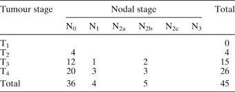
We reviewed the medical charts of 45 patients (44 men and one woman) who had undergone total laryngectomy for squamous cell carcinoma of the glottis between 1991 and 2003. The tumour–node (TN) classification of these patients is shown in Table I.
Patients' tumour–node classification
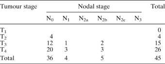
Data represent number of cases. T = tumour; N = node
The removed larynges of the 45 patients were prepared using the whole-organ serial section technique. The specimens were fixed in 10 per cent formalin, decalcified in 5 per cent hydrochloric acid, dehydrated in graded concentrations of ethanol and then embedded in paraffin. Both horizontal and coronal serial sections were made. Haematoxylin and eosin stain was applied to each section, and the sections were then observed under light microscopy. The patients' removed larynges were examined both macroscopically and microscopically. We analysed the correlations between the histopathological evidence of invasion into the paraglottic space or cricoid area and the occurrence of cervical lymph node metastasis.
Statistical analysis was undertaken, at the 0.05 level of significance using the χ2 test.
Results
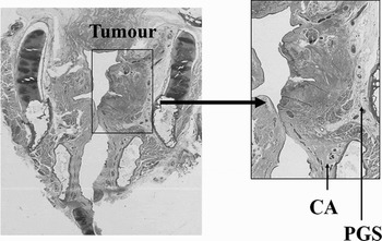
Figure 1 shows a coronal section of the larynx of a patient with T3 N0 glottic cancer and no cervical lymph node metastasis. Cancer extended to the deep portion but did not invade either the paraglottic space or the cricoid area.
Photomicrograph of a coronal laryngeal section showing no invasion of the paraglottic space (PGS) or cricoid area (CA). This patient had no cervical lymph node metastasis.

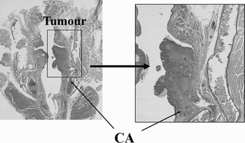
Figure 2 shows a coronal section of a larynx from a patient with T3 N0 glottic cancer who developed neck recurrence one year after surgery. Cancer extended into the deep portion and also invaded the cricoid area.
Photomicrograph of a coronal laryngeal section showing invasion of the cricoid area (CA). In this patient, neck recurrence was found one year after surgery.

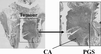
Figure 3 shows a coronal section of a larynx from a patient with T3 N1 glottic cancer and cervical lymph node metastasis. Cancer extended into the deep portion and also invaded both the paraglottic space and the cricoid area.
Photomicrograph of a coronal laryngeal section showing invasion into the cricoid area (CA) and paraglottic space (PGS). This patient had cervical lymph node metastasis.

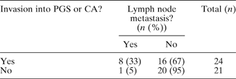
Eight of the 24 patients (33 per cent) whose glottic cancer had invaded the paraglottic space or cricoid area had cervical lymph node metastasis. In contrast, only one (5 per cent) of the 21 patients without any evidence of such invasion had cervical lymph node metastasis. This difference was statistically significant (p < 0.05) (Table II).
Cervical lymph node metastasis in glottic carcinoma patients with or without invasion into paraglottic space or cricoid area
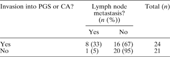
p < 0.05, for metastasis(+) vs metastasis(−). PGS = paraglottic space; CA = cricoid area
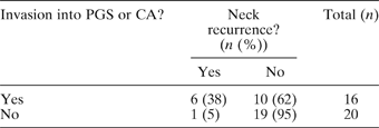
In the 36 patients free of cervical lymph node metastasis, neck recurrence occurred in six (38 per cent) of the 16 patients with histopathological evidence of paraglottic space or cricoid area extension, but in only one (5 per cent) of the 20 patients without paraglottic space or cricoid area extension (p < 0.05; Table III).
Neck recurrence in patientsFootnote * with or without initial invasion into paraglottic space or cricoid area
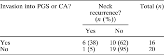
* Initially without cervical lymph node metastasis. p < 0.05, for recurrence(+) vs recurrence(−). PGS = paraglottic space; CA = cricoid area
Discussion
A space is defined as a demarcated portion of the body; it can be an area of a surface, a segment of tissue or a cavity.Reference Asimov, Bassett, Beamer, Bean and Benedict2 The chief laryngeal spaces are the pre-epiglottic space, the paraglottic space and the cricoid area.Reference Sato1, Reference Tucker and Smith3–Reference Sato, Kurita and Hirano5 These spaces are very important clinically, but they are commonly used terms and are not included among the standard anatomical terms.
The paraglottic space is enclosed by the thyroid lamina, thyroarytenoid muscle, conus elasticus and cricothyroid muscle, and is located medial to the thyroid lamina along its entire length. The paraglottic space is an area of loose, areolar tissue composed of loose elastic and collagenous fibres and adipose tissue. It contains the descending branch of the superior thyroid artery.Reference Sato1, Reference Hirano and Sato4 In patients with extension of supraglottic cancer into the paraglottic space, cervical lymph node metastasis has been found to readily occur.Reference Chijiwa, Umeno, Sato and Nakashima6
The cricoid area is one of the laryngeal connective tissue compartments initially described by Pressman et al. Reference Pressman, Simon and Monell7 The cricoid area is observed as a triangular region surrounded by the perichondrium of the cricoid cartilage, the conus elasticus and the fibrous layer of the subglottic mucosa. The cricoid area is located on the superomedial portion of the cricoid cartilage in the coronal plane and on the anteromedial portion of the cricoid cartilage in the axial plane. A blood vessel extends out of the cricoid area and pierces the conus elasticus.Reference Sato1, Reference Hirano and Sato4 In patients with laryngeal carcinoma, cancer invasion into the cricoid area and intravascular tumour invasion has been observed to facilitate metastasis to the pre-laryngeal, pre-tracheal and/or para-tracheal regions and stomal recurrence.Reference Sato, Umeno and Hirano8, Reference Umeno, Sato, Matsuda and Nakashima9
From a pathological point of view, this study revealed the paraglottic space and the cricoid area as two of the routes for lymph node metastasis from glottic carcinoma. We should therefore attempt to clarify the histopathological importance of the paraglottic space and the cricoid area, in order to better understand the aetiology of cervical lymph node metastasis in patients with glottic carcinoma. Based on these histological and anatomical findings, as well as the results of the current investigation of larynges with glottic carcinoma, it can be said that cancer invasion into the paraglottic space or cricoid area facilitates cervical lymph node metastasis.
Conclusion
These results strongly indicate the histopathological importance of the paraglottic space and the cricoid area for cervical lymph node metastasis in glottic carcinomas. In these patients, post-operative radiotherapy or a preventive neck dissection might thus be necessary to obtain better survival results.