Introduction
Attention-deficit/hyperactivity disorder (ADHD) is a common and highly heritable childhood-onset neuropsychiatric disorder, with compelling evidence of involving dopaminergic neurotransmission systems (Faraone et al. Reference Faraone, Perlis, Doyle, Smoller, Goralnick and Holmgren2005). The dopamine transporter gene (DAT1) has been consistently reported to be associated with ADHD across studies (Gizer et al. Reference Gizer, Ficks and Waldman2009; Shang et al. Reference Shang, Gau, Liu and Hwu2011). The DAT1 gene codes for the dopamine transporter (DAT), which functions to transport extracellular dopamine back into the cytoplasm of pre-synaptic dopaminergic neurons, and thereby limits the duration of synaptic activity. Although Durston et al. have suggested that DAT1 gene effects in the brain may be involved in translating the genetic risk of ADHD into a neurobiological substrate (Durston et al. Reference Durston, Fossella, Mulder, Casey, Ziermans and Vessaz2008), the exact mechanisms of the DAT1 effects on the brain function and structure of children with ADHD remain unclear (Rommelse et al. Reference Rommelse, Altink, Arias-Vasquez, Buschgens, Fliers and Faraone2008).
The pathway from gene to behavior is complex. The imaging genetic study is based on the assumption that brain structure and function are closer to gene function than trait differences in overt behaviors (Durston, Reference Durston2010). Thus, neuroimaging techniques are supposed to elucidate the influence of genes on brain structure and function in a relatively small sample, sometimes even in the absence of significant differences in behavioral measures (Wu et al. Reference Wu, Yang and Wang2014). Imaging genetics has the potential to provide a more precise and thorough understanding of the pathways and mechanisms through which the dynamic interplay of genes and environment shapes variability in the brain (Wu et al. Reference Wu, Yang and Wang2014).
Previous imaging genetic studies of the DAT1 gene in ADHD have focused on the variable number of tandem repeat (VNTR) polymorphism in 3′-untranslated region of this gene. The ventrolateral striatum has been reported to be more activated in nine-repeat carriers than 10/10 individuals during sustained reward anticipation (Aarts et al. Reference Aarts, Roelofs, Franke, Rijpkema, Fernandez and Helmich2010). In comparison with the 10/10 individuals, the nine-repeat allele carriers generate more activation in the fronto-striato-parietal regions, as well as the right putamen and the left precuneus under high working memory loads (Stollstorff et al. Reference Stollstorff, Foss-Feig, Cook, Stein, Gaillard and Vaidya2010). Besides the 3′VNTR, few additional markers in the DAT1 gene have been reported to be associated with alterations of brain function and structure in ADHD. For example, the T allele at rs37020 contributed to the activation in the frontal regions and the caudate nucleus under inhibition tasks (Cummins et al. Reference Cummins, Hawi, Hocking, Strudwick, Hester and Garavan2012). Taken together, these results have prompted the crucial role of DAT1 in the neurobiological abnormalities of ADHD. Further investigations of genetic variations of DAT1 on the brain function and structure will help to identify the gene–brain–behavior relationship.
Although resting-state functional magnetic resonance imaging (RS-fMRI) may provide a platform to investigate the gene function, only a few imaging genetics studies have used the resting state as the MRI paradigm (Wiggins et al. Reference Wiggins, Bedoyan, Peltier, Ashinoff, Carrasco and Weng2012; Gordon et al. Reference Gordon, Devaney, Bean and Vaidya2015). In a study of 39 healthy children and adolescents, those homozygous for the S allele of the serotonin transporter linked polymorphic region showed reduced resting-state functional connectivity in the superior medial frontal cortex (Wiggins et al. Reference Wiggins, Bedoyan, Peltier, Ashinoff, Carrasco and Weng2012). In another study of 50 healthy young adults, individuals with one nine-repeat DAT1 allele demonstrated greater striato-frontal connectivity than those with two 10-repeat alleles (Gordon et al. Reference Gordon, Devaney, Bean and Vaidya2015). Further studies are needed to examine the DAT1 effects on resting-state brain functioning in ADHD.
There is growing evidence for impaired visual memory in patients with ADHD (Kempton et al. Reference Kempton, Vance, Maruff, Luk, Costin and Pantelis1999; Rhodes et al. Reference Rhodes, Coghill and Matthews2004), including impairment in spatial recognition memory (SRM) and pattern recognition memory (PRM). Our previous work demonstrates that visual memory characterizes a cognitive endophenotype for ADHD (Shang & Gau, Reference Shang and Gau2011). Several studies have converged to suggest a crucial role of dopamine in visual memory (Harmer et al. Reference Harmer, McTavish, Clark, Goodwin and Cowen2001; Rhodes et al. Reference Rhodes, Coghill and Matthews2004, Reference Rhodes, Coghill and Matthews2006). For example, methylphenidate, which inhibits the action of DAT and thereby increases synaptic dopamine concentration to achieve the therapeutic effects in ADHD (Volz, Reference Volz2008), is effective in improving visual memory performance in children with ADHD (Rhodes et al. Reference Rhodes, Coghill and Matthews2004, Reference Rhodes, Coghill and Matthews2006). In addition, decreased dopaminergic function is associated with poor performance on the SRM task (Harmer et al. Reference Harmer, McTavish, Clark, Goodwin and Cowen2001). Taken together, examining the relationships between DAT1, brain imaging, and visual memory may provide an insight into the pathways from DAT1 to ADHD.
By using haplotype-tagging SNP approaches to investigate 15 polymorphisms across the DAT1 gene, our previous family-based association genetic work showed that the rs27048 (C)/rs429699 (T) haplotype was significantly associated with ADHD (Shang et al. Reference Shang, Gau, Liu and Hwu2011). However, there is a lack of data about the impact of the DAT1 rs27048 (C)/rs429699 (T) haplotype on the brain structure and function in children with ADHD. In a clinical sample of children with ADHD, the current study was designed to investigate the differential brain effects as a function of the DAT1 rs27048 (C)/rs429699 (T) haplotype by examining the regional homogeneity (ReHo) as well as gray matter volume (GMV), and the relationship between DAT1-modulated brain alterations and visual memory.
Method
Participants and procedures
We recruited 42 eligible drug-naïve children (37 males), aged 7–16, clinically diagnosed with ADHD according to the Diagnostic and Statistical Manual of Mental Disorders, Fourth Edition (DSM-IV) diagnostic criteria from the Department of Psychiatry, National Taiwan University Hospital, Taipei, Taiwan. They and their parents were interviewed by using the Chinese version of the Schedule for Affective Disorders and Schizophrenia for School-Age Children – Epidemiological Version (K-SADS-E) by the corresponding author (SSG) to confirm their diagnosis of ADHD and to exclude any other psychiatric disorder. In addition, we recruited 40 typically developing children from similar school districts to the ADHD group, with the help of principals and school teachers. Similar to the ADHD group, they and their parents were interviewed using the Chinese K-SADS-E to ensure that they did not meet DSM-IV diagnosis of ADHD or any other psychiatric disorder.
Participants were excluded if they had a serious medical illness, such as a cardiovascular disease; had a full intelligence quotient (FIQ) score <80; had a history of any psychiatric disorder (except ADHD in the ADHD group); had a history of any seizure disorder or prior electroencephalogram abnormalities related to epilepsy, or if they had ever used any psychotropic medications before the study. Informed consent was obtained from participants and their parents after they had received the detailed information of experimental purpose and administration. The informed consent procedures were approved by the Research Ethics Committee at the National Taiwan University Hospital, Taiwan before study implementation (IRB ID: 200903062R; ClinicalTrials.gov number, NCT00916851).
Measurements
ADHD Rating Scale-IV
The ADHD Rating Scale-IV (ADHDRS-IV) (DuPaul et al. Reference DuPaul, Power, Anastopoulos and Reid1998) was an investigator-based validated 18-item semi-structured interview with the parents about the participants’ ADHD symptoms over the past week. Each item, corresponding to one of the 18 DSM-IV ADHD behavioral items, was rated on a four-point scale (0, never or rarely; 1, sometimes; 2, often; 3, very often). The inattention and hyperactivity/impulsivity subscales were the sum scores of the odd-numbered and the even-numbered items, respectively. The ADHDRS-IV was a reliable and valid instrument to assess ADHD symptom severity and has been widely used in the ADHD treatment studies in Taiwan (Gau et al. Reference Gau, Huang, Soong, Chou, Chou and Shang2007).
Measures of visual memory
Visual memory was assessed by two tasks (PRM and SRM) from the Cambridge Neuropsychological Test Automated Battery (CANTAB).
Pattern recognition memory
The PRM was a test of visual recognition memory in a two-choice forced discrimination paradigm. At the center of the screen, a series of geometric patterns were presented one after the other for a 3-s viewing interval. Following the presentation of the pattern series, the screen paused for 5 s, after which the participant was presented with two geometric patterns. One of the two designs was from the previously viewed list. The other was a completely novel stimulus. The two designs differed in shape but not in color. The participant was asked to touch the design that had been presented. After each trial, the participant was given feedback as to whether their choice was correct or incorrect. Twelve trials, one containing each of the 12 target stimuli, were implemented in each of the two blocks for a total of 24 trials. Two major indices were used: (1) total correct responses across both blocks for pattern recognition score; and (2) mean latency of correct responses.
Spatial recognition memory
The SRM tested visual spatial memory in a two-choice forced discrimination paradigm to measure recognition memory for spatial locations. The participant saw empty boxes presented at different locations on the screen and was asked to remember these places. The participant then saw five stimuli in succession at different screen locations for 3 s each. Following a 5-s pause, the participants then saw two boxes, one of which was in a remembered location, and the other was novel. The participant was asked to touch the box that was previously targeted. After each trial, the participant was given feedback as to whether their choice was correct or incorrect. A total of 20 trials were presented in four blocks of five trials each. Two major indices were used: (1) total correct responses across all four blocks for computing the participant's recognition score; and (2) mean latency of correct responses.
Genotyping
The primers were designed by the platform of National Genotyping Center of Academia Sinica (http://lims.ngc.sinica.edu.tw/service/), using GenePipe (http://genepipe.ibms.sinica.edu.tw/seqtool/pages/getSeq.jsp) to retrieve SNP flanking sequences. The SNP genotyping of rs27048 and rs429699 was performed by the method of matrix-assisted laser desorption/ionization time of flight mass spectrometry (Rodi et al. Reference Rodi, Darnhofer-Patel, Stanssens, Zabeau and van den Boom2002). This genotyping method has been applied in a broad variety of clinical disorders, including ADHD (Shang et al. Reference Shang, Gau, Liu and Hwu2011; Reference Shang, Chiang and Gau2015; Shang & Gau, Reference Shang and Gau2014) and autism (Chien et al. Reference Chien, Wu, Chiu, Liu, Tsai and Lin2011).
Magnetic resonance imaging
Data acquisition
Data were obtained on a 3 T scanner (Siemens Magnetom Tim Trio) with a 32-channel phased-arrayed head coil. All participants were verbally instructed to remain still with their eyes closed to complete a 6-min RS-fMRI scan. Wakefulness was monitored and ensured at the end of each scan by checking their prompt responses to technicians’ questions. All participants denied falling into sleep during scanning. The RS-fMRI parameters were 180 echo planar imaging (EPI) volumes; TR = 2000 ms; TE = 24 ms; flip angle = 90°; field of view (FOV) = 256 × 256 mm2; matrix size = 64 × 64; 34 axial slices acquired in an interleaved descending order; slice thickness = 3 mm; voxel size = 4 mm × 4 mm × 3 mm; imaging plane being parallel to the anterior commissure–posterior commissure image plane. A high-resolution T1-weighted anatomical image was also acquired (MPRAGE, TR = 2000 ms; TE = 2.98 ms; TI = 900 ms; flip angle = 9°; FOV = 256 mm × 256 mm; matrix size = 256 × 256 × 192; isotropic voxel size = 1 mm).
RS-fMRI data preprocessing
Resting-state EPI preprocessing, including slice timing, realignment, within-participant registration of RS-fMRI data and T1 images, and spatial normalization, was performed using Data Processing Assistant for Resting-State fMRI (DPARSF) toolbox (Yan & Zang, Reference Yan and Zang2010) based on Statistical Parametric Mapping (SPM8, Wellcome Trust Centre for Neuroimaging, London, UK). The first five volumes of the scanning sessions were removed to allow for scanner calibration and participants’ adaptation to the scanning environment. For each participant, each volume was slice time corrected and realigned to the first image volume using a least-squares minimization and a six-parameter (rigid-body) spatial transformation. Realigned EPI image was then registered to structural scans, normalized to a Montreal Neurological Institute (MNI) template in isotropic 3 mm voxel via the gray matter (GM) segment. The normalized fMRI data were denoised by implementing Independent Component Analysis-based strategy for Automatic Removal of Motion Artifacts (ICA-AROMA) (Pruim et al. Reference Pruim, Mennes, Buitelaar and Beckmann2015a, Reference Pruim, Mennes, van Rooij, Llera, Buitelaar and Beckmannb). After motion denoising, the EPI data were further denoised using the CONN toolbox v.15c (Whitfield-Gabrieli & Nieto-Castanon, Reference Whitfield-Gabrieli and Nieto-Castanon2012) to remove other resources of non-neuronal noises through a component-based (anatomical CompCor) approach (Behzadi et al. Reference Behzadi, Restom, Liau and Liu2007). The first three principal components of the signals from the white matter (WM) and cerebral spinal fluid (CSF) regions-of-interest (ROIs) (anatomical masks derived from the prior segmentation steps), and linear detrending were included as regressors in the first-level denoising regression model. All of the regressors were filtered before performing the denoising regression (Hallquist et al. Reference Hallquist, Hwang and Luna2013). Band-pass filtering (0.01–0.1 Hz) was then conducted to decrease the impact of high-frequency physiological noise and the very low-frequency drift.
Head motion
In-scanner head motion during EPI acquisition was indexed by mean frame-wise displacement (FD) derived from Jenkinson's relative root mean square algorithm (Jenkinson et al. Reference Jenkinson, Bannister, Brady and Smith2002). Mean FD was used due to its consideration of voxel-wise differences in motion in its derivation (Yan et al. Reference Yan, Cheung, Kelly, Colcombe, Craddock and Di Martino2013). Participants with mean FD exceeding one standard deviation (s.d.) above the sample mean (0.19 ± 0.18 mm) were excluded from further analysis. Using this criterion, eight participants (five in the ADHD group and three in the neurotypical group) were excluded, yielding a final sample of 74 participants. There was no significant difference in mean FD (p = 0.059) between the ADHD (n = 37) and neurotypical (n = 37) groups. We visually inspected the structural image data to confirm no anatomical lesions, nor imaging artifacts, and ensure data quality without excessive in-scanner head motion in all of these 74 participants.
ReHo analyses
The ReHo analyses were conducted by using DPARSF (Yan & Zang, Reference Yan and Zang2010). Individual ReHo map was generated by calculating the Kendall coefficient of concordance (KCC) of the time series of a given voxel with those of its neighbors (26 voxels) in a voxel-wise way (Zang et al. Reference Zang, Jiang, Lu, He and Tian2004). Afterward, a whole-brain mask was adopted to remove the non-brain tissues. For standardization purposes, the individual ReHo maps were divided by their own global mean KCC within the whole-brain mask. Then spatial smoothing was performed on the standardized individual ReHo maps with a Gaussian kernel of 4.5 mm FWHM.
Structural imaging preprocessing (voxel-based morphometry)
Individual T1-weighted images were segmented by the New Segment toolbox in SPM8 to produce native space GM, WM, and CSF images. During segmentation, age- and sex-matched study-specific tissue probability maps generated from the Template-O-Matic toolbox (using the ‘matched-pair’ approach) were used for all the participants (Wilke et al. Reference Wilke, Holland, Altaye and Gaser2008). The native-space GM and WM images of all participants were then registered to a study-specific template using a high-dimensional non-linear diffeomorphic registration algorithm (Ashburner, Reference Ashburner2007), with modulation (preserve volume). The modulated GM maps were smoothed with a 4 mm FWHM Gaussian kernel. Individual total GM, WM, and CSF volumes were estimated by summing up the partial volume estimates throughout each class of segmented image in the native space. Total brain volumes were estimated by summing up the total GM and WM volumes. Total intracranial volumes were calculated by summing up the total GM, WM, and CSF volumes.
Statistical analyses
SAS 9.4 software (SAS Institute, Cary, NC, USA) was used to conduct non-neuroimaging data analyses. The mean scores and s.d. were presented for continuous variables, and the percentage was used for categorical variables for the demographics and neurocognitive assessments.
The genotypes of the DAT1 variant were divided into two groups according to the presence status of the rs27048 (C)/rs429699 (T) haplotype: the CT group and the non-CT group. We used non-parametric Friedman rank tests to determine whether there were statistically significant differences in the PRM and SRM performance for the four groups (ADHD-CT, ADHD-Non-CT, Control-CT, and Control-Non-CT). To investigate the differential genotype effects on the ReHo and GMV, we performed a two-way analysis of variance including the factors diagnosis (ADHD, neurotypical children) and genotype (CT, non-CT) based on the full factorial design in SPM12 to illuminate the effects of DAT1 and ADHD on the ReHo and GM maps. We included age, gender, FIQ, and individual mean FD as nuisance covariates (Yan et al. Reference Yan, Cheung, Kelly, Colcombe, Craddock and Di Martino2013) for the ReHo analysis; while FIQ and total GMV were included as covariates in the voxel-based morphometry analysis. To reduce type I errors, all significant clusters in neuroimaging-related statistical analyses were corrected for multiple comparisons at the cluster level by controlling topological family-wise error (FWE) calculated based on Gaussian Random Field theory, using a cluster-forming voxel-level height threshold of p < 0.005 and a spatial extent threshold that ensured a cluster-wise FWE at p < 0.05. The GM group comparisons were constrained within the GM segment of the study-specific template image with a threshold of partial volume estimates >0.15. Owing to the finite spatial coverage of the EPI scan, we excluded cerebellum in the RS-fMRI group analysis by generating the group-wise GM mask which included voxels present in all of the participants. The significant GM clusters were localized based on the Automated Anatomical Labelling atlas (Tzourio-Mazoyer et al. Reference Tzourio-Mazoyer, Landeau, Papathanassiou, Crivello, Etard and Delcroix2002) and visual inspection.
To determine whether DAT1-modulated alterations in ReHo and GMV were associated with visual memory function, the clusters that showed significant effects of the CT haplotype or diagnosis × CT haplotype interaction in ReHo and GMV analyses were identified as ROIs. Mean ReHo and GMV values of these ROIs were extracted for further Spearman's rank correlation with the measures of SRM and PRM. To accommodate multiple testing in the correlation analyses, we used false discovery rate (FDR) procedure and an FDR-adjusted q value threshold of 0.05 to determine statistical significance.
Results
Demographic and clinical characteristics
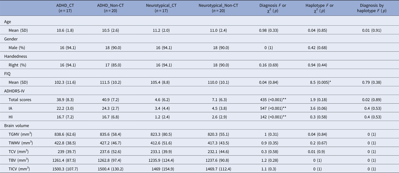
These 74 participants were classified as either carriers of the DAT1 rs27048 (C)/rs429699 (T) haplotype (CT, n = 17 in each of the ADHD and neurotypical groups) or non-carriers of this CT haplotype (non-CT, n = 20 in each of the ADHD and neurotypical groups). As shown in Table 1, there was no difference in age, gender, handedness, FIQ, total GMV, total WM volume, total CSF volume, total brain volume, and total intracranial volume between the ADHD and neurotypical groups. Similarly, the CT carriers did not differ from non-carriers on any of the demographic variables except that children with the CT haplotype had lower FIQ than those without the CT haplotype. By using non-parametric statisitcs, online Supplementary Table S1 showed no difference in the measures of PRM and SRM tasks in terms of diagnosis or the CT haplotype.
Demographics, ADHD symptomatology, and brain volume among the four groups

ADHD, attention-deficit/hyperactivity disorder; FD, frame-wise displacement; FIQ, full intelligence quotient; ADHD-RS-IV, ADHD Rating Scale-IV – Parent version: Investigator-Administered and Scored; IA, inattention subscale; HI, hyperactivity/impulsivity subscale; TGMV, total gray matter volume; TWMV, total white matter volume; TCV, total cerebral spinal fluid volume; TBV, total brain volume; TICV, total intracranial volume.
*p < 0.01, **p < 0.001.
Effects of ADHD on ReHo and GMV
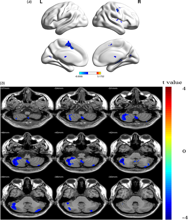
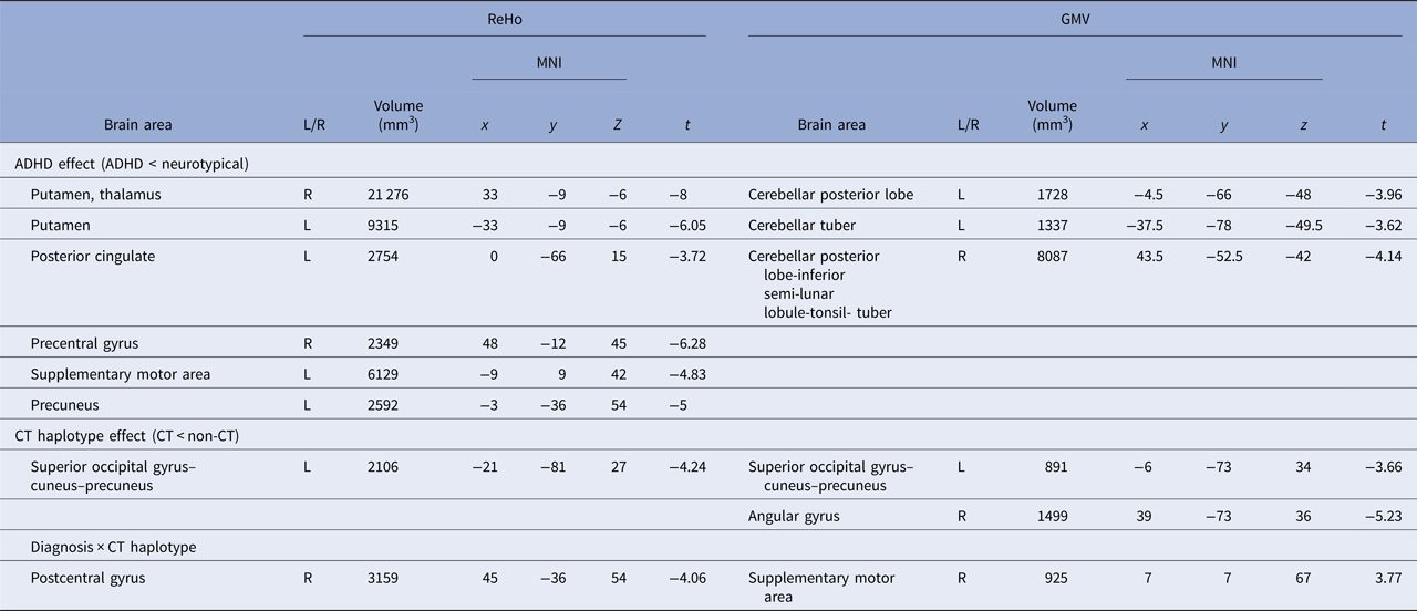
Compared with the neurotypical group, the ADHD group showed decresed ReHo in bilateral putamen, left posterior cingulate, supplementary motor area (SMA), precuneus, and right precentral gyrus (Table 2, Fig. 1a); and decreased GMV in bilateral cerebellar posterior lobes extending to the inferior semi-lunar lobule, tonsil, and tuber (Table 2, Fig. 1b).
Main effects of ADHD on ReHo and GMV. Relative to neurotypical children, children with ADHD demonstrate (a) decreased ReHo in bilateral putamen, left posterior cingulate, SMA, and precuneus, and right precentral gyrus and (b) decreased GMV in bilateral cerebellar posterior lobe, inferior semi-lunar lobule, tonsil, and tuber. Cold colors indicate lower ReHo and GMV in the ADHD group than in the neurotypical group; p < 0.05, Gaussian Random Field corrected. GMV, gray matter volume; ReHo, regional homogeneity; SMA, supplementary motor area.

Significant clusters show the effects of ADHD and the CT haplotype on regional homogeneity and gray matter volume

ADHD, attention-deficit/hyperactivity disorder; GMV, gray matter volume; ReHo, regional homogeneity.
Effects of CT haplotype on ReHo and GMV
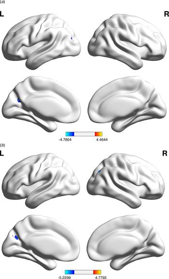
Compared with the non-carriers, the CT carriers showed significantly decreased ReHo in a cluster encompassing the left superior occipital gyrus, cuneus, and precuneus (Table 2, Fig. 2a); and significantly decreased GMV in a cluster encompassing the left superior occipital gyrus, cuneus, and precuneus, as well as the right angular gyrus (Table 2, Fig. 2b).
Main effects of the CT haplotype on ReHo and GMV. Relative to participants without the CT haplotype, those with this haplotype demonstrate (a) decreased ReHo in left superior occipital gyrus, cuneus, and precuneus and (b) decreased GMV in left superior occipital gyrus, cuneus, and precuneus, as well as in the right angular gyrus. Cold colors indicate lower ReHo and GMV in the CT group than in the non-CT group; p < 0.05, Gaussian Random Field corrected. GMV, gray matter volume; ReHo, regional homogeneity.

Effects of diagnosis × CT haplotype interaction on ReHo and GMV
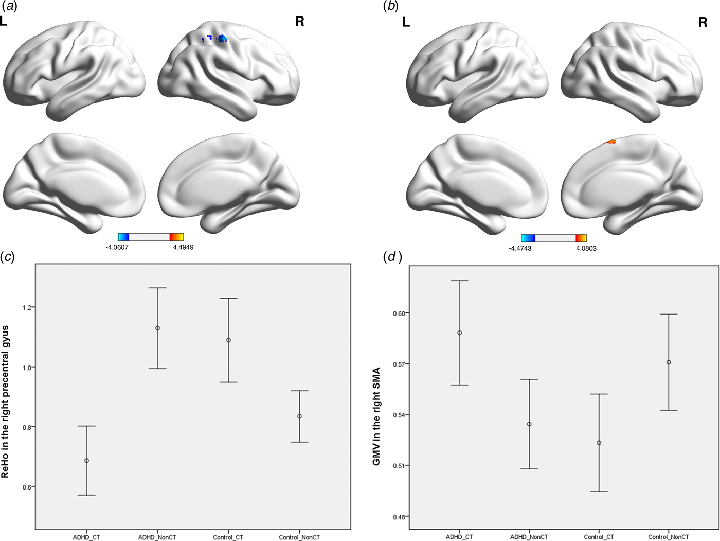
Significant diagnosis by CT haplotype interactions were found in the right postcentral gyrus for ReHo (Table 2, Fig. 3a) and in the right SMA for GMV (Table 2, Fig. 3b). In the ADHD group, ReHo in the right postcentral gyrus decreased (Fig. 3c) and GMV in the right SMA increased (Fig. 3d) with the CT haplotype, while in the neurotypical group the reverse pattern was found.
Effects of interaction of diagnosis × CT haplotype on ReHo and GMV. Significant interactions are found (a) in the right postcentral gyrus for ReHo and (b) in the right SMA for GMV; (c) ReHo in the right postcentral gyrus across the four groups; and (d) GMV in the right SMA across the four groups. p < 0.05, Gaussian Random Field corrected. Each error bar indicates 95% confidence interval. GMV, gray matter volume; ReHo, regional homogeneity, SMA, supplementary motor area.

Spearman's correlations between visual memory and DAT1-modulated differences in ReHo and GMV
Average ReHo and GMV values were extracted from the four clusters showing significant effects of the CT haplotype (the left superior occipital gyrus–cuneus–precuneus cluster in ReHo and GMV, respectively) or diagnosis × CT haplotype interaction (the right postcentral gyrus in ReHo, and the right SMA in GMV). For the ADHD-CT group (online Supplementary Table S2), there were negative correlations of total correct responses in PRM (Spearman's ρ = −0.61, q = 0.02) and SRM (Spearman's ρ = −0.56, q = 0.04) and positive correlations of mean latency of correct responses in PRM (Spearman's ρ = 0.56, q = 0.03) with the GMV of the left superior occipital gyrus–cuneus–precuneus cluster; negative correlations of total correct responses in SRM with the GMV in the right SMA (Spearman's ρ = −0.61, q = 0.01). For the ADHD-Non-CT group, there were positive correlations of mean latency of correct responses in PRM with the GMV in the right SMA (Spearman's ρ = 0.47, q = 0.05). We did not identify any significant correlations between visual memory and ReHo in the left superior occipital gyrus–cuneus–precuneus cluster and the right postcentral gyrus.
Discussion
This study is the first to demonstrate that ReHo and GMV in specific brain regions are affected by the DAT1 rs27048 (C)/rs429699 (T) haplotype in children with ADHD, and also the first to demonstrate that these DAT1-affected structural brain alterations are related to visual memory function in ADHD. Our results provide evidence that the DAT1 rs27048 (C)/rs429699 (T) haplotype influences intrinsic neural activity and morphometry in the posterior cortical areas, which are implicated in the pathophysiology of ADHD (Castellanos & Proal, Reference Castellanos and Proal2012). Despite possible advancements of disambiguating heterogeneity of ADHD, before interpreting our findings, we acknowledge that the present imaging-genetic-behavioral approach in a cross-sectional design is still inherently observational and correlational, and this caveat may limit the causal inference. Another major caveat is lack of replicated findings, which not only may be the case in our work, but also is the critical concern in the imaging genetic community. Future studies with additional levels of methodologies are needed, such as longitudinal follow-up cohorts, developing multimodal research strategies, and integrating results from different types of molecular studies (Vilor-Tejedor et al. Reference Vilor-Tejedor, Caceres, Pujol, Sunyer and Gonzalez2016; Klein et al. Reference Klein, Onnink, van Donkelaar, Wolfers, Harich and Shi2017).
Regardless of DAT1 genotypes, we identify reduced ReHo in bilateral putamen, the left posterior cingulate, SMA, precuneus, and right precentral gyrus, and reduced GMV in bilateral cerebellar posterior lobes, the inferior semi-lunar lobules, tonsils, and tuber in children with ADHD. Consistent with the previous literature (Alonso Bde et al. Reference Alonso Bde, Hidalgo Tobon, Dies Suarez, Garcia Flores, de Celis Carrillo and Barragan Perez2014; Gaddis et al. Reference Gaddis, Rosch, Dirlikov, Crocetti, MacNeil and Barber2015; Sutcubasi Kaya et al. Reference Sutcubasi Kaya, Metin, Tas, Buyukaslan, Soysal and Hatiloglu2016), neurobiological alterations of these regions are implicated in the pathophysiology of ADHD. These regions are also functionally involved in neuropsychological difficulties in patients with ADHD, especially during sensory processing (Ghanizadeh, Reference Ghanizadeh2011), movement organization (D'Agati et al. Reference D'Agati, Casarelli, Pitzianti and Pasini2010), information integration (Metin et al. Reference Metin, Roeyers, Wiersema, van der Meere, Thompson and Sonuga-Barke2013), and emotional control (Wehmeier et al. Reference Wehmeier, Schacht and Barkley2010).
Our imaging genetic findings highlight differential brain function and structure related to the DAT1 rs27048 (C)/rs429699 (T) in the posterior cortical regions. Compared with the non-CT group, the CT group shows decreased ReHo and GMV in the left superior occipital gyrus–cuneus–precuneus area. Our present study implies that the DAT1 rs27048 (C)/rs429699 (T) haplotype is associated with functional and structural alterations in the cuneus that express DATs (Ciliax et al. Reference Ciliax, Drash, Staley, Haber, Mobley and Miller1999), as well as in the precuneus and superior occipital gyrus that are sparse in DATs (Ciliax et al. Reference Ciliax, Drash, Staley, Haber, Mobley and Miller1999). Although the exact function of this CT haplotype is unknown, it may influence neural activity and structure through a combination of the direct effects on dopaminergic function and the indirect effects on the function of the posterior brain regions, resulting in the presentation of different ReHo and GMV values between genotypes. For a full understanding of the impact of the DAT1 rs27048 (C)/rs429699 (T) haplotype on ADHD, our next step is to examine genetic effects on the individual differences in response to methylphenidate for ADHD, and in the dose of methylphenidate required to bring about improvement.
Occipital cortex interacts with the dorsal attentional network to maintain visual attention (Shulman et al. Reference Shulman, Astafiev, Franke, Pope, Snyder and McAvoy2009) and suppress attention to irrelevant visual stimuli (Capotosto et al. Reference Capotosto, Babiloni, Romani and Corbetta2009). Moreover, the cuneus and precuneus have been reported to be affected in ADHD and to be involved in vision and the default mode network (Castellanos et al. Reference Castellanos, Margulies, Kelly, Uddin, Ghaffari and Kirsch2008; Alonso Bde et al. Reference Alonso Bde, Hidalgo Tobon, Dies Suarez, Garcia Flores, de Celis Carrillo and Barragan Perez2014). A previous study has suggested that patients with ADHD display significant alterations in connectivity in the left cuneus during working memory processing compared with healthy controls (Wolf et al. Reference Wolf, Plichta, Sambataro, Fallgatter, Jacob and Lesch2009). Another report also demonstrates that individuals with ADHD have altered ReHo during the resting state in the bilateral cuneus and precuneus (Wang et al. Reference Wang, Jiao, Tang, Wang and Lu2013). Our present study extends these findings to show the association of DAT1 genotypes with the resting-state and structural alterations in the left superior occipital gyrus, cuneus, and precuneus in children with ADHD.
Despite comparable overall performance in PRM and SRM across the four groups in our present imaging genetic study, previous neuropsychological studies with larger sample sizes have demonstrated that children with ADHD make fewer correct responses in PRM and SRM than healthy controls (Rhodes et al. Reference Rhodes, Coghill and Matthews2005; Shang & Gau, Reference Shang and Gau2011). For the ADHD-CT group, we find that the GMV in the left superior occipital gyrus, cunues, and precuneus is positively correlated with the reaction time in PRM, and negatively correlated with the accuracy in PRM and SRM. Our results suggest that decreased GMV in the posterior cortical regions are associated with increased accuracy and faster reaction time of visual memory in children with ADHD who are the carriers of the CT haplotype. A recent study has demonstrated positive correlations between increased cortical thickness in the left occipital cortex and inattention severity in adults with ADHD (Soros et al. Reference Soros, Bachmann, Lam, Kanat, Hoxhaj and Matthies2017). Structural imaging studies have also revealed WM abnormalities in the left occipital lobe and cuneus which modulate visual information processing and attention in children with ADHD (Lei et al. Reference Lei, Ma, Du, Shen, Jin and Gong2014). Taken together, our findings suggest that genetically determined differences in the posterior brain regions may contribute to visual memory performance in ADHD children with the CT haplotype.
Combining our findings of gene–brain–behavior linkage within one study and several previous studies describing dopamine-posterior brain regions (Nakama et al. Reference Nakama, Chang, Fein, Shimotsu, Jiang and Ernst2011; Albrecht et al. Reference Albrecht, Roberts, Price, Lee, Iyyalol and Martin-Iverson2016), posterior brain regions-visual memory (Simons et al. Reference Simons, Graham, Owen, Patterson and Hodges2001; Guggenmos et al. Reference Guggenmos, Thoma, Cichy, Haynes, Sterzer and Richardson-Klavehn2015), and dopamine-visual memory (Rhodes et al. Reference Rhodes, Coghill and Matthews2004; Reference Rhodes, Coghill and Matthews2006) relationships, three major conclusions are made. First, dexamphetamine, a dopamine-releasing agent, altered the resting-state electroencephalogram of healthy participants in the posterior brain regions, including parietal, occipital–temporal, and occipital regions (Albrecht et al. Reference Albrecht, Roberts, Price, Lee, Iyyalol and Martin-Iverson2016). In addition, methamphetamine users showed altered GMV in the occipital regions (Nakama et al. Reference Nakama, Chang, Fein, Shimotsu, Jiang and Ernst2011). Second, visual memory involves a network of the posterior brain regions, including parietal and occipital lobes (Simons et al. Reference Simons, Graham, Owen, Patterson and Hodges2001; Guggenmos et al. Reference Guggenmos, Thoma, Cichy, Haynes, Sterzer and Richardson-Klavehn2015). Third, the deficits in visual memory performance were restored by treatment with methylphenidate in children with ADHD (Rhodes et al. Reference Rhodes, Coghill and Matthews2004, Reference Rhodes, Coghill and Matthews2006), which inhibited the action of DAT to achieve the therapeutic effects. By demonstrating the linkages between DAT1, structural alterations in the posterior brain regions, and visual memory performance, our present work may contribute to a model of visual memory enabled by dopamine-related brain regions in children with ADHD.
A significant diagnosis × CT haplotype interaction is observed in the right SMA on GMV, with the ADHD-CT group having the greatest GMV among the four groups (Fig. 3d). Moreover, the GMV in the right SMA is correlated with several measures of PRM and SRM in the ADHD-CT and ADHD-Non-CT groups. Previous morphological studies have demonstrated that the cortical thickness of the SMA is increased in ADHD patients (Duerden et al. Reference Duerden, Tannock and Dockstader2012), and the SMA has been found to be involved in visual–spatial memory in adults with ADHD (Ko et al. Reference Ko, Yen, Yen, Chen, Lin and Wang2013). Our findings suggest that the DAT1-related GMV alterations in the right SMA may be associated with the visual memory performance in children with ADHD.
Results from this study should be interpreted in the context of some methodological limitations. First, the sample size of the current study is modest relative to samples in genetic studies, but it is comparable to other studies that have examined imaging in the context of genetics (Bedard et al. Reference Bedard, Schulz, Cook, Fan, Clerkin and Ivanov2010; Wiggins et al. Reference Wiggins, Bedoyan, Peltier, Ashinoff, Carrasco and Weng2012). Second, we include dADHD children with a wide age range, potentially confounding our data with multiple stages of brain development and gene expression. Future studies with a narrower age range are warranted.
Several features of this study constitute its strengths. First, only drug-naïve children with ADHD were recruited to avoid the potential confounding effects of medication history. Second, this study consists of an ethnically homogeneous sample, which is optimal for examining genetic differences in neural structure and activity, since allelic frequencies of the DAT1 polymorphisms may differ widely across ethnic populations (Banoei et al. Reference Banoei, Chaleshtori, Sanati, Panahi, Majidizadeh and Rostami2007; Bhaskar et al. Reference Bhaskar, Thangaraj, Mulligan, Wasnik, Nandan and Sharma2009). Third, the combination of neuropsychological and both structural and functional neuroimaging assessments in this study provides detailed and specific information for our understanding of the gene–brain–behavior relationship in ADHD.
In conclusion, our present study indicates that alterations of ReHo and GMV in the posterior brain regions are related to the DAT1 haplotype rs27048 (C)/rs429699 (T). Although more research is definitely needed in this area, our findings add to those already published in describing the differential effects of DAT1 genotype on the neuroanatomy and neurophysiology in ADHD (Wu et al. Reference Wu, Yang and Wang2014) with illustrating the functional and structural consequences of the DAT1 rs27048 (C)/rs429699 (T) haplotype.
Supplementary material
The supplementary material for this article can be found at https://doi.org/10.1017/S0033291718000144
Acknowledgements
This study was supported by grants NSC96-2628-B-002-069-MY3, NSC98-2314-B-002-051-MY3, and NSC99-2321-B-002-037 from National Science Council (Taiwan) and grants NHRI-EX98-9407PC and NHRI-EX100-0008PI from National Health Research Institute (Taiwan).
Declaration of interest
None.