Introduction
Histological examination of nasal mucosal tissue is essential when studying the aetiology of conditions such as chronic sinusitis, polyposis, allergic rhinitis and asthma. Tissue remodelling is an important feature of these diseases, often being associated with increased severity or chronicity, although the mechanisms involved are unclear and require investigation.Reference Watelet, Van Zele, Gjomarkaj, Canonica, Dahlen and Fokkens1 Multiple labelling of different molecules on the same section is highly desirable, as it allows co-localisation of different factors involved in remodelling. However, most established protocols use paraffin-embedded human tissue.Reference Mumbuc, Karakok, Baglam, Karatas, Durucu and Kibar2 This restricts the tissue markers available, as many antibodies will not recognise epitopes after paraffin-embedding. Thus, research into airway remodelling is currently limited to studying a relatively small set of proteins.
Using fresh tissue allows whole mount staining (using relatively large tissue samples); the sample can then be examined with confocal microscopy and analysed in three dimensions. This approach could yield a wider range of information, avoiding problems with heterogeneity of structure by examining a larger mass of tissue, and correcting for the large changes in tissue volume typically seen in the nasal mucosa due to the nasal cycle. This approach has been successfully applied to the examination of blood vessels with fluorescent lectins in animal modelsReference Williams, Cartland, Hussain and Egginton3 and in a proof-of-principle human study.Reference Hamid, Daly and Campbell4 However, the use of lectins can be problematic in large human studies, as no single lectin will stain a population with heterogeneous blood groups.Reference Capaldi, Dunn, Sewry and Dubowitz5 Antibody staining, such as cluster of differentiation (CD) 31 or CD34, is poor at consistently staining contiguous structures such as blood vessels.
As different lectins bind to different sugar moieties, we reasoned that a mixture of the three different lectins reported to stain human blood vesselsReference Capaldi, Dunn, Sewry and Dubowitz5 would successfully stain a far greater proportion of patient samples, and would therefore provide a more robust staining protocol for visualising capillary beds in three dimensions, allowing further investigation of vasculature changes during tissue remodelling in patients.
Materials and methods
Subjects
Patients were recruited from University Hospitals Birmingham NHS Trust, UK, with local ethical committee approval (numbers 0692 and 06/Q2702/15). Patients' informed consent was obtained. Polyp and ipsilateral inferior turbinate specimens, 1 cm back from the anterior edge, were obtained from patients undergoing routine polypectomy. Previous studies have found no difference in vascularity between the different areas of the lateral nasal wall.Reference Philpott, Wild, Guzail and Murty6 Inferior turbinate specimens from the same anatomical site were also taken from patients undergoing routine septoplasty or septorhinoplasty.
Immunohistochemical staining
Samples taken for confocal analysis were immediately fixed in 4 per cent buffered formalin and left for 24 hours. Samples were then washed in three changes of phosphate-buffered saline for a further 24 hours, before being stained whole with dilutions of either fluorescein-conjugated Ulex europeaus lectin (Vector Labs, Peterborough, U.K.) or a fluorescein-conjugated lectin mixture (1:1:1 Ulex europeaus, Griffonia simplicifolia and Euonymus europeaus; Vector Labs), in order to account for different affinities associated with different blood groups.Reference Capaldi, Dunn, Sewry and Dubowitz5 Samples were then washed and stored in phosphate-buffered saline until viewed under a confocal microscope (DM IRE2; Leica, Milton Keynes). A total of 60 samples were stained with the single lectin protocol, and 50 different samples were stained with the triple lectin protocol.
Samples taken for immunostaining were immediately snap-frozen in liquid nitrogen cooled isopentane in the operating theatre, and 8 µm sections were subsequently cut on a cryostat. These were allowed to air-dry before being fixed in 4 per cent buffered formalin (for 1, 5, 10, 20 or 60 minutes) or ice-cold acetone (for 10, 30, 60 or 300 seconds). Antigen retrieval was performed in citrate buffer (10 mM citric acid adjusted to pH 6.0 with NaOH, 0.05 per cent Tween® 20) heated to 95°C for 5 minutes then allowed to cool to room temperature. Slides were blocked for 30 minutes at room temperature in wash buffer (either 5 per cent fetal bovine serum or 1.5 per cent weight/volume bovine serum albumin, 0.6 per cent volume/volume Triton® X-100 in phosphate-buffered saline, or 5 per cent fetal bovine serum in phosphate-buffered saline), then incubated for 2 hours with primary antibodies (Ki-67 and proliferating cell nuclear antigen; Dako, Santa Cruz, California U.S.A.) diluted in wash buffer to label proliferating nuclei. Sections were then washed in phosphate-buffered saline and incubated for 1 hour with secondary antibody (Tetramethyl Rhodamine Iso-Thiocyanate polyclonal rabbit anti-mouse; Dako) diluted in wash buffer. Sections were rinsed in phosphate-buffered saline and then incubated for 30 minutes with lectin to stain blood vessels. Sections were washed in phosphate-buffered saline, rinsed with dH20 and mounted using Vectashield containing 4′,6-diamidino-2-phenylindole, in order to label all nuclei and enable calculation of a proliferation index (Ki67 count/4′,6-diamidino-2-phenylindole count), based on counts of four 500 µm × 500 µm sampling squares, using inclusion/exclusion lines. All counts were made either on the same section or on a serial section from the same sample.
Results and analysis
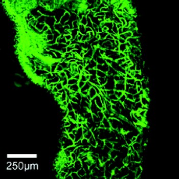
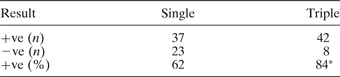
Samples from a total of 60 patients were stained using Ulex europeaus lectin, and compared to samples from 50 patients stained with a combination of Ulex europeaus, Griffonia simplicifolia and Euonymus europeaus. Samples that showed stained blood vessels were designated as positive, and samples in which blood vessels were not visualised were restained. Repeated failure to stain capillaries was seen in 23 of the single-lectin samples and in eight of the triple-stained samples (p < 0.005; Pearson's chi-square test), representing a significant difference between single and triple lectin methods (see Table I and Figure 1).
Inferior turbinate mucosa stained with whole mount protocol using three lectins.

Comparison of single Ulex europeaus lectin staining vs triple lectin protocol
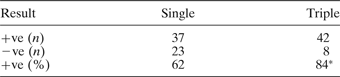
* Staining was interpreted as +ve = samples stained well after up to 3 attempts; −ve = samples repeatedly failed to stain
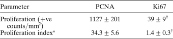
Both proliferating cell nuclear antigen and Ki67 staining gave the most consistent results when sections were attached to poly-L-lysine coated slides and fixed in 4 per cent buffered formalin for 20 minutes. Both stains gave best results with a wash buffer containing Triton X-100. Staining with Ki67 required antigen retrieval, and the optimal antibody concentration was 1:100 for both the primary and secondary antibodies. Staining with proliferating cell nuclear antigen required antibody concentrations of 1:50 for primary and 1:100 for secondary antibodies. A significant difference was seen in the level of proliferation marked by proliferating cell nuclear antigen and Ki67 staining (p < 0.05), with both the count per mm2 and the proliferation index being significantly higher when measured using proliferating cell nuclear antigen staining compared with Ki67 (Table II).
Comparison of PCNA and Ki67 as measures of proliferation
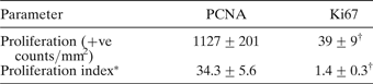
Data are expressed as mean ± standard error of the mean. *Proliferation index expressed as percentage of total nuclei; n = 12. †p < 0.05; Student's t-test. PCNA = proliferating cell nuclear antigen; +ve = positive
Discussion
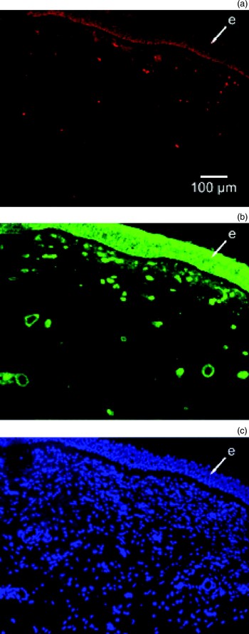
The objective of this study was to develop a robust multiple staining protocol using samples that had not been embedded in paraffin, so that a broader range of antibodies could be used. The use of lectins to stain blood vessels is well established in animal models, but it is less widely used in humans due to variability in staining between individuals with different blood groups.Reference Capaldi, Dunn, Sewry and Dubowitz5 This study found a marked improvement when using a mixture of the three most commonly used human-specific lectins (which bind to different sugar moieties present on the endothelium), compared with use of a single lectin. Lectins were also seen to bind to nasal mucosa epithelium (Figure 2), as previously described;Reference Hassid, Salmon, Brugmans, Dawance, Kiss and Gabius7, Reference Huang, Jing, Li, Zhou, Xiao and Ma8 however, as this layer is avascular, it does not interfere with capillary visualisation. Lectins have several advantages over other immunohistochemical methods in the staining of blood vessels. They can be readily conjugated to several fluorophores, allowing multiple stains to be used on the same section. The one-step protocol reduces the signal degradation associated with other multiple labelling methods, as well as saving time. Lectins are relatively inexpensive, allowing whole-mount stains to be performed economically. Finally, using lectins avoids the problems commonly associated with the use of two primary antibodies raised in the same host species.
Triple stain of a single section of inferior turbinate mucosa under different fluorescent illuminations. (a) Rhodamine-conjugated Ki67 stain; (b) fluorescein- conjugated lectin stain; (c) 4′,6-diamidino-2-phenylindole stain for nuclei. e = epithelial layer.

Proliferating cell nuclear antigen (also termed cyclin) is a protein that acts as a processivity factor for deoxyribonucleic acid polymerase delta in eukaryotic cells, and the staining of this antigen is well documented for paraffin-embedded sections. However, this study shows that staining for proliferating cell nuclear antigen is not appropriate for frozen sections, as the calculated proliferation index is unreasonably high. Normal mucosal tissue is highly unlikely to have 34 per cent of total nuclei undergoing mitosis at any one time (Table II). We surmise that this antibody stains proliferating cell nuclear antigen epitopes in fresh human cells that are not in their biologically active form, as we have also seen unrealistically high labelling indices in serum-starved Human Umbilical Vein Endothelial Cells monolayers where mitosis is arrested (J Williams et al., unpublished data). Staining for Ki67 is far more successful after a brief formalin fixation with subsequent antigen retrieval. Results for proliferation indices for fresh tissue using this method broadly agree with those published for tissue prepared with paraffin-embedding.Reference Hiroshima, Iyoda, Shibuya, Hoshino, Haga and Toyozaki9
Conclusions
This study demonstrates a novel and robust method for analysing the capillary bed and the extent of cellular proliferation in unfixed samples of respiratory mucosa. This method will allow further investigation into the vascular response during tissue remodelling in human biopsies, and could thus help elucidate the aetiology and pathogenesis of tumours, chronic sinusitis, nasal polyposis, allergic rhinitis and asthma. The protocols used are detailed below.
Protocols
Lectin staining protocol
Place samples in standard hospital pathology formalin pots for 24 hours at 4°C. Stain with a 1:100 dilution, in phosphate-buffered saline, of a 1:1:1 stock of fluorescein-conjugated Ulex europeaus, Griffonia simplicifolia and Euonymus europeaus lectins for 24 hours at 4°C. Wash three times with phosphate-buffered saline for 1 hour each wash, before examining under confocal microscopy.
Triple staining protocol
Snap-freeze samples in liquid nitrogen cooled isopentane and store at −80°C, before cutting 8 µm sections on a cryostat. Air-dry the sections, perform an antigen retrieval with citrate buffer (10 mM citric acid adjusted to pH 6.0 with NaOH, 0.05 per cent Tween 20) heated to 95°C for 5 minutes then allowed to cool to room temperature. Fix for 20 minutes in 4 per cent buffered formalin, then block for 30 minutes using a wash buffer consisting of 1.5 per cent weight/volume bovine serum albumin and 0.6 per cent volume/volume Triton X-100 in phosphate-buffered saline. Incubate for 2 hours with primary antibody (Ki-67, Dako) diluted 1:100 in wash buffer. Wash with three changes of phosphate-buffered saline for 5 minutes each, then incubate for 1 hour with secondary antibody (Tetramethyl Rhodamine Iso-Thiocyanate polyclonal rabbit anti-mouse; Dako) diluted 1:100 in wash buffer. Rinse with phosphate-buffered saline, then incubate for 30 minutes with 1:100 of stock 1:1:1 fluorescein-conjugated Ulex europeaus, Griffonia simplicifolia and Euonymus europeaus lectins. Wash with three changes of phosphate-buffered saline for 5 minutes each, rinse with dH20 and mount with VectaShield containing 4′,6-diamidino-2-phenylindole.
Acknowledgements
This study received financial support from the ‘Get A Head’ charity (UK).