Kawasaki disease is an acute multisystemic vasculitis of unknown aetiology that occurs in infants and children.Reference Kawasaki 1 , Reference Kawasaki, Kosai, Okawa, Shigematsu and Yanagawa 2 Coronary arterial lesions seriously affect patient prognosis.Reference Kuo, Wang and Liang 3 Although studies have shown that some indicators are associated with coronary artery lesions in Kawasaki disease, there are no reliable indicators for predicting coronary artery lesions in the early stage of disease.Reference Wang, Wang and Gong 4 – Reference Kobayashi, Inoue and Takeuchi 8
Red blood cell distribution width, representing the size of circulating erythrocytes, is a component of the complete blood count. Red blood cell distribution width has been used for the diagnosis of differential anaemia due to its increase in cases of haemolysis, blood transfusion, or ineffective erythropoiesis; however, recent studies have shown that elevated red blood cell distribution width is associated with adverse outcome in various clinical conditions, including thromboembolic events, cardiovascular diseases, and respiratory diseases.Reference Aung, Ling and Cheng 9 – Reference Zorlu, Bektasoglu and Guven 12 The predictive value of red blood cell distribution width for patients with Kawasaki disease has not been studied yet. In the present study, we aimed to investigate the relationship between red blood cell distribution width and coronary artery lesions in patients with Kawasaki disease.
Materials and methods
Materials
Between August, 2009 and December, 2012, 422 patients with Kawasaki disease – 267 boys and 155 girls – treated at the Children’s Hospital, Zhejiang University School of Medicine, were enrolled into this study. These patients had a median age of 2.17 years (range 2 months to 14.7 years). All the patients were diagnosed with Kawasaki disease according to the criteria established by the Kawasaki Disease Research Committee.Reference Ayusawa, Sonobe and Uemura 13
The present study was approved by the ethics committee of the Children’s Hospital, Zhejiang University School of Medicine, and was based on the institution’s guidelines for human studies. The investigation conformed to the principles outlined in the Declaration of Helsinki. Informed consent was obtained from the patients’ parents.
Methods
Echocardiography was used to detect coronary artery lesions during the course of the disease, and left ventricular ejection fraction was also measured. Patients were divided into two groups: coronary artery lesions group, which included patients who had coronary artery lesions within 3 months after the diagnosis, and without coronary artery lesions group, which included patients who did not develop coronary artery lesions within 3 months after the diagnosis. Coronary artery lesions were defined according to internal lumen diameter of >2.5 mm in children <3 years of age, >3 mm in children of 3–9 years of age;, and >3.5 mm in children of 9–14 years of age; internal diameter of a segment measuring ⩾1.5 times that of an adjacent segment; and lumen that is clearly irregular.Reference Fu, Gong and Xie 7 All the children received intravenous immunoglobulin treatment. In addition to aspirin, dipyridamole and warfarin were included for patients with coronary artery lesions depending on the severity of the coronary artery lesions.
Venous blood samples were collected from the patients with Kawasaki disease at the acute stage of disease before intravenous immunoglobulin treatment; red blood cell distribution width levels were measured on admission; and clinical and laboratory parameters were recorded for each subject. Baseline red blood cell distribution width, haemoglobin levels, and counts of lymphocytes, neutrophils, monocytes, platelets, and white blood cells were measured. Concentrations of C-reactive protein, aspartate aminotransferase, alanine aminotransferase, fibrin, serum Th1 and Th2 cytokines, including interferon-γ, tumour necrosis factor-α, interleukin-10, interleukin-6, interleukin-4, and interleukin-2, erythrocyte sedimentation rate, and activated partial thromboplastin time were measured at the same time.
Statistics
Continuous variables were expressed as mean±standard deviation or median with interquartile range (25–75% percentile); categorical variables were expressed as number and percentage. A χ2 test or Fisher’s exact test was performed to compare categorical variables. The normality of distribution of variables was assessed using the Kolmogorov–Smirnov test. Student’s t-test was used for normally distributed continuous variables; the Mann–Whitney U-test was used when the distribution was skewed. The discrimination of red blood cell distribution width for coronary artery lesions was evaluated using the area under the receiver operating characteristic curve. The optimal cut-off point for receiver operating characteristic curves was determined for maximising the sensitivity and specificity of the red blood cell distribution width values. Patients with Kawasaki disease were categorised into two groups based on the cut-off value. Logistic regression analysis was used to identify the independent predictor of coronary artery lesions. Variables that had a p-value <0.1 in the univariate analysis were included in this model; two-tailed p-values <0.05 were considered to be significant and the confidence interval was 95%. All the statistical analyses were performed using SPSS software version 16.0 (SPSS Inc., Chicago, Illinois, United States of America).
Results
Baseline characteristics of patients with Kawasaki disease
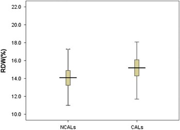
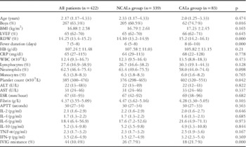
A total of 422 patients, who met the inclusion criteria, with boys comprising 63% of the cohort, were enrolled into our study; 83 patients of the coronary artery lesions group (62 boys and 21 girls) were found to have coronary artery lesions during the disease process within 3 months after the diagnosis, and 339 patients of the without coronary artery lesions group (205 boys and 134 girls) did not develop coronary artery lesions within 3 months after the diagnosis. Table 1 shows the differences between the coronary artery lesions group and without coronary artery lesions group regarding demographic, clinical, and echocardiographic properties. There were no significant differences in age, body mass index, haemoglobin levels, counts of lymphocytes, neutrophils, monocytes, and white blood cells, concentrations of C-reactive protein, aspartate aminotransferase, alanine aminotransferase, serum Th1 and Th2 cytokines, and fibrin, activated partial thromboplastin time, erythrocyte sedimentation rate, and left ventricular ejection fraction. In patients with coronary artery lesions, the levels of platelet count and red blood cell distribution width were higher, boys and patients with intravenous immunoglobulin resistance were more common, and fever duration was longer. Figure 1 shows the median red blood cell distribution width values in the coronary artery lesions and without coronary artery lesions groups.
Comparison of red blood cell distribution width (RDW) values between CALs group and NCALs group. CALs=patients with coronary artery lesions; NCALs=patients without coronary artery lesions.

Baseline characteristics of the patients with Kawasaki disease.

ALT=alanine aminotransferase; APTT=activated partial thromboplastin time; AST=aspartate aminotransferase; BMI=body mass index; CALs=patients with coronary artery lesions; CRP=C-reactive protein; ESR=erythrocyte sedimentation rate; HB=haemoglobin; IFN-γ=interferon-γ; IL=interleukin; IVIG=intravenous immunoglobulin; LVEF=left ventricular ejection fraction; NCALs=patients without coronary artery lesions; RDW=red blood cell distribution width; TNF-α=tumour necrosis factor-α; WBC=white blood cell count
Comparison of the characteristics of patients with Kawasaki disease according to red blood cell distribution width values
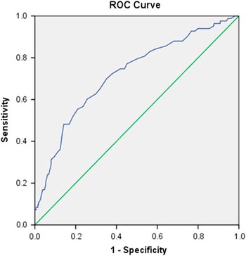
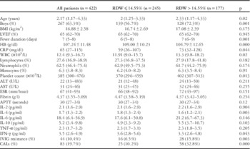
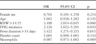
According to the receiver operating characteristic curve analysis, the optimal cut-off value of red blood cell distribution width level on admission for predicting coronary artery lesions was >14.55%, with a sensitivity of 70% and specificity of 65% (area under the curve was 0.721, 95% confidence interval was 0.658–0.784; p=0.000) (Fig 2). To evaluate the relationship between red blood cell distribution width and clinical outcome, patients were divided into two groups according to this red blood cell distribution width cut-off value (⩽14.55 versus >14.55%). The properties were compared between the two groups (Table 2). White blood cell counts, C-reactive protein levels, and platelet counts were higher in patients with higher red blood cell distribution width, but haemoglobin, interleukin-4, and interferon-γ levels were lower in patients with lower red blood cell distribution width. In addition, fever duration was longer, and boys and patients with intravenous immunoglobulin resistance were more common among patients with higher red blood cell distribution width. Moreover, patients were younger in the lower red blood cell distribution width group. According to the logistic regression analysis, red blood cell distribution width >14.55%, fever duration >14 days, and intravenous immunoglobulin resistance were independent predictors of coronary artery lesions in patients with Kawasaki disease (Table 3).
The receiver operating characteristic (ROC) curve analysis for red blood cell distribution width levels in predicting CALs. AUC=0.721 (0.658–0.784). AUC=area under the curve; CALs=patients with coronary artery lesions.

Comparison of the characteristics of the patients with Kawasaki disease according to red blood cell distribution width values.

ALT=alanine aminotransferase; APTT=activated partial thromboplastin time; AST=aspartate aminotransferase; BMI=body mass index; CALs=coronary artery lesions; CRP=C-reactive protein; ESR=erythrocyte sedimentation rate; HB=haemoglobin; IFN-γ=interferon-γ; IL=interleukin; IVIG=intravenous immunoglobulin; LVEF=left ventricular ejection fraction; RDW=red blood cell distribution width; TNF-α=tumour necrosis factor-α; WBC=white blood cell count
Logistic regression analysis of Kawasaki disease with CALs.

CI=confidence interval; IVIG=intravenous immunoglobulin; OR=odds ratio; RDW=red blood cell distribution width
Discussion
Our study showed that 83 (19.6%) patients had coronary artery lesions during the disease process, within 3 months after the diagnosis, and the most remarkable finding was the relationship between red blood cell distribution width on admission with coronary artery lesions in patients with Kawasaki disease. Red blood cell distribution width was significantly higher in patients with coronary artery lesions than those without coronary artery lesions. Logistic regression analysis revealed that higher red blood cell distribution width on admission was an indicator of coronary artery lesions in patients with Kawasaki disease before and after adjusting for other potential confounders. In addition to its relationship with coronary artery lesions, boys and patients with intravenous immunoglobulin resistance were more likely to have higher red blood cell distribution width. We found that the patients with red blood cell distribution width >14.55% had a higher proportion of intravenous immunoglobulin resistance (15.8%) than those with red blood cell distribution width ⩽14.55% (6.5%); they also had a longer fever duration, higher levels of C-reactive protein, platelet count, and white blood cell count, and lower levels of haemoglobin. It is possible that the patients with intravenous immunoglobulin resistance had severe inflammatory reaction, leading to higher red blood cell distribution width values. To the best of our knowledge, this is the first study to evaluate the relationship between red blood cell distribution width and coronary artery lesions in patients with Kawasaki disease.
Red blood cell distribution width is a parameter that demonstrates variations in the dimensions of circulating erythrocytes – that is, anisocytosis. Up until now, red blood cell distribution width values have been assessed as haematological indications for malnutritional anaemias and diseases leading to the destruction of erythrocytes; however, numerous studies have reported that higher red blood cell distribution width is an independent predictor of mortality in a broad spectrum of cardiovascular diseases such as heart failure, stable coronary disease, acute coronary syndrome, acute pulmonary embolism, and pulmonary hypertension.Reference Aung, Ling and Cheng 9 , Reference Veeranna, Zalawadiya, Panaich, Patel and Afonso 11 , Reference Zorlu, Bektasoglu and Guven 12 , Reference Rhodes, Wharton, Howard, Gibbs and Wilkins 14 – Reference Núňez, Núňez and Rizopoulos 16 Besides mortality, red blood cell distribution width can also predict morbidity, such as recurrent hospital admissions or recurrent infarctions in patients with acute coronary syndromes.Reference Wang, Hua, Bai and Tang 15 , Reference Felker, Allen and Pocock 17 Nevertheless, no study has investigated the relationship between red blood cell distribution width and coronary artery lesions in patients with Kawasaki disease.
Although the mechanism of the relationship between increased red blood cell distribution width and poor outcome is unclear, some relevant studies related to oxidative stress and inflammatory reactions have led the way.Reference Semba, Patel and Ferrucci 18 – Reference Lippi, Targher, Montagnana, Salvagno, Zoppini and Guidi 20 High red blood cell distribution width reflects the presence of immature and larger red blood cells in the circulation, and can be found in any condition when erythrocytes are destructed or produced ineffectively. As inflammation and oxidative stress also play a role in the progression of cardiovascular diseases, red blood cell distribution width is thought to be an important marker for cardiovascular diseases.Reference Tsuboi, Miyauchi and Kasai 21 A number of studies have also demonstrated that higher red blood cell distribution width level is a potential predictive and prognostic marker for coronary and peripheral arterial diseases.Reference Ye, Smith and Kullo 22 – Reference Akyel, Çelik and Öksüz 24 Although the pathogenesis of Kawasaki disease is not completely understood, multiple mechanisms are suggested to be involved and inflammation may play a role. Inflammation may alter erythropoiesis, iron homoeostasis, the proliferation of erythroid progenitor cells, lifespan of red blood cells, and red cell membrane deformability, which might increase anisocytosis.Reference Weiss and Goodnough 25 We found that the red blood cell distribution width in this group of Kawasaki disease patients was 14.25 (13.4–15.2), significantly higher than the levels (13.1±1.0) in normal children (data from Park et al).Reference Park and Kim 26 Our data suggest that red blood cell distribution width might be regarded as a valuable and sensitive marker for a high level of inflammatory activity in patients with Kawasaki disease, and it is independent of haemoglobin levels. Lippi et al found a correlation between high red blood cell distribution width and elevated indices of inflammation, such as elevated erythrocyte sedimentation rate and C-reactive protein levels. This correlation was independent of concomitant diseases and was also demonstrated in anaemic patients.Reference Lippi, Targher, Montagnana, Salvagno, Zoppini and Guidi 20 According to our findings, C-reactive protein level and white blood cell counts were higher in patients with higher red blood cell distribution width. This suggests that elevated red blood cell distribution width on admission indicates a severe inflammatory process, which may be an early indicator of poor prognosis in patients with Kawasaki disease.
Patel et al found an inverse correlation between red blood cell distribution width and erythrocyte deformability in a community population. Red blood cell distribution width >14% was shown to decrease RBC deformability.Reference Patel, Mohanty, Kanapuru, Hesdorffer, Ershler and Rifkind 27 Increased red blood cell distribution width may be accompanied by a loss of red blood cell deformability, which could jeopardise red blood cell transit across the microvasculature, compromising not only tissue oxygen delivery but also its vascular “antioxidant” function.Reference Minetti, Agati and Malorni 28 Lower erythrocyte elongation index can potentially result in impaired blood flow in the coronary microcirculation, resulting in hypoxia and increased cardiovascular risks.Reference Patel, Mohanty, Kanapuru, Hesdorffer, Ershler and Rifkind 27 Red blood cell distribution width may be an integrative marker, reflecting multiple biological imbalances contributing to higher coronary artery lesion risks in Kawasaki disease patients, such as oxidative stress, tissue hypoxia, neuro-humoral hyperactivity, endothelial dysfunction, and chronic inflammation.Reference Lippi, Targher, Montagnana, Salvagno, Zoppini and Guidi 20 , Reference Minetti, Agati and Malorni 28 – Reference Patel, Ferrucci, Ershler, Longo and Guralnik 31 Although more comprehensive studies that can explain the mechanism of the relationship between coronary artery lesions and red blood cell distribution width values are needed, our results suggest that increased red blood cell distribution width is an independent predictor of coronary artery lesions in patients with Kawasaki disease. The exact cut-off value of red blood cell distribution width for identifying high-risk patients is different in the literature, including the median value of the study cohorts or receiver operating characteristic curves for optimum cut-off levels. In light of the data from studies carried out so far, red blood cell distribution width approximately above 15% is the threshold.Reference Zorlu, Bektasoglu and Guven 12 , Reference Felker, Allen and Pocock 17 According to our findings, red blood cell distribution width >14.55% is an optimal threshold that may alert clinicians to take more aggressive treatment measures in order to prevent poor outcome.
Acknowledgement
None.
Financial Support
This work was supported, in part, by grants from The National Natural Science Foundation of China (81270177), Ministry of Health Research Foundation of China (WKJ-ZJ-020), and Special Major Science and Technology Project of the Zhejiang Province (2013C03043-1).
Conflicts of Interest
None.
Ethical Standards
The authors assert that all procedures contributing to this work comply with the ethical standards of the relevant national guidelines on human experimentation (Children’s Hospital, Zhejiang University School of Medicine) and with the Helsinki Declaration of 1975, as revised in 2008, and has been approved by the institutional committees (Children’s Hospital, Zhejiang University School of Medicine). The authors assert that all procedures contributing to this work comply with the ethical standards of the relevant national guides on the care and use of laboratory animals (Children’s Hospital, Zhejiang University School of Medicine) and has been approved by the institutional committee (Children’s Hospital, Zhejiang University School of Medicine).