Leuconostocs are heterofermentative lactic acid bacteria frequently used in dairy industry. Some species of Leuconostoc are very important for fermented dairy products, as they contribute to the formation of openings in blue cheeses and the organoleptic characteristics of butter and cream as well. Additionally, as with the majority of lactic acid bacteria (LAB), Leuconostoc may have a role in the prevention of growth and activity of spoilage and pathogenic microorganisms. This feature has been attributed to diverse metabolic end products including organic acids (lactic and citric), bacteriocins, hydrogen peroxide, diacetyl and ethanol (Alakomi et al. Reference Alakomi, Skytta, Saarela, Mattila-Sandholm, Latva-Kala and Helander2000; Cleveland et al. Reference Cleveland, Montville, Nes and Chikindas2001; Hemme & Focaud-Scheunemann Reference Hemme and Foucaud-Scheunemann2004).
Bacteriocins are ribosomally synthesised antimicrobial peptides (Cotter et al. Reference Cotter, Hill and Ross2005) antagonistic against microorganisms that are related or not to the producer bacterium.
Theoretically, purified bacteriocins could be prepared as an ingredient and incorporated afterwards in the food. However, in a practical way, this is difficult to accomplish since it requires regulatory approval. An alternative could be the inclusion of bacteriocin-producing strains to the starter culture, so the antimicrobial compound will be produced in situ while the strain is developing, which is a regulatory-free action and has the advantage of localised and continuous release of the antibacterial compound while the culture is multiplying (Deegan et al. Reference Deegan, Cotter, Hill and Ross2006; Trias et al. Reference Trias, Badosa, Montesinos and Bañeras2008). Many bacteriocins produced by Leuconostoc strains belong to subclass IIa, are pediocin-like, small heat-stable non-modified peptides and they are all active against Listeria (Hastings et al. Reference Hastings, Sailer, Johnson, Roy, Vederas and Stiles1991; Stiles, Reference Stiles1994; Papathanasopoulos et al. Reference Papathanasopoulos, Krier, Revol-Junelles, Lefebvre, Le Caer, von Holy and Hastings1997; Blom et al. Reference Blom, Katla, Holck, Sletten, Axelsson and Holo1999; Ennahar et al. Reference Ennahar, Sashihara, Sonomoto and Ishizaki2000).
Listeria monocytogenes is an opportunistic pathogen responsible of listerioris, one of most significant food-borne diseases. Immunodepressed individuals including pregnant women, infants and aged persons, are more susceptible to suffer from infections. List. monocytogenes is ubiquitous and has the ability of surviving and growing under adverse environments, especially at refrigeration temperatures, acid conditions and high salt concentrations. These features makes it a potential health threat to consumers of foods that have long, refrigerate shelf lives and are eaten without further cooking prior to consumption, as milk and dairy products (Cataldo et al. Reference Cataldo, Conte, Chiarini, Seganti, Ammendolia, Superti and Longhi2007; Mellefont et al. Reference Mellefont, McMeekin and Ross2008).
Most of Leuconostoc bacteriocins characterised are produced by Leuc. mesenteroides (Daba et al. Reference Daba, Pandian, Gosselin, Simard, Huang and Lacroix1991; Héchard et al. Reference Héchard, Dérijard, Letellier and Cenatiempo1992, Reference Héchard, Berjeaud and Cenatiempo1999; Revol-Junelles et al. Reference Revol-Junelles, Mathis, Krier, Fleury, Delfour and Lefebvre1996; Papathanasopoulos et al. Reference Papathanasopoulos, Krier, Revol-Junelles, Lefebvre, Le Caer, von Holy and Hastings1997; Mataragas et al. Reference Mataragas, Metaxopoulos, Galiotou and Drosinos2003; Trias et al. Reference Trias, Badosa, Montesinos and Bañeras2008; Xiraphi et al. Reference Xiraphi, Georgalaki, Rantsiou, Cocolin, Tsakalidou and Drosinos2008), followed by those produced by Leuc. gelidum (Hastings et al. Reference Hastings, Sailer, Johnson, Roy, Vederas and Stiles1991), Leuc. carnosum (Felix et al. Reference Felix, Papathanasopoulos, Smith, von Holy and Hastings1994) and Leuc. pseudomesenteroides (Sawa et al. Reference Sawa, Okamura, Zendo, Himeno, Nakayama and Sonomoto2010). According to our knowledge, there is only one reported Leuc. citreum strain carrying a bacteriocin gene (Kim et al. Reference Kim, Jeong, Lee, Choi, Ha, Hur, Kim, Lee, Park, Park and Oh2008). In a previous study carried out by our group (Cardamone et al. Reference Cardamone, Quiberoni, Mercanti, Fornasari, Reinheimer and Guglielmotti2011), specific antilisterial activity was evidenced in the supernatant of Leuc. citreum MB1. Preliminary studies demonstrated the proteinaceous, heat-stable, bacteriocin-like trait of the antimicrobial component. The aim of the present work was to evaluate the ability of Leuc. citreum MB1 to control the List. monocytogenes growth by the production of antilisterial bacteriocin. In addition, a preliminary identification of the bacteriocin/s produced by this strain was carried out.
Materials and methods
Bacterial strains and growth conditions
The bacteriocin-producing strain Leuc. citreum MB1 isolated from soft Cremoso Argentino cheese was previously characterised by our group (Cardamone et al. Reference Cardamone, Quiberoni, Mercanti, Fornasari, Reinheimer and Guglielmotti2011). List. monocytogenes ATCC 15313 was used as pathogenic strain. Leuc. citreum MB1 was routinely grown in MRS broth (32 °C, 16–18 h), while List. monocytogenes ATCC 15313 was grown in BHI broth (37 °C, 16–18 h).
Preliminary identification of the antilisterial compound
PCR amplification and sequencing of the bacteriocin gene
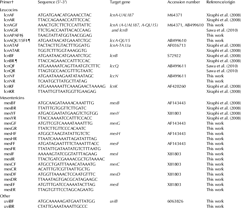
To determine the genes encoding bacteriocins, PCR and DNA sequencing were performed using the primers listed in Table 1. Leucocins A, B, K, Q, N, A-TA33a, mesentericins B, Y, G, H, F, I, C, D, E and UviB were considered. A number of primers previously reported (Xiraphi et al. Reference Xiraphi, Georgalaki, Rantsiou, Cocolin, Tsakalidou and Drosinos2008; Sawa et al. Reference Sawa, Okamura, Zendo, Himeno, Nakayama and Sonomoto2010) were applied with some modifications. Based on available sequences of bacteriocins from Leuconostoc in GeneBank database, new specific primers were designed. A standard reaction containing PCR buffer 10×, 200 μm dNTPs, 2·25 U/100 μl Taq-polymerase (Life Technologies™, Invitrogen Argentina S.A.) and 0·210 μm of each primer was used. One nanogram of DNA, extracted from Leuc. citreum MB1 using a commercial kit (Gen Elute Bacterial Genomic DNA, Sigma) was loaded into the PCR reaction. Amplifications were carried out in a GeneAmp PCR System 2400 (Applied Biosystem) thermalcycler. The PCR conditions included an initial denaturation at 95 °C for 5 min, followed by 35 amplification cycles at 95 °C for 1 min (denaturation), 45 °C for 45 s (annealing) and 72 °C for 1 min and 30 s (extension), and a final extension step at 72 °C for 7 min. Each PCR reaction was analysed by electrophoresis agarose gel (1·8% w/v). Positive PCR products of the expected molecular weight were purified using GenElute™ PCR Clean-Up Kit (Sigma) and sent for sequencing (Macrogen Korea, Seoul, Korea). Sequences were aligned using GeneBank tools for identification purposes.
Primers used for PCR amplification of genes responsible for bacteriocin production in Leuconostoc citreum MB1

† F: forward primer; R: reverse primer
‡ For these two forward primers, lcnBR was used as reverse primer
§ Primer targeted to active peptide
¶ Primer targeted to leader peptide
SDS-PAGE analysis
Sodium dodecyl sulphate polyacrylamide gel electrophoresis (SDS-PAGE) on a discontinuos gradient gel was performed according to Laemmli procedure (Laemmli, Reference Laemmli1970) modificated by Candioti et al. (Reference Candioti, Hynes, Perotti and Zalazar2002). A 4% and 20% (w/v) of acrylamide concentration of stacking and separating gels were respectively used. Separation was carried out at alkaline pH (8·3) with a Tris-Glycine carrier buffer, using a Mini Protean II electrophoretic chamber (Bio-Rad Laboratories, California, USA). The working conditions were 150 V and 45 mA. A wide-range (2·5–200 kDa) molecular weight marker was used (Mark12™ Unstained Standard, Novex, Life Technologies™). Cell-free supernatant (CFS) was obtained by centrifugation (12 000 g, 5 min, 5 °C) of overnight Leuc. citreum MB1 culture and sterilisation by filtration (0·22-μm pore diameter, Millipore). Two identical gels were prepared, in which 10 and 20 μl CFS from Leuc. citreum MB1 were subjected to the electrophoresis. Both gels were afterwards stained/fixed with Coomasie blue and destained with a distilled water/ethanol/acetic acid (1 : 1 : 0·4) solution. Afterwards, one of gels was used for direct detection of antimicrobial activity. Briefly, the gel was boiled (100 °C for 5 min) in distilled water, washed in sterilised bidistilled water and finally revealed by a double-layer method: a BHI agar plate was overlaid with BHI soft agar containing 107 CFU/ml List. monocytogenes 15313 as indicator strain; the gel was then put onto the soft agar and finally covered by another BHI soft agar layer also containing the pathogenic strain. Incubation was carried out for 24 h at 37 °C, and bacteriocin activity was revealed by a transparent halo of growth inhibition.
Inactivation kinetics of List. monocytogenes ATCC 15313 with Leuc. citreum MB1 concentrated cell-free supernatant
Bacteriocins can be added as additives in fermented food products, thus avoiding negative interactions between primary starter and bacteriocin-producing strains. The present test was aimed to evaluate the antilisterial ability of Leuc. citreum MB1 concentrated supernatant which contains antilisterial bacteriocins. Following this purpose, concentrated cell-free supernatant (CCFS) from a fresh Leuc. citreum MB1 culture (16–18 h, 32 °C) was obtained after centrifugation (12 000 g, 5 min, 5 °C), concentration (5×, in a rotary evaporator) and filter-sterilisation (0·22 μm pore diameter, Millipore). Aliquots of a fresh List. monocytogenes ATCC 15313 culture (16–18 h, 37 °C) were inoculated in 10 ml BHI broth, at a low (7·3×103 CFU/ml) and a high initial cell load (8·7×107 CFU/ml). Then, the CCFS was added in volumes of 50 or 500 μl, and incubated for 48 h at 37 °C. Additionally, other aliquots of the pathogenic strain inoculated at the same low and high initial concentrations, were pre-grown at 37 °C until OD560 ∼ 0·15 (for low initial cell load) or OD560 ∼ 0·5 (for high initial cell load), subsequently added of 50 or 500 μl CCFS and incubated at 37 °C for 48 h. The latter condition emulates a post-processing contamination. Tubes of BHI broth inoculated only with List. monocytogenes ATCC 15313 were used as controls. Growth of the pathogenic strain was followed by measuring OD560 at certain intervals of time.
Co-cultures experiments
Optimal Leuc. citreum MB1 initial concentration for List. monocytogenes ATCC 15313 biocontrol
Diverse initial cell loads of Leuc. citreum MB1 and List. monocytogenes ATCC 15313 were used to find maximum pathogen inhibition. With this aim, overnight cultures (16–18 h, 32 °C) of Leuc. citreum MB1 and the pathogenic strain (16–18 h, 37 °C) were co-inoculated in 10 ml reconstituted sterilised skim milk at: (i) 106 and 104 CFU/ml; (ii) 104 and 104 CFU/ml or (iii) 106 and 102 CFU/ml of Leuconostoc and Listeria initial concentrations, respectively. Tubes of skim milk inoculated only with Leuc. citreum MB1 or List. monocytogenes ATCC 15313 at each initial concentration were used as growth controls. Co-culture and control tubes were incubated at 34 °C for 48 h in a water bath. This temperature was selected to allow growth of both pathogenic and bacteriocinogenic strains. At 0, 24 and 48 h, pH values were measured and viable cell counts in BHI agar (Listeria) or MRS agar (Leuconostoc) of both controls and co-cultures were performed.
Biocontrol of List. monocytogenes by Leuc. citreum MB1 in milk at refrigeration temperature
The ability of Leuc. citreum MB1 to control List. monocytogenes ATCC 15313 growth was studied in a fermented-milk model. Tubes of skim milk were co-inoculated with Leuc. citreum MB1 and List. monocytogenes ATCC 15313 at 107 and 102 CFU/ml, respectively. Tubes of skim milk inoculated only with Leuconostoc or Listeria were used as controls. One set of inoculated tubes was immediately incubated at 8 °C, while the other was pre-incubated at 30 °C for 5 h and then kept at 8 °C during 28 d. Pre-incubation step was intended to accelerate the growth of the strains before incubation at 8 °C. Viable cell counts in BHI (Listeria) or MRS (Leuconostoc) agar were carried out at the beginning and throughout the experience.
Results and discussion
Preliminary identification of the antilisterial compound
PCR amplification and sequencing of the bacteriocin gene
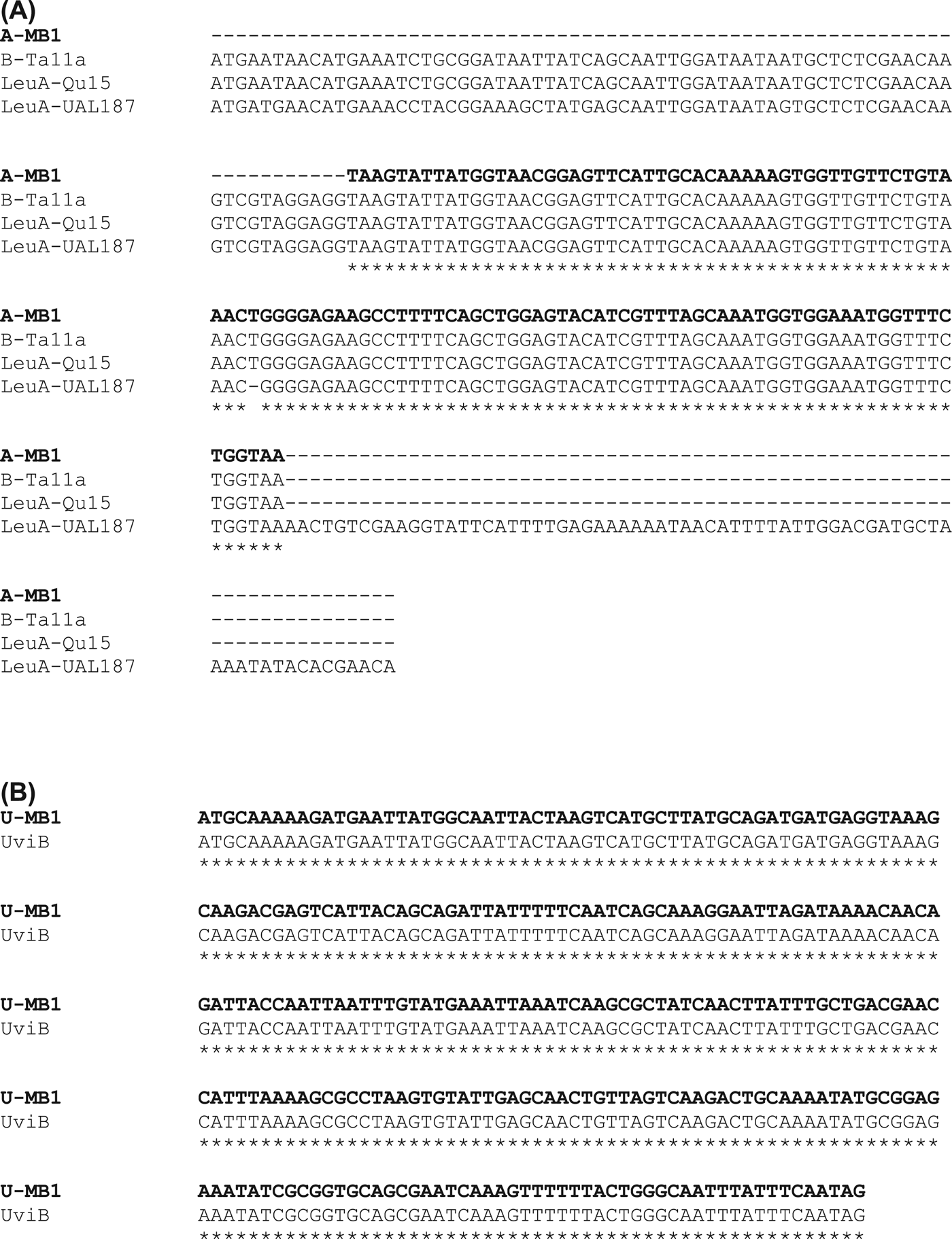
From all bacteriocin-specific primers tested, those for leucocin A (lcnAPAF and lcnBR) and UviB (uviBF and uviBR) showed amplification products with the expected size (∼130 and ∼300 bp, respectively). Amplification products of expected size were sequenced and compared with nucleotide sequences of the corresponding bacteriocins deposited in GenBank (Fig. 1). Amplification product of ∼130 bp (named a-mb1, putative leucocin A-MB1) exhibited high homology (>98%) with genes codifying for active peptide from leucocin A: A-QU 15 (accession no. AB499610), A-UAL 187 (accession no. M64371) and B-Ta11a (accession no. S72922). No amplification was obtained with primers designed on the basis of the leader peptide from leucocins described above. In comparison with previous findings (Sawa et al. Reference Sawa, Okamura, Zendo, Himeno, Nakayama and Sonomoto2010), putative leucocin A-MB1 found in Leuc. citreum MB1 would share the same nucleotide sequence as that codifying for active peptide. However, structural genes (including immunity) and nucleotide sequence codifying for leader peptide could be different. Similarly leucocin A is produced by diverse Leuconostoc species, such as Leuc. pseudomesenteroides QU 15 (leucocin A-QU 15, Sawa et al. Reference Sawa, Okamura, Zendo, Himeno, Nakayama and Sonomoto2010), Leuc. gelidum UAL 187 (leucocin A-UAL 187, Hastings et al. Reference Hastings, Sailer, Johnson, Roy, Vederas and Stiles1991), Leuc. carnosum Ta11a (leucocin B-Ta11a, Felix et al. Reference Felix, Papathanasopoulos, Smith, von Holy and Hastings1994). Even though their structure is highly homologous, they present some differences principally in their leader peptides or structural genes (Sawa et al. Reference Sawa, Okamura, Zendo, Himeno, Nakayama and Sonomoto2010).
Alignment of nucleotide sequences of the products obtained by PCR amplification using specific primers, with sequences of bacteriocins deposited in GenBank. (A) A-MB1, ∼130 bp-product, highly homologous with sequences from “leucocin A” group, (B) U-MB1, ∼300 bp-product, highly homologous with bacteriocin UviB sequence.
PCR product of ∼300 bp (named u-mb1, putative bacteriocin U-MB1) showed complete (100%) homology with bacteriocin UviB sequence (accession no. YP_001728928). This is the first report of a gene with high homology with bacteriocin UviB, which is also codified in the genome of a Leuc. citreum strain (Kim et al. Reference Kim, Jeong, Lee, Choi, Ha, Hur, Kim, Lee, Park, Park and Oh2008).
SDS-PAGE analysis
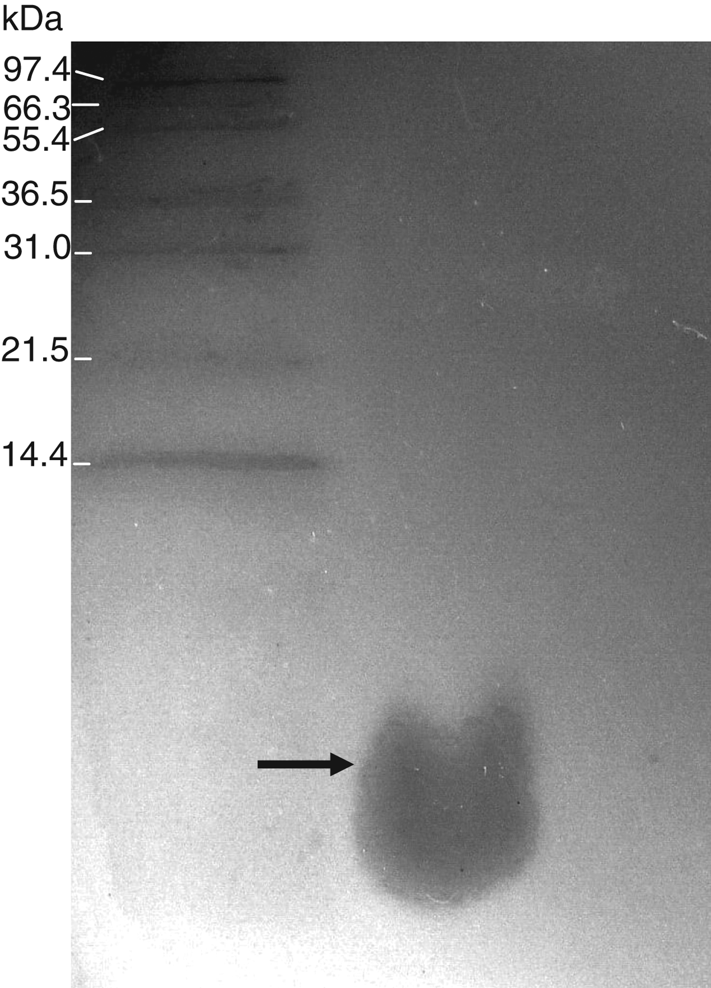
SDS-PAGE was used to estimate the molecular mass of bacteriocins present in CFS obtained from Leuc. citreum MB1. However, the antimicrobial compound did not give definite bands in the Coomasie Blue-stained gel, possibly due to the low mass of these peptides and thus the complexity of a proper resolution of the bands. On the other hand, the gel subjected to direct detection of the antimicrobial activity by using List. monocytogenes ATCC 15313 as indicator strain, revealed a clear zone of inhibition (Fig. 2). The molecular mass was estimated to be much lower than 14·4 kDa, which is the last band resolved in the gel. Molecular masses reported for leucocins produced by Leuconostoc strains studied ranged between 2500 and 4000 kDa (Hastings et al. Reference Hastings, Sailer, Johnson, Roy, Vederas and Stiles1991; Papathanasopoulos et al. Reference Papathanasopoulos, Krier, Revol-Junelles, Lefebvre, Le Caer, von Holy and Hastings1997; Xiraphi et al. Reference Xiraphi, Georgalaki, Rantsiou, Cocolin, Tsakalidou and Drosinos2008; Sawa et al. Reference Sawa, Okamura, Zendo, Himeno, Nakayama and Sonomoto2010). In the present work, estimation of molecular masses by SDS-PAGE was not certain. However, their assessment from products obtained by PCR amplification lead to molecular masses in the range of previously reported bacteriocins.
SDS-PAGE gel subjected to direct detection of antimicrobial activity present in Leuc. citreum MB1 cell-free supernatant containing bacteriocin. List. monocytogenes ATCC 15313 was used as indicator strain. Inhibition zone is indicated with a black arrow. A wide-range (2·5–200 kDa) weight molecular marker was used.
Inactivation kinetics of List. monocytogenes ATCC 15313 with Leuc. citreum MB1 concentrated cell-free supernatant
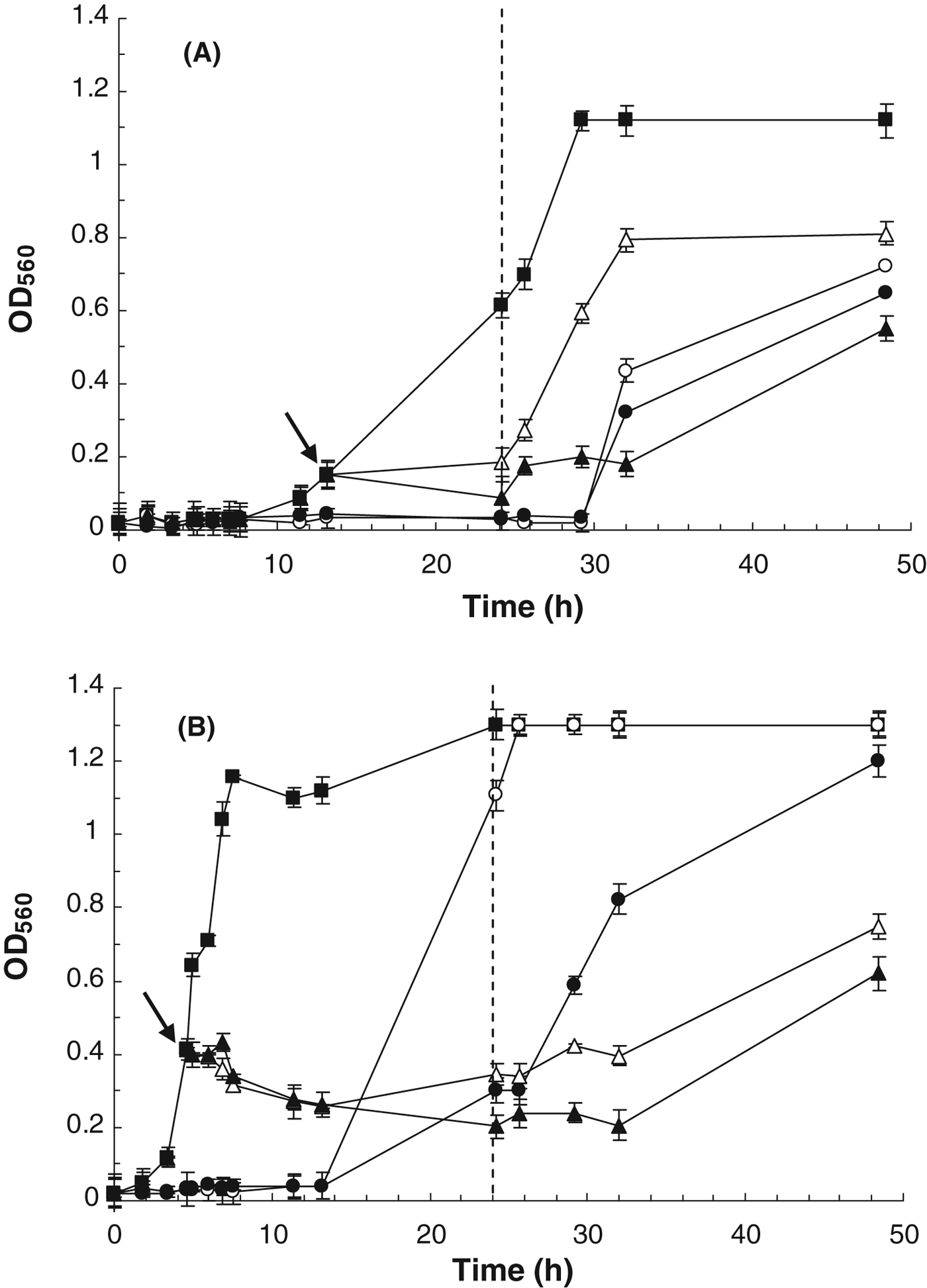
Results are shown in Fig. 3. Leuc. citreum MB1 concentrated cell-free supernatant (CCFS) containing antilisterial bacteriocins was generally able to delay the growth of List. monocytogenes ATCC 15313 in BHI broth for approximately 24 h at 37 °C. The effectiveness of the bacteriocin appeared to be dependent on both bacteriocin concentration and initial number of pathogen bacteria. In Fig. 3a, growth kinetics of List. monocytogenes at 7·3×103 CFU/ml initial cell load, added with 50 or 500 μl CCFS are shown. In general, the addition of CCFS at the beginning of the experience, inhibit the growth of List. monocytogenes ATCC 15313 during 30 h. After this period, the pathogen started to grow exponentially, although much slower than the control. On the other side, the addition of 50 or 500 μl of CCFS after pre-incubation of the pathogen until DO560 ∼ 0·15, held up the development of List. monocytogenes for the subsequent 12 h of incubation. For the pathogen inoculated with the highest volume of CCFS (500 μl), the inhibitory effect was even longer. In all cases, the addition of CCFS controlled and even inhibited the growth of List. monocytogenes for at least 24 h. Figure 3b shows growth kinetics of List. monocytogenes at a higher initial cell load (8·7×107 CFU/ml). In this case, the addition of 50 or 500 μl at the beginning of the experience, led to a 12 h-delay in List. monocytogenes growth. Moreover, inoculation with 500 μl CCFS led to a very slow growth of the pathogen until 24 h of incubation. Pre-incubation of the pathogen until DO560 ∼ 0·5 before the addition of 50 or 500 μl of CCFS, allowed a rapid decrease in pathogen numbers during approximately 24 h, followed by a reduced development of List. monocytogenes during the next 32 h of incubation. The latter results showed that efficiency of CCFS in List. monocytogenes inhibition is more remarkable when the supernatant was added on the pre-incubated pathogen. Recently, it has been shown that a set of bacteriocins Class IIa produced by Gram-positive species can employ the membrane components of the mannose phosphotransferase system (Man-PTS) on sensitive cells as receptor molecules (Kjos et al. Reference Kjos, Nes and Diep2011). In our work, inhibitory effect of the CCFS was more pronounced when added to the pre-incubated pathogen cells. This effect could be attributable to an increased expression of those receptors linked to a high metabolism of the pathogenic strain.
Inactivation kinetics at 37 °C in BHI broth of List. monocytogenes ATCC 15313 by Leuc. citreum MB1 concentrated cell-free supernatant (CCFS). List. monocytogenes ATCC 15313 initial cell loads of 7·3×103 CFU/ml (A) and 8·7×107 CFU/ml (B); added of 50 μl (○) or 500 μl (●) CCFS at the beginning, and 50 μl (△) or 500 μl (▲) CCFS after reaching OD560 ∼ 0·15 (104 CFU/ml initial cell load) or OD560 ∼ 0·4 (107 CFU/ml initial cell load). List. monocytogenes ATCC 15313 without addition of CCFS (control) (■). Dotted vertical lines indicate 24 h incubation; black arrows indicate the addition of CCFS. Values are the mean of three determinations±standard determination.
Our results showed that the inhibitory effect of the CCFS against List. monocytogenes ATCC 15313 was greater at the lowest initial cell load tested (∼104 CFU/ml) of the pathogenic strain. Vignolo and co-workers (1996) arrived at similar conclusions studying the effect of lactocin 705 on the growth of List. monocytogenes in meat at 20 °C. They observed a partial reduction in pathogen cell loads only by 40% at 24 h when the initial concentration of the pathogenic bacteria was 1×10⁷, and a reduction by 44% when the initial cell load of pathogen bacteria was 1×10⁴. Pucci et al. (Reference Pucci, Vedamuthu, Kunka and Vandenbergh1988) evaluated the lysis of List. monocytogenes by PA-1 bacteriocin during 7 h. They observed a greater reduction in number of pathogen bacteria when the maximal bacteriocin concentration was used. The addition of higher concentrations of bacteriocin should confer a protective effect against Listeria over longer periods (Pucci et al. Reference Pucci, Vedamuthu, Kunka and Vandenbergh1988; Motlagh et al. Reference Motlagh, Holla, Johnson, Ray and Field1992; Trias et al. Reference Trias, Badosa, Montesinos and Bañeras2008). The extent of inhibitory effect will depend on many factors, principally of the initial number of pathogen bacteria and the bacteriocin concentration.
Co-cultures experiments
Optimal Leuc. citreum MB1 initial concentration for List. monocytogenes ATCC 15313 biocontrol
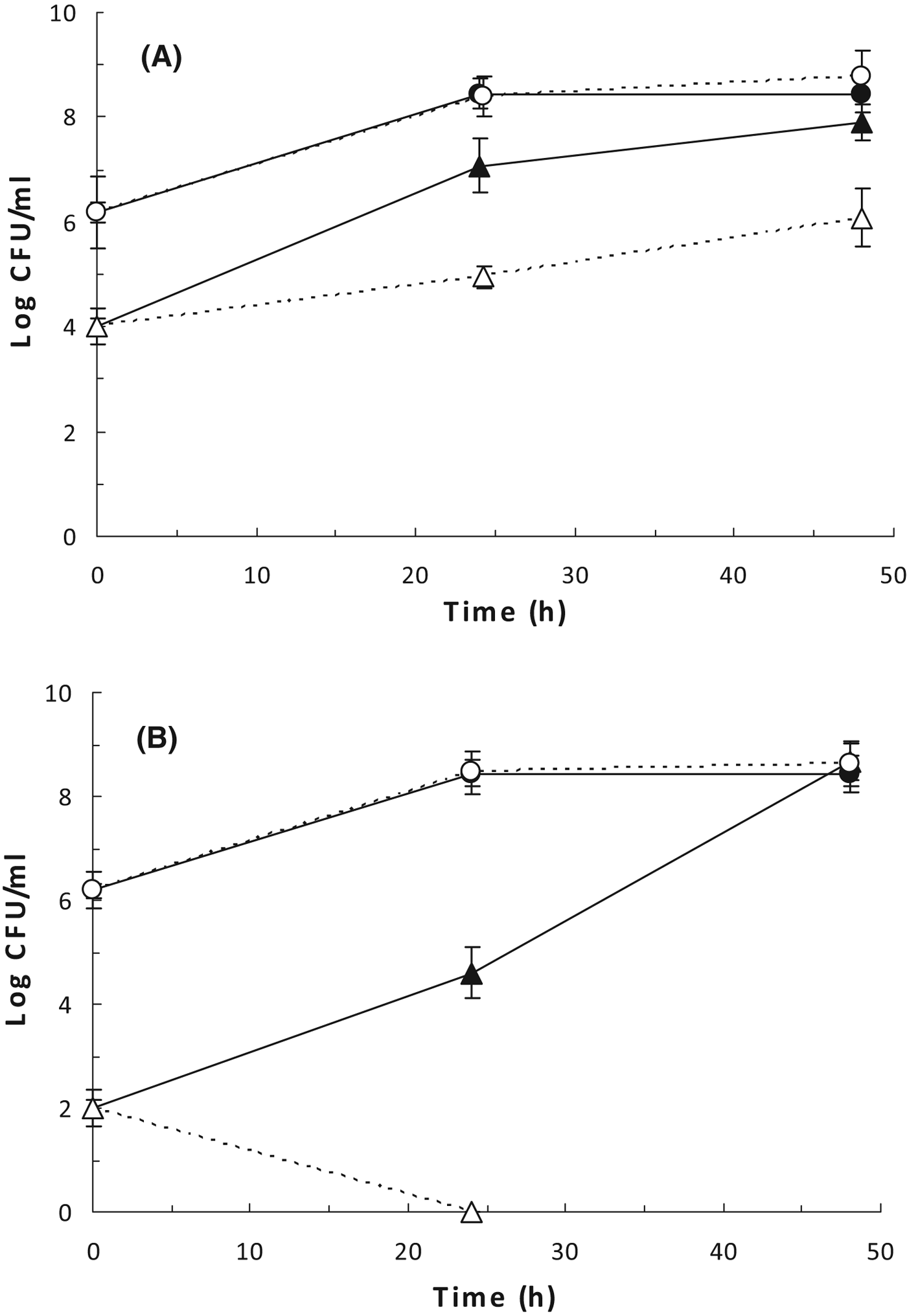
To determine the range of pathogen concentrations that the bacteriocinogenic strain is able to control, diverse combinations of bacteriocinogenic and pathogenic strains were tested. Initial List. monocytogenes cell loads added (102 and 104 CFU/ml) were selected on the basis of most probable pathogen levels in postprocessing contamination (Pucci et al. Reference Pucci, Vedamuthu, Kunka and Vandenbergh1988). Our results showed that equal Leuconostoc and Listeria initial cell loads (104 CFU/ml), allowed normal growth of the pathogenic strain (data not shown). However, when Leuc. citreum MB1 initial concentration was higher than that of List. monocytogenes ATCC 15313, partial or total growth inhibition of the pathogenic strain was observed. Specifically, at 106 CFU/ml Leuconostoc and 104 CFU/ml Listeria initial cell loads, the growth of the pathogenic strain in co-culture was maintained two log orders lower than the control (Fig. 4a) throughout the experiment. Moreover, at 106 CFU/ml Leuconostoc and 102 CFU/ml Listeria initial concentrations, viable cell counts of the pathogenic strain were undetectable after 48 h of incubation at 34 °C (Fig. 4b). Efficacy of the bioprotective effect depends on initial relative concentrations of both Leuc. citreum MB1 and List. monocytogenes ATCC 15313, being higher when the bacteriocinogenic strain is in a predominant number from the start. Similar behaviour was obtained by Trias et al. (Reference Trias, Badosa, Montesinos and Bañeras2008) when inoculating wounds of apples and lettuce leaf cuts with different doses of pathogenic and bacteriocinogenic Leuc. mesenteroides strains: the highest List. monocytogenes inhibition was revealed in co-culture with any of the three Leuc. mesenteroides strains tested at the highest initial concentration tested (109 CFU per wound or per gram).
Effect of Leuc. citreum MB1 initial cell loads on List. monocytogenes 15313 growth, in co-culture (skim milk, 34 °C, 48 h). Relative Leuconostoc and Listeria initial inocula: (A) 106 and 104 CFU/ml; (B) 106 and 102 CFU/ml. Symbols correspond to Leuc. citreum MB1 control (●), Leuc. citreum MB1 in co-culture (○), List. monocytogenes 15313 control (▲) and List. monocytogenes 15313 in co-culture (△).Values are the mean of three determinations±standard determination.
Comparison of our results are difficult since other reported in vitro co-culture experiments were carried out using different bacteriocinogenic LAB strains, for example, Lactococcus lactis (Liu et al. Reference Liu, O'Conner, Cotter, Hill and Ross2008) and Lactobacillus casei CRL 705 (Vignolo et al. Reference Vignolo, Fadda, de Kairuz, de Ruiz Holgado and Oliver1996). In any case, the consensus result is that bacteriocinogenic strains reached their maximum effectiveness when inoculated at higher initial numbers than that of the pathogenic strain, which indicates the importance of maintaining a high standard manufacturing process together with strict sanitation.
Biocontrol of List. monocytogenes ATCC 15313 by Leuc. citreum MB1 in milk at refrigeration temperature
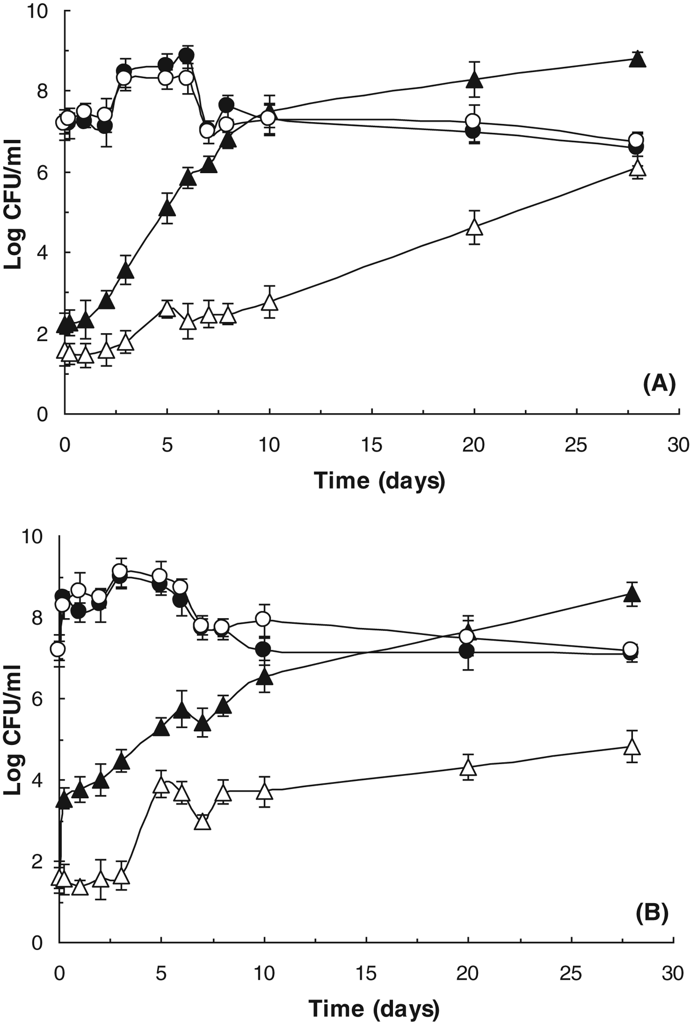
Results are shown in Fig. 5. In general, growth of List. monocytogenes ATCC 15313 was clearly delayed when it was co-inoculated in skim milk with Leuc. citreum MB1, as viable cell counts of the pathogen were below 102 CFU/ml during the first 3 d for both directly incubated at 8 °C and pre-incubated at 30 °C co-cultures. Afterwards and for 10 d, direct incubation at 8 °C allowed a very low growth rate in co-cultured List. monocytogenes 15313. After 28 d of refrigerated storage, viable cell counts of the pathogenic strain were raised more pronouncedly, although they were maintained at least two log orders lower than the control (Fig. 5a).
Co-culture growth of Leuc. citreum MB1 and List. monocytogenes ATCC 15313 in skim milk, at 106 CFU/ml Leuconostoc and 102 CFU/ml Listeria initial cell loads. (A) incubation at 8 °C for 28 d; (B) pre-incubated at 30 °C for 5 h, followed by incubation at 8 °C for 28 d. Symbols correspond to Leuc. citreum MB1 control (●), Leuc. citreum MB1 in co-culture (○), List. monocytogenes 15313 control (▲) and List. monocytogenes 15313 in co-culture (△).Values are the mean of three determinations±standard determination.
Pre-growth at 30 °C for 5 h and further incubation at 8 °C of List. monocytogenes 15313 together with Leuc. citreum MB1, allowed considerable delay of the pathogen growth and even after 28 d, the number of pathogenic cells was extremely inferior to that of the control (6·7×104 and 3·8×108 CFU/ml, respectively) (Fig. 5b). Leuconostoc growth in skim milk was not affected by the presence of the pathogenic strain as shown by similar counts in co-culture and control tubes.
Leuc. citreum MB1 ability to control List. monocytogenes 15313 growth at 8 °C was greater in the pre-incubated (30 °C, 5 h) culture. This previous treatment at higher temperature certainly allowed rapid development of Leuc. citreum MB1. Superiority in cell number and the production of high amounts of antimicrobial compounds at an earlier stage facilitated Leuc. citreum MB1 to control the growth of the pathogenic strain. Similar behaviour was obtained by Vignolo et al. (Reference Vignolo, Fadda, de Kairuz, de Ruiz Holgado and Oliver1996) when studying the effect of in situ-produced bacteriocin by Lb. casei CRL 705 in meat. They observed no changes in the number of inoculated Listeria cells during 24 h, indicating a partial inhibition and bacteriostatic effect of the produced bacteriocin.
Since bacteriocins may not be considered as inhibitors by themselves, for optimal effectiveness they should be used together with other preservation methods, as part of a general multihurdle food preservation system, in which the combined effects of pH, temperature and oxygen availability are studied together with the added bacteriocinogenic LAB (Ennahar et al. Reference Ennahar, Sashihara, Sonomoto and Ishizaki2000; Castellano et al. Reference Castellano, Belfiore, Fadda and Vignolo2008).
Conclusions
Leuc. citreum MB1 belongs to a group of adventitious Leuconostoc strains previously isolated from dairy environment. This strain has previously been shown to grow well in milk and produce antibacterial bacteriocin-like compounds specific against Listeria.
Our results showed that Leuc. citreum MB1 harbours two different genes (a-mb1 and u-mb1) compatible with the active fraction of two known bacteriocins: leucocin A and bacteriocin UviB. It is unquestionable that at least one bacteriocin is active in Leuc. citreum MB1, since important antilisterial activity was verified in its cell-free supernatant. We verified that the addition of Leuc. citreum MB1 concentrated cell-free supernatant conferred a protective effect against the pathogenic strain during 24 h. Moreover, high initial concentrations of Leuc. citreum MB1 are able to control List. monocytogenes growth in milk during refrigerated storage. This feature, together with its ability to grow at low temperatures, may be very advantageous to control the growth of Listeria.