Introduction
Mechanical suture devices in the upper aerodigestive tract were first described in 1969, by Hoehn and Payne,Reference Hoehn and Payne1 who used mechanical suturing to repair mucosal defects after the excision of Zenker's diverticula. In the early 1970s, Paches et al.,Reference Paches, Ogoltsova, Petrova and Tsybyrne2 and Lukyanchenko and Knowles,Reference Lukyanchenko and Knowles3 reported the first clinical case of using stapling devices during a laryngectomy in Russian medical literature. In the 2000s, Sofferman and Voronetsky,Reference Sofferman and Voronetsky4 and Agrawal and Schuller,Reference Agrawal and Schuller5 published the first series of results, with a detailed description of the surgical technique. Since then, several authors have published their results of using stapling devices in total laryngectomies.Reference Altissimi and Frenguelli6–Reference Paddle, Husain, McHugh and Franco18
Two techniques for mechanically suturing the pharynx have been described. In the open technique, the device is used to suture the pharyngeal mucosa after a conventional open laryngectomy. In the closed technique, the stapling device is applied when the skeletonised larynx is attached to the pharynx only by the mucosa. The specimen is then removed and sutured simultaneously. Sometimes it is difficult to keep the suprahyoid epiglottis outside the jaws of the stapler. In these cases, some authors propose a mixed approach, consisting of a small, controlled pharyngeal opening made in the midline at the valleculaReference Altissimi and Frenguelli6 or at the interarytenoid area,Reference Zhang, Liu, Li, Liu, Li and Liu7 in order to facilitate retraction of the epiglottis. Closed or mixed reconstructions are the most widely reported techniques related to mechanical suturing of the pharynx.
The advantages of mechanically suturing the pharyngeal mucosa, compared to manual suturing, are the decrease in surgical time, the absence of contamination of the operating field by endopharyngeal secretions when using the closed technique, and minimal mucosal trauma.
One of the limitations of mechanical suturing is that the tumour is not visualised during resection and so the resection margins cannot be tailored to the tumour; thus, the method harbours the potential for oncological compromise if applied in inappropriately selected cases.
Since 2008, we have employed mechanical suturing using a closed stapling technique in selected cases of total laryngectomy in our institution. The present study aimed to evaluate the results obtained with this type of technique, compared to the conventional manual suturing technique.
Materials and methods
We performed a retrospective review of all total laryngectomies carried out in our institution between January 2008 and April 2018. Patients treated with an extended total laryngectomy were excluded from the study, because these patients were not candidates for mechanical suturing of the pharynx.
The choice of suturing technique depends on the location and extension of the tumour, and on the surgeon's preferences. The indication for the mechanical suturing technique was an endolaryngeal tumour without involvement of: the aryepiglottic folds, suprahyoid epiglottis, inter-arytenoid and retro-cricoid areas, or the medial wall of pyriform sinuses. Careful pre-operative assessment is necessary to select appropriate patients. In case of doubt, we carried out a direct laryngoscopy before initiation of the total laryngectomy.
Surgical technique
Mechanical suturing
After subplatysmal flap elevation and cervical neck dissection, the laryngeal framework is skeletonised according to the traditional technique. The pharyngeal constrictor muscles are sectioned following the posterior margin of the thyroid cartilage. Next, the external side of the piriform sinus mucosa is separated from the internal perichondrium of the thyroid cartilage in order to preserve as much mucosa as possible. The hyoid bone is released from the suprahyoid musculature and its connections with the suprahyoid epiglottis, to reduce tension after closure and to keep the suprahyoid epiglottis away from the suture line. At this point, it is important to avoid accidental opening of the pharynx.
After division of the thyroid isthmus, the trachea is sectioned at the required level depending on the degree of subglottic extension, and the tracheoesophageal membrane is dissected to expose the posterior cricoarytenoid muscles. At this point, the larynx is completely mobilised and remains connected to the pharynx only by the mucosa. The free edge of the epiglottis is pulled into the larynx using a haemostatic clamp, passed through the tracheal lumen, to avoid including the epiglottis in the suture line.
At this stage, the linear stapler (75 mm linear cutter; Ethicon Endo-Surgery, Cincinnati, Ohio, USA) is placed as close to the larynx as possible, to preserve a maximum amount of pharyngeal mucosa. The device is locked and fired, creating two double suture lines that seal the hypopharynx and release the laryngectomy sample.
Finally, the suture is checked to ensure complete and watertight closure. If any leak points are discovered, the suture is reinforced with manual absorbable stitches. Constrictor musculature is sutured as reinforcement, by following the usual technique. The suture line of the specimen is opened to visually check the resection margins. Frozen section margins may be sent at this stage if needed.
As the pharynx is automatically closed, the placement of a primary voice prosthesis requires a modified technique as follows. After fixation of the tracheal stoma to the inferior skin flap, a direct oesophagoscopy is performed. The tracheoesophageal membrane is punctured, as conducted in the secondary voice prosthesis technique, and the voice prosthesis is inserted in a retrograde fashion.
Manual suturing
Once the laryngeal specimen is removed, the hypopharyngeal mucosa is sutured with absorbable material (Vicryl size 4-0). In most cases, a horizontal suture line is created, connecting the lateral and inferior resection borders to the base of tongue mucosa. However, in patients previously treated with radiotherapy, the remaining mucosa loses elasticity, and a T-shape suture is performed to avoid tension in the suture line. Care is taken to assure inversion of the mucosa during the suture. Routinely, a second layer of suturing is performed, in order to decrease the tension in the first suture line. Finally, the constrictor musculature is placed above as a reinforcement layer.
Post-suturing procedure
In both types of reconstruction, once the pharyngeal suture is completed, the haemostasis is checked and the neck wound is closed with suction drains.
All patients received antibiotic prophylaxis 1 hour before the incision and during the procedure.
Enteral nutrition by nasogastric tube was initiated on the 1st post-operative day. In the absence of wound complications, the nasogastric tube was usually removed on the 7th post-operative day in non-irradiated patients and on the 12th post-operative day in patients who had previously received radiotherapy. The oral feeding initiation protocol was maintained regardless of the type of pharyngeal suturing performed.
Patients
During the study period, a total of 126 simple total laryngectomies were carried out. The surgery was indicated as the initial treatment of the tumour in 61 cases, as a salvage surgery after radiotherapy or chemoradiotherapy treatment in 44 cases, and because of a second laryngeal neoplasm in 18 patients previously treated with radiotherapy. The remaining three patients presented with a non-functional larynx following radiotherapy. Sixty-five patients (51.5 per cent) had previously been treated with radiotherapy.
A pharyngocutaneous fistula was defined as any documented clinical suspicion or evidence of a salivary leak. All suspected pharyngocutaneous fistulas were confirmed by the oral administration of methylene blue dye. In cases with a confirmed fistula, the initial treatment was conservative management. The pharyngocutaneous fistulas were classified as major or minor. A major fistula was defined as a fistula persisting for more than four weeks, requiring surgical treatment or associated with peri-operative mortality.
A multivariate analysis was conducted, with the incidence rates of pharyngocutaneous fistula and major fistula as dependent variables. The independent variables assessed included age, gender, previous radiotherapy, American Society of Anesthesiologists physical status classification, diabetes mellitus, tumour location, neck dissection, category of local tumour extension, pre-operative haemoglobin and type of pharyngeal suture.
In order to verify that the type of suture did not compromise the oncological outcome, the surgical margin status, local recurrence-free survival rate and cancer-specific survival rate were assessed in relation to the type of suture. Those patients for whom the indication for surgery was a non-functional larynx were excluded from this part of the study.
The study was approved by the institutional review committee of our centre (approval number: IIBSP-CCC-2014-93), and was carried out in accordance with the principles indicated in the Declaration of Helsinki.
Statistical techniques
The chi-square test or Fisher's exact test were used to compare qualitative variables, and the student's t-test was used to compare quantitative variables. Multivariate analyses were carried out using the binary logistic regression model. Survival was calculated with the Kaplan–Meier actuarial method, using the log-rank test to compare survival curves.
Results
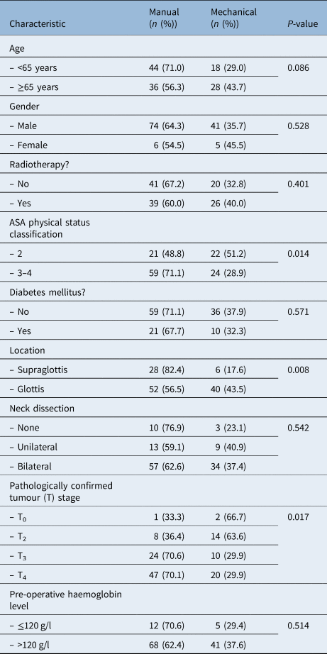
Closure of the pharyngeal mucosa was performed with the manual suturing technique in 80 cases (63.5 per cent) and with the mechanical suturing technique in 46 cases (36.5 per cent). Table 1 shows the characteristics of the patients in the study according to the type of suturing utilised. Mechanical suturing was used significantly more often in patients with: glottic tumours (p = 0.008), less local tumour extension (p = 0.017) and less pre-operative morbidity measured according to the American Society of Anesthesiologists physical status classification (p = 0.014).
Patient characteristics according to pharyngeal suturing technique

ASA = American Society of Anesthesiologists
In the cohort of patients studied, the percentage of pharyngocutaneous fistulas was 15.1 per cent (n = 19) and the percentage of major fistulas was 8.7 per cent (n = 11). The rates of pharyngocutaneous fistulas and major fistulas were 16.3 per cent (n = 13) and 8.8 per cent (n = 7) for the patients reconstructed with manual suturing (n = 80), and were 13.0 per cent (n = 6) and 8.7 per cent (n = 4) for patients reconstructed with stapler suturing (n = 46), respectively. There was no significant relationship between the type of pharyngeal closure and the rate of pharyngeal fistula (p = 0.628) or major fistula (p = 0.992).
The incidence of pharyngocutaneous fistulas and major fistulas was evaluated based on previous radiotherapy treatment and the type of pharyngeal suturing (Table 2). There was a non-significant tendency for non-irradiated patients treated with mechanical suturing to have a lower percentage of fistulas compared to those treated with manual suturing (5.0 per cent vs 19.5 per cent respectively). However, that trend reversed in the irradiated patients, showing a higher percentage of fistulas in the mechanical closure group (19.2 per cent vs 12.8 per cent), but the differences did not reach statistical significance. The percentage of major fistulas did not vary significantly according to the type of suture, regardless of previous radiotherapy.
Pharyngocutaneous fistula and major fistula rates according to previous radiotherapy and pharyngeal suturing technique

RT = radiotherapy
A multivariate analysis was performed that included the rates of pharyngocutaneous fistula and major fistula as dependent variables. The independent variables assessed included age, gender, American Society of Anesthesiologists physical status score, diabetes, tumour location and extension, prior radiotherapy, pre-operative haemoglobin value, suture type, and neck dissection (Table 1 in the supplementary material, available on The Journal of Laryngology & Otology website). None of the variables assessed correlated significantly with the frequency of pharyngocutaneous fistula or major fistula.
In 14 patients treated with mechanical suturing, a primary voice prosthesis was placed. No complications were reported in any of these patients.
The median hospital stay was 14 days (range, 8–74 days) for patients who underwent manual suturing of the pharynx, and was 12 days (range, 7–85 days) for patients who underwent mechanical suturing. There were no significant differences between the length of hospital stay and type of suturing (p = 0.998). If we only consider cases that did not present a pharyngocutaneous fistula, the median hospital stay was 13 days for the manual suture group and was 10.5 days for the mechanical suture group. The patients treated with mechanical suturing had a significantly shorter hospital stay (p = 0.040). Patients with a pharyngocutaneous fistula had a median hospital stay of 30 days if treated with manual suturing, compared to 55.5 days if treated with stapler closure; this was a non-significant difference (p = 0.183).
Surgical margins were analysed in the 123 oncological patients. Among patients who underwent manual suturing (n = 79), 6.3 per cent (n = 5) had positive margins and 6.3 per cent (n = 5) had close surgical margins of less than 5 mm. In those patients who underwent mechanical suturing (n = 44), none had positive margins and only one patient (2.3 per cent) had close surgical margins. There were no significant differences in terms of resection margin status based on the type of pharyngeal suturing (p = 0.165).
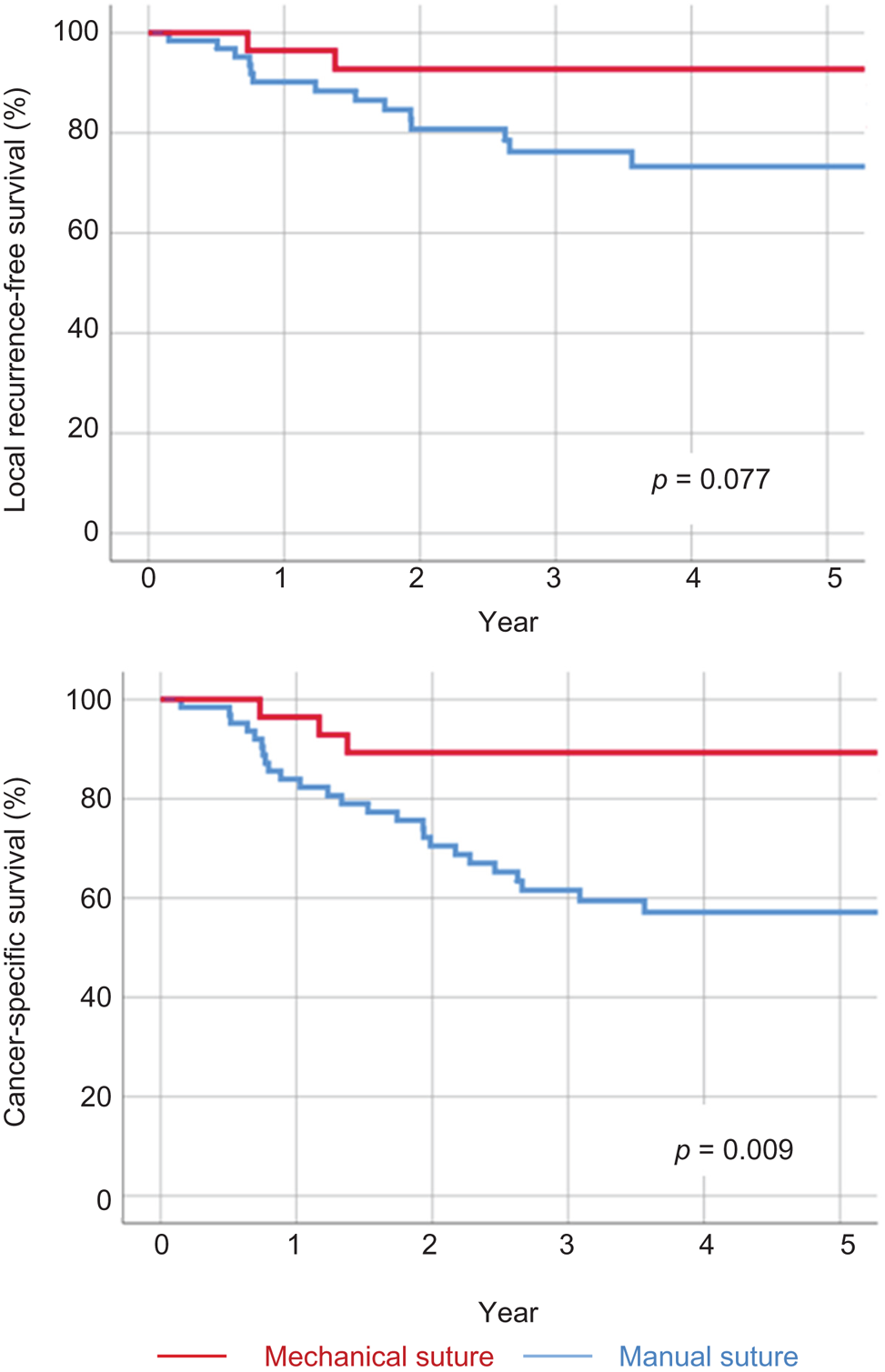
Ninety-two patients (64 who underwent manual suturing and 28 who received mechanical suturing) had a follow up of more than two years. Figure 1 shows the curves for local recurrence-free survival (Figure 1a) and cancer-specific survival (Figure 1b) for these patients. The mechanical suture group tended to have a lower percentage of local recurrence, but the differences did not reach statistical significance (p = 0.077). The cancer-specific survival rate for patients treated with mechanical suturing was significantly higher than that for patients treated with manual suturing (p = 0.009).
(a) Local recurrence-free survival and (b) cancer-specific survival according to the pharyngeal suturing technique.

Discussion
Mechanical sutures are routinely used in many surgical procedures. However, the use of such sutures applied to the pharynx after total laryngectomy is limited. The main advantages of mechanical sutures are their ease of use and the speed of pharyngeal closure. The regular spacing and pressure exerted uniformly by a double row of staples minimises tissue damage, allowing watertight closure and optimal healing. Additional benefits of mechanical sutures with the closed technique are minimal trauma of the mucosa and minimal exposure of the wound to pharyngeal secretions, which decreases the incidence of infections and fistulas in the post-operative period.
The main drawback of mechanical sutures with a closed technique is that the tumour is not visualised during the resection, compromising the oncological outcome if used in unsuitable cases. The mechanical suture must only be used in cases of endolaryngeal tumours without extension to the aryepiglottic folds, piriform sinuses or retro-cricoid area. The strict observance of this principle is reflected in our results, which show an excellent rate of negative margins. Mechanical suturing was used significantly more often in patients who had glottic tumours with limited extension.
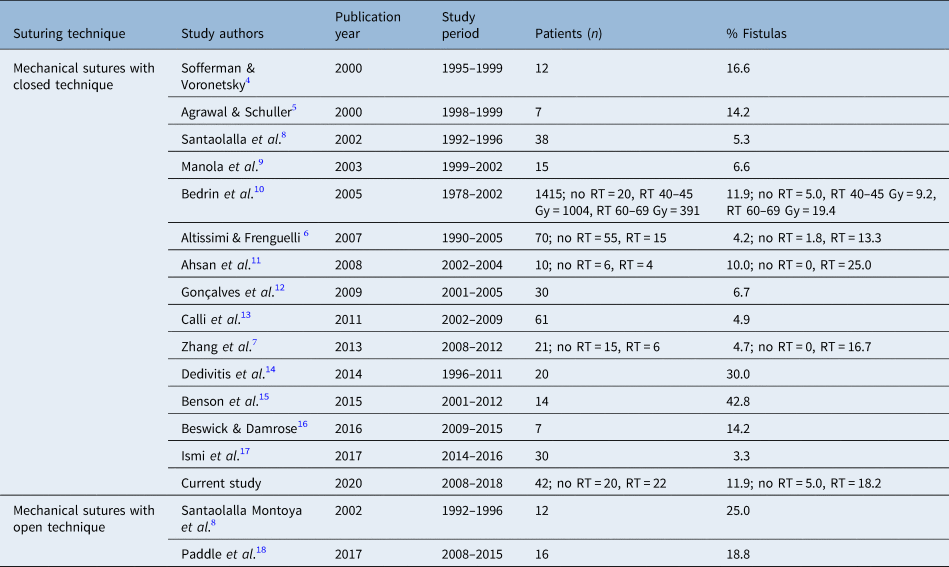
Table 3 shows the percentages of pharyngocutaneous fistulas reported by several authors using mechanical pharyngeal suturing with an open or a closed technique. The incidence of fistula ranges from 3.3 per centReference Ismi, Unal, Vayisoglu, Yesilova, Helvaci and Gorur17 to 42.8 per cent.Reference Benson, Hirata, Thompson, Ha, Fakhry and Saunders15
Incidence of pharyngocutaneous fistula in patients treated with mechanical suturing of pharynx

RT = radiotherapy
Bedrin et al.Reference Bedrin, Ginsburg, Horowitz and Talmi10 published findings for an extensive series of patients who underwent laryngectomy using mechanical sutures. The authors reported the experience of more than 1400 patients treated with mechanical pharyngeal sutures using a closed technique, carried out in two institutions in Russia and Israel. Of the patients, 98.6 per cent had previously been treated with radiotherapy. Fistula occurred in 12 per cent of cases, and there was a correlation between the fistula risk and the radiotherapy dose. Some authors have reported a higher percentage of post-operative fistulas in patients treated with mechanical sutures who had previously received radiotherapy.Reference Altissimi and Frenguelli6,Reference Zhang, Liu, Li, Liu, Li and Liu7 Regarding our results, the rate of fistula after mechanical suturing was higher in the irradiated patient group (5.0 per cent vs 19.2 per cent), although the difference did not reach statistical significance (p = 0.212).
Many authors have compared the presence of fistula in patients treated with total laryngectomy according to the type of suture used, and the results were contradictory. In most studies, mechanical sutures were associated with a significantly lower fistula rate than manual sutures.Reference Santaolalla Montoya, Ruiz de Galarreta, Sánchez del Rey, Martínez Ibargüen and Zabala López de Maturana8,Reference Gonçalves, de Souza, Menezes, Kavabata, Suehara and Lehn12,Reference Calli, Pinar and Oncel13,Reference Ismi, Unal, Vayisoglu, Yesilova, Helvaci and Gorur17 For other studies, the results were comparable,Reference Dedivitis, Aires, Pfuetzenreiter, Castro and Guimarães14 or the fistula rate was higher in patients treated with mechanical sutures although there was no significant difference.Reference Benson, Hirata, Thompson, Ha, Fakhry and Saunders15 According to the results of a meta-analysis carried out by Aires et al., the overall incidence of fistula in patients who underwent the mechanical suturing technique was 8.7 per cent, compared to 22.9 per cent in patients with manual sutures.Reference Aires, Dedivitis, Castro, Bernardo, Cernea and Brandão19 Assuming a 15 per cent reduction in the fistula risk, the treatment of seven patients would be required to achieve the benefit associated with the use of mechanical sutures.
Our results revealed no significant differences in the incidence of pharyngocutaneous fistulas or major fistulas based on the suture types used for the pharynx. However, we should be careful when comparing both suturing techniques. The mechanical suturing technique is only indicated in patients with endolaryngeal tumours without extension to the piriform sinuses, epilarynx or retro-cricoid area, in which pharyngeal mucosa excision should not be carried out, to reduce the risk of suture failure. There is a lack of randomised clinical trials on patients with tumours potentially suitable for mechanical suturing. Thus, there is no evidence that pharyngeal closure type is a predisposing factor in the risk of developing fistulas after total laryngectomy.
All the research indicates that the use of mechanical sutures achieves a reduction in surgical time.Reference Zhang, Liu, Li, Liu, Li and Liu7,Reference Santaolalla Montoya, Ruiz de Galarreta, Sánchez del Rey, Martínez Ibargüen and Zabala López de Maturana8,Reference Calli, Pinar and Oncel13,Reference Ismi, Unal, Vayisoglu, Yesilova, Helvaci and Gorur17 Regarding the results of the meta-analysis by Aires et al.,Reference Aires, Dedivitis, Castro, Bernardo, Cernea and Brandão19 the average difference in surgical time was 80 minutes (95 per cent confidence interval = 23–136 minutes, p < 0.006). Considering the retrospective nature of our study, we cannot provide information about the surgical time reduction. However, our perception is that surgical time when performing the mechanical suturing technique was reduced by at least 30 minutes compared to manual suturing.
• The use of mechanical sutures for pharyngeal closure after total laryngectomy remains limited
• Advantages of mechanical sutures are: easy use, faster pharyngeal closure, minimal mucosal trauma and minimal wound exposure to pharyngeal secretions
• The main drawback of mechanical sutures is that the tumour is not visualised during resection
• Mechanical sutures must only be used for endolaryngeal tumours without extension to the aryepiglottic folds, piriform sinuses or retro-cricoid area
• Mechanical suturing of the pharynx after total laryngectomy is oncologically safe if used in select cases
• Mechanical suturing could help decrease the incidence of post-operative complications
An additional benefit of using mechanical sutures is the possibility of early oral feeding, which could mean a shorter hospital stay.Reference Santaolalla Montoya, Ruiz de Galarreta, Sánchez del Rey, Martínez Ibargüen and Zabala López de Maturana8,Reference Calli, Pinar and Oncel13 In our institution, oral feeding was initiated regardless of pharyngeal closure type. Nevertheless, our future aim is to start oral feeding earlier in the mechanical suture group in patients who have not undergone previous radiotherapy.
The placement of a primary voice prosthesis seems to be a limitation for using mechanical sutures with a closed technique. Several authors have proposed technical modifications for the primary placement of a voice prosthesis, such as using endotracheal tubes at the hypopharyngeal level,Reference Beswick and Damrose16 or flexible oesophagoscopes.Reference Leahy and Tufano20 In our centre, primary prostheses have been routinely placed without any problem by performing a direct oesophagoscopy once mechanical closure of the pharynx has been completed.
As a consequence of non-visually controlled tumour resection, a possible drawback of mechanical suturing with a closed technique could be an increase in local recurrences because of positive surgical margins. The studies reported until now do not provide information regarding the oncological outcomes based on the suture type. Regarding our results, none of the patients treated with mechanical sutures had positive surgical margins, and only one patient (2.3 per cent) had close surgical margins of less than 5 mm. There were no significant differences in the resection margin status in terms of the suture type. Local recurrence-free survival and cancer-specific survival rates were higher in the mechanical suture group. We consider that these increases in local control and cancer-specific survival are not a direct consequence of suture type, but a result of the strict selection criteria for mechanical suturing, with a higher proportion of patients with glottic tumours and less tumour extension.
In conclusion, mechanical suturing of the pharynx after total laryngectomy is an oncologically safe technique if used in suitable cases. Its use could help to decrease the incidence of post-operative complications, especially in patients who have not previously undergone radiotherapy.
Supplementary material
The supplementary material for this article can be found at https://doi.org/10.1017/S0022215120001371.
Competing interests
None declared