Introduction
Parasitic infections represent a significant concern for both human and animal health, particularly in regions where consumption of raw or undercooked seafood is common (Abd Elraheem et al. Reference Abd Elraheem, Dyab, Darwish and Mohamed2025; Kassem and Bowashia Reference Kassem and Bowashia2015; Roca-Geronès et al. Reference Roca-Geronès, Segovia, Godínez-González, Fisa and Montoliu2020). Among marine parasites, nematodes of the genus Hysterothylacium are highly prevalent in teleost fish worldwide (Ali and Mizher Reference Ali and Mizher2023). Hysterothylacium thalassini, in particular, has been reported in a variety of marine fish, including the greater lizardfish (Saurida tumbil Bloch 1795), which is widely distributed in the Red Sea and other tropical waters (Abdel-Gaber et al. Reference Abdel-Gaber, Alojayri, Al-Quraishy, Al-Shaebi and Mohammed2024). These nematodes exhibit complex life cycles involving invertebrate and vertebrate intermediate hosts, with third-stage larvae (L3) representing the infective form capable of establishing infection in definitive or accidental mammalian hosts (Bao et al. Reference Bao, Levsen, Giulietti, Wiech, Ferter, Karlsbakk and Cipriani2025; Shih and Jeng Reference Shih and Jeng2002).
The L3 stage is of particular concern due to its ability to survive outside the intermediate host and its potential to cause tissue damage upon transmission (Lopes et al. Reference Lopes, Pimpão, Takemoto, Malta and Varella2011; Poblete Reference Poblete1998). When ingested by mammals, these larvae may invade internal organs, with the liver being a primary target. The liver is central to metabolic regulation, detoxification, and immune surveillance, making it highly susceptible to parasite-induced injury (Ying et al. Reference Ying, Fang, Wang, Yang, Xu, Liu and Yin2022). Parasitic invasion can disrupt hepatocyte function, induce oxidative stress, and impair antioxidant defence systems, leading to lipid peroxidation, protein oxidation, and cellular apoptosis. Oxidative imbalance is often accompanied by inflammation, which can exacerbate tissue damage and contribute to long-term organ dysfunction (Pawłowska et al. Reference Pawłowska, Mila-Kierzenkowska, Szczegielniak and Woźniak2023; Ren et al. Reference Ren, Liu, Huang, Cao, Cui and Dou2019).
Immune responses to L3 larval infection are complex, involving both innate and adaptive mechanisms (Cho and Lee Reference Cho and Lee2006). Pro-inflammatory mediators such as inducible nitric oxide synthase (iNOS) and tumour necrosis factor-alpha (TNF-α) are critical for parasite clearance but may also contribute to tissue pathology when excessively activated (Hesse et al. Reference Hesse, Dörger, Kupatt and Krombach2004; Parameswaran and Patial Reference Parameswaran and Patial2010). At the same time, regulatory cytokines, including interleukin-10 (IL-10), and Th2-associated humoral responses, such as elevated immunoglobulin E (IgE), play roles in controlling inflammation and promoting tissue repair (Rasquinha et al. Reference Rasquinha, Sur, Lasrado and Reddy2021). The balance between pro-inflammatory and immunoregulatory responses is therefore essential for determining the outcome of infection, influencing both parasite survival and host tissue integrity (Al-Qahtani et al. Reference Al-Qahtani, Alhamlan and Al-Qahtani2024).
Food safety interventions, particularly thermal processing and freezing, are widely employed to reduce parasite viability in seafood (Siddiqui et al. Reference Siddiqui, Singh, Bahmid and Sasidharan2024; White et al. Reference White, Jackson-Davis, Gordon, Morris, Dudley, Abdallah-Ruiz, Allgaier, Sharpe, Yenduri, Green and Santos2025). Heat treatment denatures larval proteins and disrupts metabolic activity, whereas freezing can arrest larval development and reduce infectivity. While these methods are effective in limiting transmission, their influence on the host’s pathological response—specifically in terms of liver injury, oxidative stress, and immune modulation—remains poorly characterised. Understanding how larval viability affects host pathology is critical for elucidating mechanisms of host-parasite interactions and for developing strategies to mitigate the health risks posed by marine nematodes (Cooper and Eleftherianos Reference Cooper and Eleftherianos2016; Saood et al. Reference Saood, Shehab and Alahmed2025).
Despite increasing recognition of the pathogenic potential of H. thalassini L3 larvae, there is a limited understanding of how physical treatments of larvae, such as heat or freezing, influence the subsequent physiological, oxidative, and immunological responses in mammalian hosts. Addressing this knowledge gap is essential for evaluating both the risk associated with consuming infected seafood and the effectiveness of common larval inactivation strategies. Therefore, the present study was designed to investigate the impact of fresh, thermally treated, and frozen Hysterothylacium thalassini L3 larval crude extract on liver function, oxidative stress, and immune responses in a murine model, contributing to a better understanding of larval pathogenicity and host responses.
Materials and methods
Fish and parasite collection
Thirty specimens of the greater lizardfish Saurida tumbil (Family: Synodontidae) were obtained from the Red Sea coast of Jeddah, Saudi Arabia, and transferred to the Parasitology Laboratory for examination. After dissection, the visceral cavity and digestive tract of each fish were examined visually for third-stage larvae (L3), following the methodology mentioned in Abdel-Gaber et al. (Reference Abdel-Gaber, Alojayri, Al-Quraishy, Al-Shaebi and Mohammed2024).
The recovered larvae were washed several times with phosphate-buffered saline (PBS) to remove debris and then allocated into three treatment groups: (i) fresh L3 larvae, kept at room temperature (24±2°C) for 60 minutes; (ii) heat-treated L3 larvae, subjected to 100°C in a water bath for 60 minutes; and (iii) frozen L3 larvae, stored at −20°C for 24 hours.
Crude larval extracts were prepared following procedures similar to those described by Alotaibi et al. (Reference Alotaibi, Al Quraishy, Almohawis, Santourlidis, Al-Shaebi and Abdel-Gaber2026), with minor adaptations for L3 larvae. Twenty portions (approximately 10 larvae per sample) from each treatment group were homogenised using microtube pestles, followed by sonication on ice at 100 W for five cycles of 30 seconds each with an ultrasonic homogenizer (Iglesias et al. Reference Iglesias, Leiro, Ubeira, Santamarina, Navarrete and Sanmartín1996). The homogenates were then centrifuged at 1000× g for 15 minutes at 4°C, and the resulting supernatant was collected and stored for later experimental use.
Mice animals
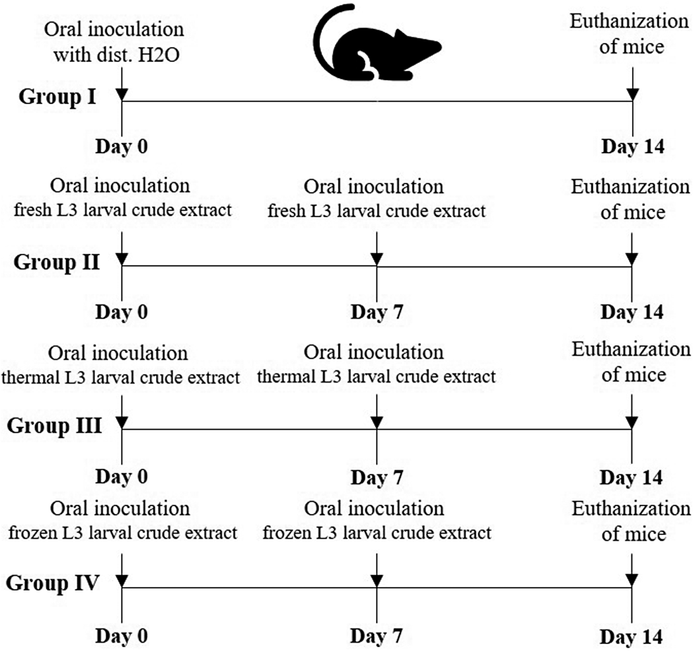
Twenty male C57BL/6 mice were included in this study. They were sourced from the animal facility at the College of Pharmacy, King Saud University. At the beginning of the experiment, the mice were 9–12 weeks old and weighed between 20 and 25 g. Animals were housed in polypropylene cages, five per cage, under controlled conditions at 24 ± 2°C with a 12-hour light/dark cycle, and were allowed a one-week acclimation period. Standard laboratory diet and water were provided freely. The mice were randomly assigned to four experimental groups, each containing five animals, and inoculated orally (per os) with 200 μL of purified crude larval extracts per mouse twice, on days 0 and 7, as shown in Figure 1. The study was conducted over 14 days, encompassing two larval inoculations.
Timeline of the experiment showing preparation of purified crude larvae extracts of H. thalassini, inoculation, and euthanization days.

Body weight of experimental mice
In the study by Rebin et al. (Reference Rebin, Dyary, Harseen, Rozha and Hassan2020), mice’s body weight (BW) was tracked over the course of the experiment. The change in BW was determined by subtracting the initial weight recorded on day 0 from the final weight measured on day 14.
Sample collection
On day 14, the mice were humanely euthanized and dissected. Blood samples were drawn from the heart into heparinized tubes. The liver was carefully removed, cut into small pieces, and preserved according to the intended analyses: (i) fixed in neutral buffered formalin (NBF) for histopathology; (ii) placed in microtubes for oxidative status assays and stored at −80°C; and (iii) immersed in RNA later for gene and protein expression studies, also stored at −80°C.
Biochemical analysis
Plasma biochemical markers of liver function were evaluated using commercial kits from Biodiagnostic Co., Egypt. Glutamic oxaloacetic transaminase (AST) and glutamic-pyruvic transaminase (ALT) levels were determined following the methods of Reitman and Frankel (Reference Reitman and Frankel1957), while alkaline phosphatase (ALP) activity was measured according to Belfield and Goldberg (Reference Belfield and Goldberg1971). Direct bilirubin was quantified using kits from Human Gesellschaft für Biochemica und Diagnostica mbH, Germany, based on Thomas (Reference Thomas1998). Plasma glucose and total protein levels were assessed with kits from Bio-Diagnostic Co., Egypt, following the procedures of Trinder (Reference Trinder1969) and Gornall et al. (Reference Gornall, Bardawill and David1949), respectively. Absorbance was measured using an Ultrospec 2000 UV spectrophotometer (Amersham Pharmacia Biotech, Cambridge, UK).
Histopathological examination
Liver samples were fixed in 10% neutral buffered formalin (NBF) for 24 hours, then dehydrated through a graded ethanol series, cleared in xylene, and embedded in paraffin. Sections of 5 μm thickness were cut with a microtome and stained with hematoxylin and eosin (H&E) following standard procedures. The stained sections were examined and imaged using an Olympus BX61 microscope (Tokyo, Japan). Liver histology was scored based on the criteria described by Ishak et al. (Reference Ishak, Baptista, Bianchi, Callea, De Groote, Gudat, Denk, Desmet, Korb, Macsween, Phillips, Portmann, Poulsen, Scheuer, Schmid and Thaler1995).
Biochemical investigations
Liver tissues were homogenised at 10% (w/v) in ice-cold 0.1 M phosphate buffer (pH 7.4). The homogenates were centrifuged at 3000 rpm for 15 minutes at 4°C, and the supernatants were collected and stored at −20°C for subsequent analysis of oxidative stress markers (Tsakiris et al., Reference Tsakiris, Schulpis, Marinou and Behrakis2004). Levels of oxidative stress parameters were measured using commercial kits from Bio-Diagnostic Co., Egypt, according to the manufacturer’s instructions. The assessed markers included Catalase (CAT) (Aebi Reference Aebi1984), Superoxide Dismutase (SOD) (Nishikimi et al. Reference Nishikimi, Appaji and Yagi1972), Nitric Oxide (NO) (Green et al. Reference Green, Wagner, Glogowski, Skipper, Wishnok and Tannenbaum1982), and Hydrogen Peroxide (H₂O₂) (Aebi Reference Aebi1984). Absorbance was measured using a SpectraMax 190 microplate reader, and the data were analysed with SoftMax Pro software version 6.3.1.
Gene expression
Total RNA was extracted from liver tissues using TRIzol reagent (Invitrogen, USA) according to the manufacturer’s protocol. The RNA was treated with DNase I (Applied Biosystems, Darmstadt, Germany) for at least 1 hour to eliminate genomic DNA contamination, and then reverse-transcribed into cDNA using a commercial reverse transcription kit (Qiagen, Hilden, Germany). Quantitative real-time PCR (qRT-PCR) was conducted on an ABI Prism 7500HT system (Applied Biosystems, Darmstadt, Germany) with SYBR Green PCR master mix (Qiagen, Hilden, Germany).
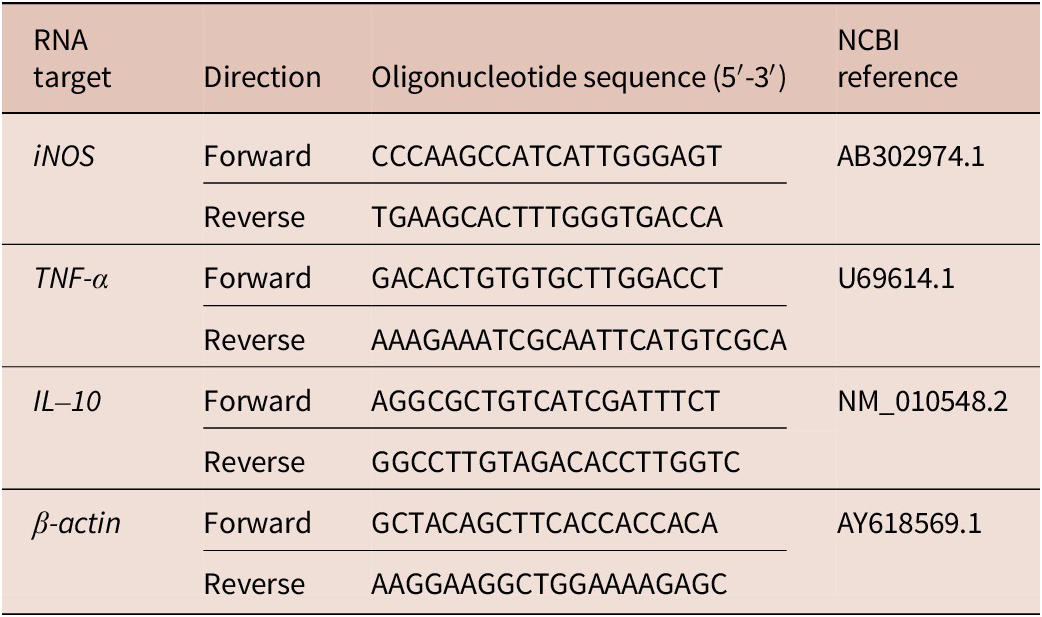
Expression levels of inducible nitric oxide synthase (iNOS), tumour necrosis factor-alpha (TNF-α), and interleukin-10 (IL-10) were quantified using primers from Qiagen (Hilden, Germany) (Table 1). Amplification and data analysis were performed with Applied Biosystems software version 3.1 (StepOne™, USA). Relative gene expression was calculated using the comparative Ct (2−ΔΔCT) method (Livak and Schmittgen, Reference Livak and Schmittgen2001), with β-actin as the internal control.
Oligonucleotide primer sequences for reverse transcription PCR amplification in the experiment

Sandwich enzyme-linked immunosorbent assay (ELISA)
Protein levels of iNOS, IL-10, and immunoglobulin E (IgE) were quantitatively measured using mouse-specific ELISA kits according to the manufacturer’s instructions. Optical densities (OD) of liver tissue samples were read with a Bio-Rad iMark Microplate Reader (version SW 1.04.02.E). Protein concentrations were determined against a standard curve and reported as μg/mg for iNOS, ng/mg for IL-10, and ng/mL for IgE.
Statistical analysis
Data are presented as mean ± standard deviation (SD) to indicate variability within each group. Comparisons among groups were conducted using one-way analysis of variance (ANOVA). Statistical analyses were carried out with SPSS software, version 18 (SPSS Inc., Chicago, IL, USA), and a p-value ≤0.05 was considered statistically significant.
Results
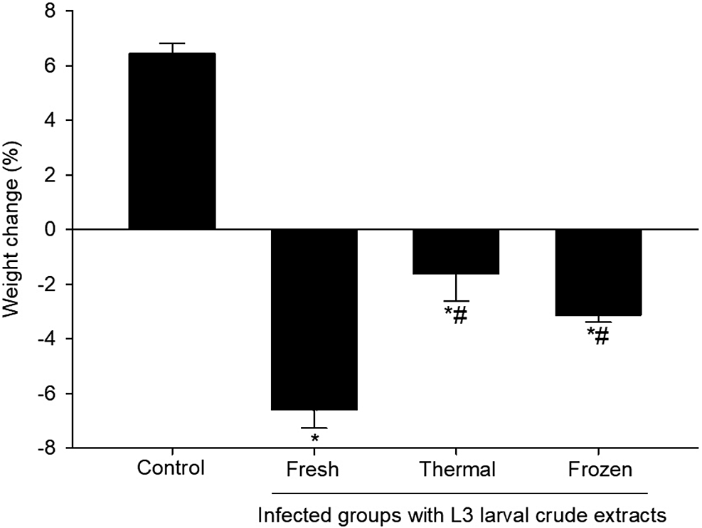
Mice inoculated with L3 purified crude larvae extract showed a significant decrease in body weight compared to the non-inoculated control group (p ≤0.05, Figure 2). While control mice gained approximately 6% of their initial body weight, those inoculated with fresh L3 larval crude extract exhibited a marked weight loss of about 7% (p ≤0.05 vs. control). Likewise, inoculation with thermally treated and frozen L3 larval crude extract resulted in significant weight reductions of approximately 3% and 5%, respectively (p ≤0.05). These results suggest that inoculation with purified crude larvae extracts of H. thalassini impairs host growth, with the greatest detrimental effect observed in mice inoculated with fresh L3 larval crude extract.
Effect of temperature variations on the body weight of mice due to inoculation with purified crude larvae extracts of H. thalassini in mice. Values are means ± SD. * significance (p ≤0.05) against the non-inoculated control group, # significance (p ≤0.05) against the inoculated group with fresh L3 larvae.

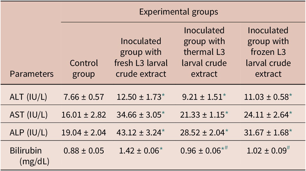
Inoculation of mice with L3 purified crude larvae extract resulted in significant alterations in liver function markers (p ≤0.05, Table 2). Mice inoculated with fresh L3 larval crude extract showed markedly elevated levels of ALT (12.50 ± 1.73 IU/L), AST (34.66 ± 3.05 IU/L), ALP (43.12 ± 3.24 IU/L), and bilirubin (1.42 ± 0.06 mg/dL) compared with the non-inoculated control group. Similarly, inoculation with thermally treated or frozen L3 larval crude extract led to significant increases in these parameters relative to controls, although the magnitude of elevation was less pronounced. Notably, bilirubin concentrations in mice inoculated with thermally treated (0.96 ± 0.06 mg/dL) or frozen (1.02 ± 0.09 mg/dL) L3 larval crude extract were significantly lower than those observed in the fresh L3-inoculated group (p ≤0.05), indicating that larval thermal treatment partially alleviated liver injury. Overall, these findings suggest that the extent of liver dysfunction induced by inoculation of purified crude larvae extracts of H. thalassini is closely associated with larval viability and treatment temperature.
Clinico-chemical parameters of liver functions of mice inoculated with L3 larval crude extracts

* p ≤0.05, significant change with respect to control.
# p ≤0.05, significant change with respect to inoculated group with fresh L3 larval crude extract.
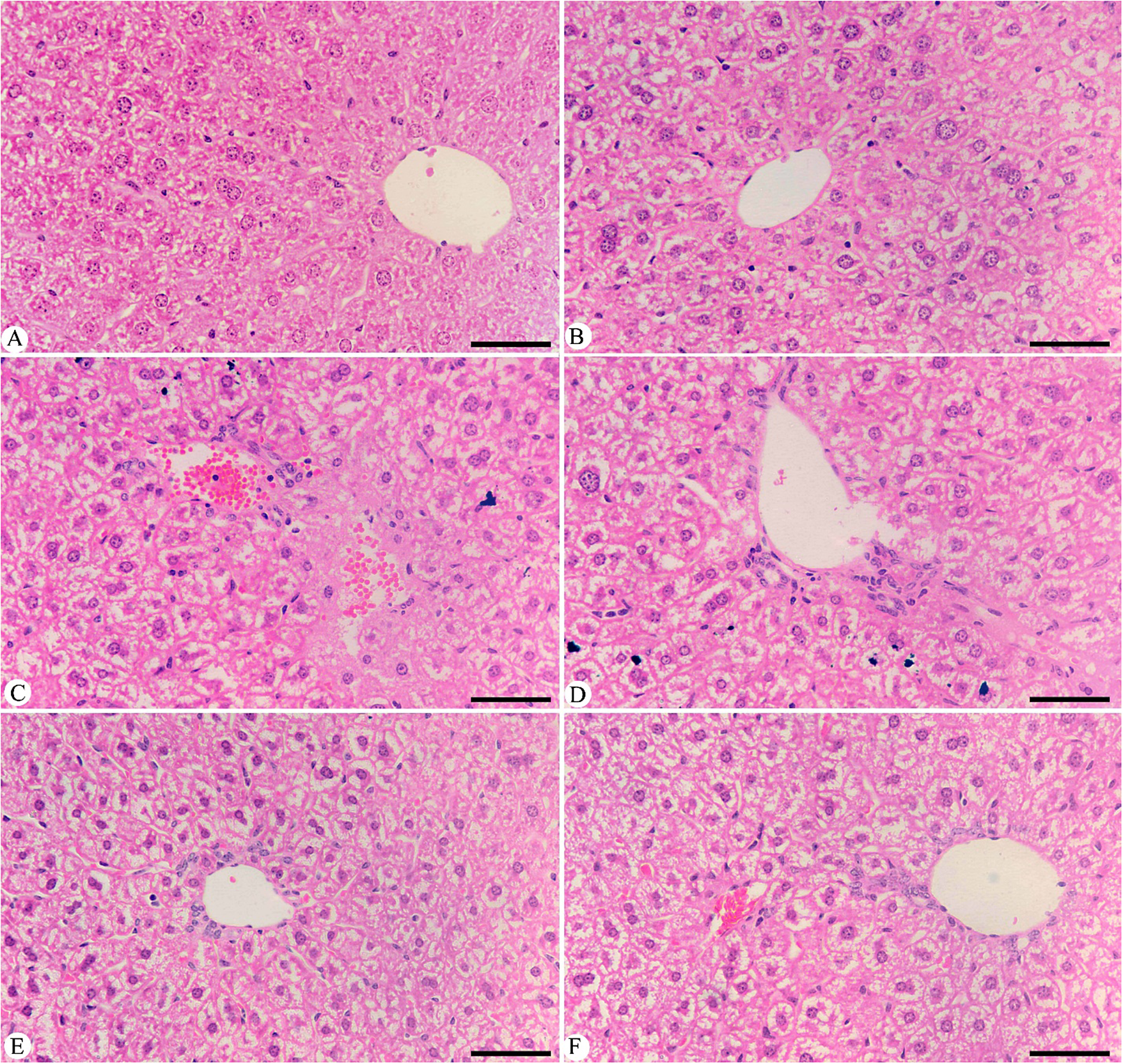
Histological sections of the mouse liver stained with hematoxylin and eosin (H&E) are presented in Figure 3 (A-F) to evaluate the liver alterations resulting from inoculation with L3 larval crude extracts. The control group exhibited normal liver architecture characterised by well-organised hepatocytes with distinct nuclei and clearly defined central veins (Figure 3A). In contrast, mice inoculated with fresh L3 larval crude extract exhibited severe liver pathology, characterised by vacuolar degeneration, hepatocellular necrosis, inflammatory cell infiltration, and congestion of central veins (Figure 3B–D). Liver sections from mice inoculated with thermally treated L3 larval crude extract demonstrated noticeably milder histopathological alterations compared to those inoculated with fresh L3 larval crude extract (Figure 3E). Similarly, inoculation with frozen L3 larval crude extract resulted in reduced tissue damage relative to the fresh L3-inoculated group (Figure 3F).
Sections stained with hematoxylin and eosin (H&E) showed the effect of L3 larval crude extracts in the liver tissue of mice. (A) Control mouse liver. (B–D) Inoculated with fresh L3 larval crude extract. (E) Inoculated the mouse liver with thermal L3 larval crude extract. (F) Inoculated the mouse liver with frozen L3 larval crude extract. Scale bar = 50 μm.

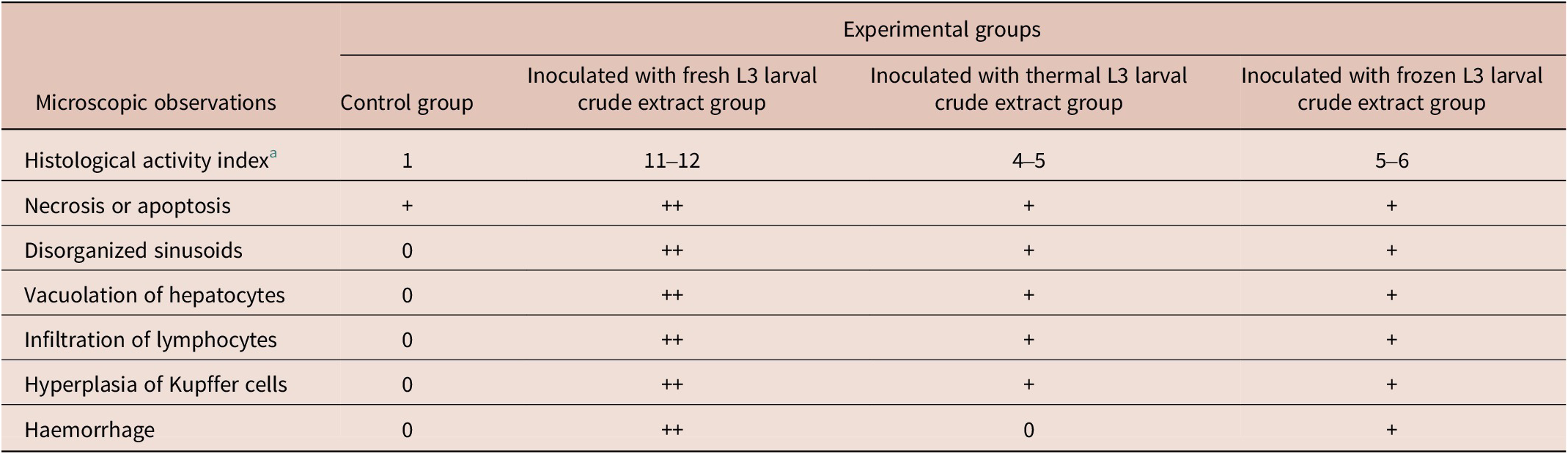
These qualitative observations were supported by total histological scores of liver tissues, evaluated using a modified Ishak scoring system (Table 3). Control mice exhibited minimal histological alterations, reflected by a histological activity index (HAI) of 1, with normal liver architecture and only slight or absent changes in necrosis, sinusoidal organisation, hepatocyte vacuolation, lymphocyte infiltration, Kupffer cell hyperplasia, and haemorrhage. Mice inoculated with fresh L3 larval crude extract displayed moderate liver injury, with HAI values ranging from 11 to 12 and moderate alterations across most microscopic parameters, indicating marked hepatocellular degeneration and inflammatory infiltration. Inoculation with thermally treated or frozen L3 larval crude extract resulted in noticeably milder histopathological changes (HAI 4–6), with only mild degrees of necrosis, vacuolation, lymphocyte infiltration, and Kupffer cell hyperplasia. Haemorrhage was absent in the thermal L3 group but mild in the frozen L3 group. These findings demonstrate that inoculation with purified crude larvae extracts of H. thalassini induces significant liver injury, while thermal or freezing treatment of L3 larvae substantially attenuates the severity of liver damage.
Total histological scores in the liver tissue of mice inoculated with L3 larval crude extract

a Modified Ishak et al. (Reference Ishak, Baptista, Bianchi, Callea, De Groote, Gudat, Denk, Desmet, Korb, Macsween, Phillips, Portmann, Poulsen, Scheuer, Schmid and Thaler1995) score. Score, 1–3 (minimal); 4-8 (mild); 9–12 (moderate); 13–18 (severe); 0 (absent), + (mild), ++ (moderate), +++ (severe).
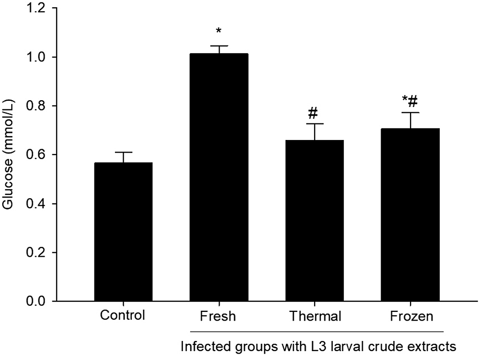
The impact of L3 larval crude extract inoculation on blood plasma glucose levels in mice is shown in Figure 4. Mice inoculated with fresh L3 larval crude extract exhibited a significant increase in plasma glucose compared to the non-inoculated control group (p ≤0.05), indicating a hyperglycaemic response. Inoculation with thermally treated L3 larval crude extract resulted in lower glucose levels than the fresh L3 group (p ≤0.05). In contrast, inoculation with frozen L3 larval crude extract caused an intermediate glucose elevation, significantly higher than controls (p ≤0.05) but lower than in the fresh L3 group (p ≤0.05). These findings suggest that larval viability and thermal treatment modulate the metabolic effects of L3 inoculation on host glucose homeostasis.
Glucose content in the blood plasma of mice inoculated with L3 larval crude extracts. *p ≤0.05, significant change with respect to control, #p ≤0.05, significant change with respect to inoculated group with fresh L3 larval crude extract.

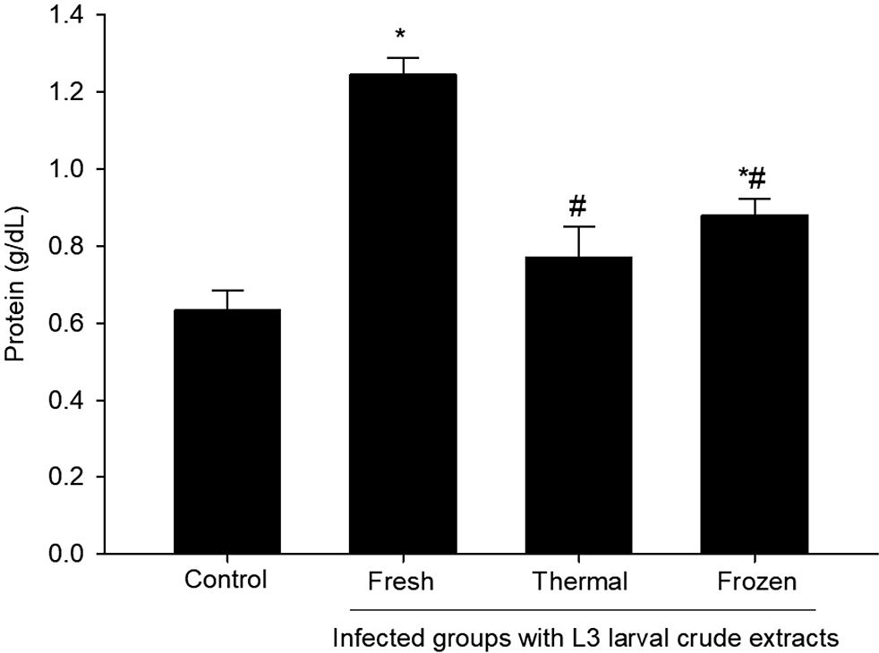
Total plasma protein levels are presented in Figure 5 to illustrate the significant reduction observed in mice following inoculation with L3 larval crude extracts compared to the control group (p ≤0.05), reflecting the adverse effects of the extracts on liver function and protein metabolism. In contrast, mice inoculated with thermally treated or frozen larval crude extract exhibited significantly higher plasma protein levels than those inoculated with fresh L3 larval crude extract (p ≤0.05). This partial restoration of protein content suggests that thermal or freezing treatment of larvae mitigates the impact of inoculation, helping to preserve normal protein synthesis and liver function.
Protein content in the blood plasma of mice inoculated with L3 larval crude extracts. *p ≤0.05, significant change with respect to control, #p ≤0.05, significant change with respect to inoculated group with fresh L3 larval crude extract.

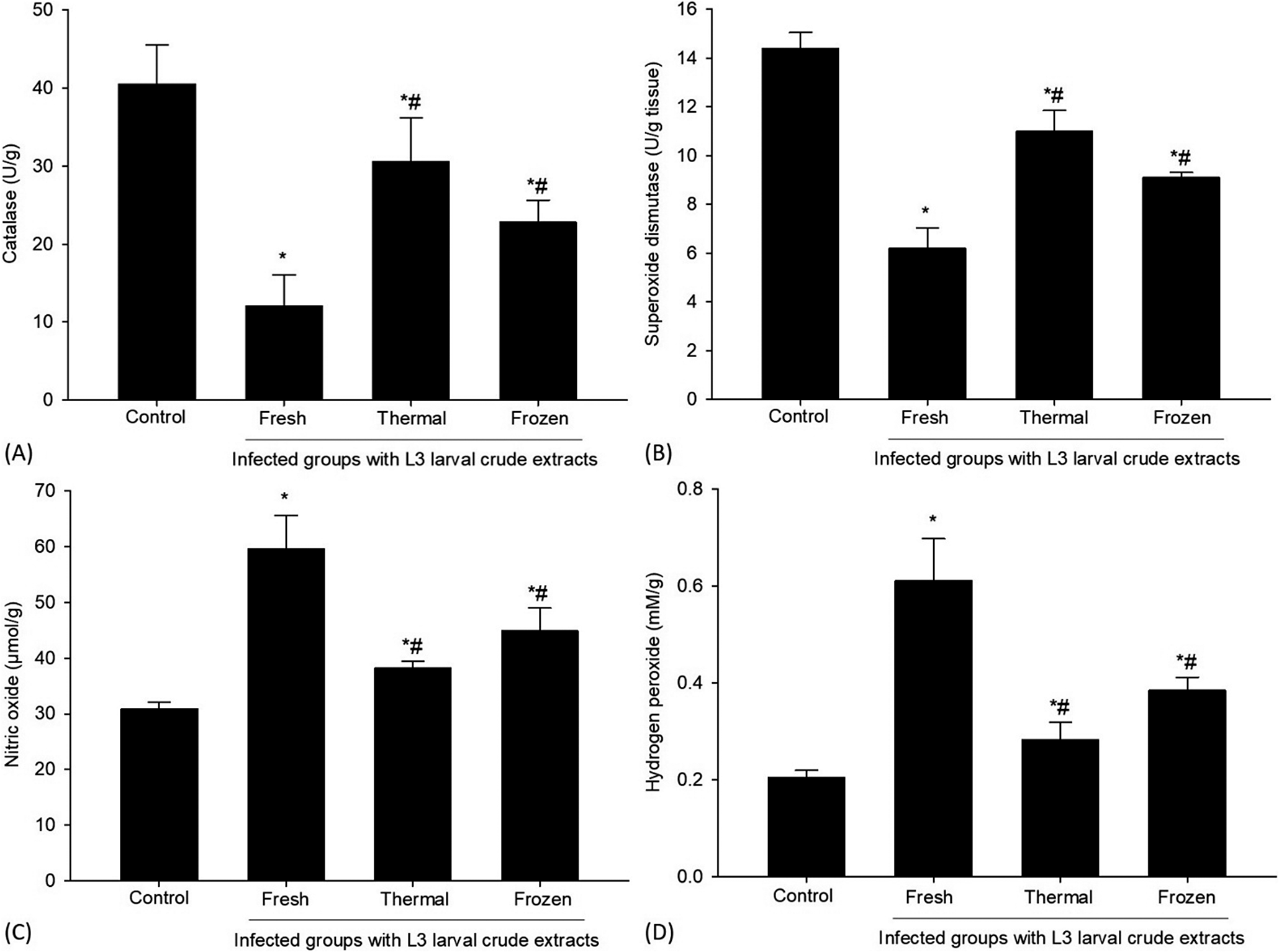
Liver antioxidant enzyme activities and oxidative stress markers are presented in Figure 6 to show the significant effects induced by inoculation with purified crude larval extracts relative to controls (p ≤0.05). Mice inoculated with fresh L3 larval crude extract exhibited a marked decrease in CAT and SOD activities, indicating a compromised hepatic antioxidant defence. Concurrently, NO and H₂O₂ levels were significantly elevated, reflecting enhanced oxidative stress and cellular damage associated with the inoculation. In contrast, inoculation with thermally or frozen-treated larval crude extract resulted in improved hepatic antioxidant profiles, with higher CAT and SOD activities and lower NO and H₂O₂ concentrations compared with the fresh L3 group (p ≤0.05). These findings suggest that temperature treatments reduce larval infectivity or metabolic activity, thereby attenuating oxidative damage and partially restoring the antioxidant balance in liver tissue.
Effect of temperature variations on the level of catalase (CAT), superoxide dismutase (SOD), nitric oxide (NO), and hydrogen peroxide (H2O2) activities of the liver of mice inoculated with purified crude larvae extracts of H. thalassini. *p ≤0.05, significant change with respect to control, #p ≤0.05, significant change with respect to inoculated group with fresh L3 larval crude extract.

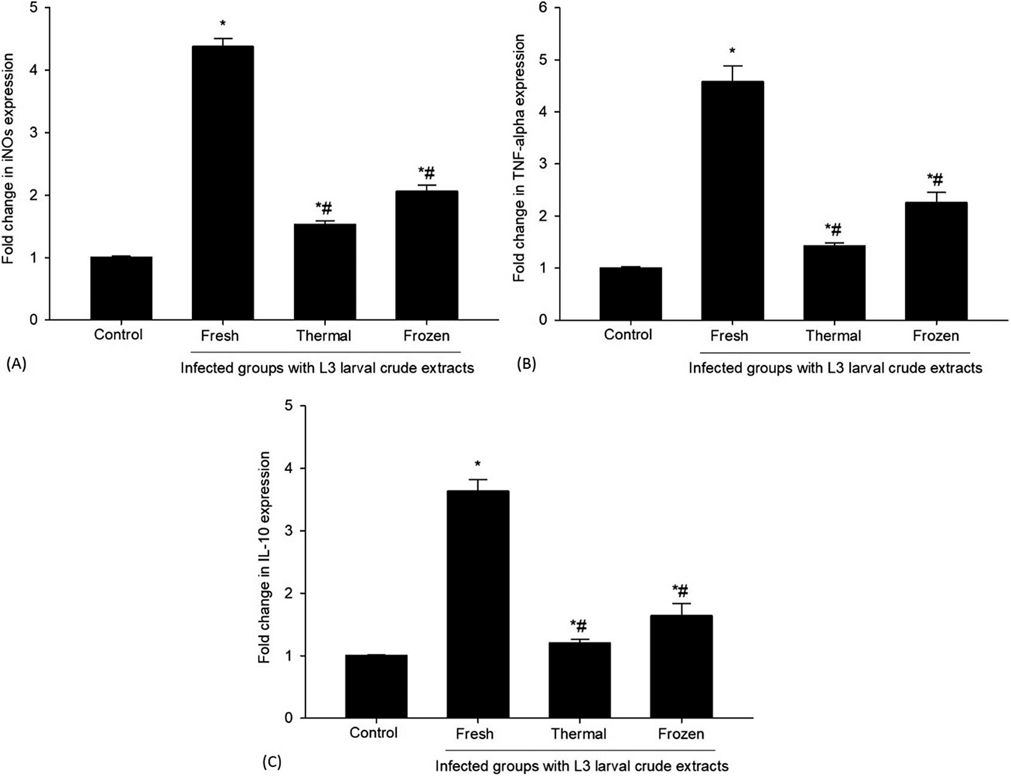
Hepatic gene expression associated with inflammation and immune responses is presented in Figure 7 to show the significant effects of inoculation with L3 larval crude extracts. Mice inoculated with fresh L3 larval crude extract exhibited marked upregulation of iNOS, TNF-α, and IL-10 mRNA compared to controls (p ≤0.05), indicating an enhanced pro-inflammatory response and increased NO production, likely linked to tissue injury and oxidative stress caused by the inoculation. In contrast, mice inoculated with temperature-treated larval crude extract showed significantly lower expression of these genes than those inoculated with fresh L3 larval crude extract (p ≤0.05), suggesting that temperature treatment attenuated larval pathogenicity, resulting in reduced inflammation and a shift towards immune regulation and tissue repair.
mRNA expression of (A) iNOS, (B) TNF-α, and (C) IL-10 in the liver tissue from the different experimental groups. The expression values obtained by RT-PCR analysis were normalised to the reference gene β-actin mRNA level and are shown as fold induction (in log 2 scale) relative to the mRNA level in the control. *p ≤0.05, significant change with respect to control, #p ≤0.05, significant change with respect to inoculated group with fresh L3 larval crude extract.

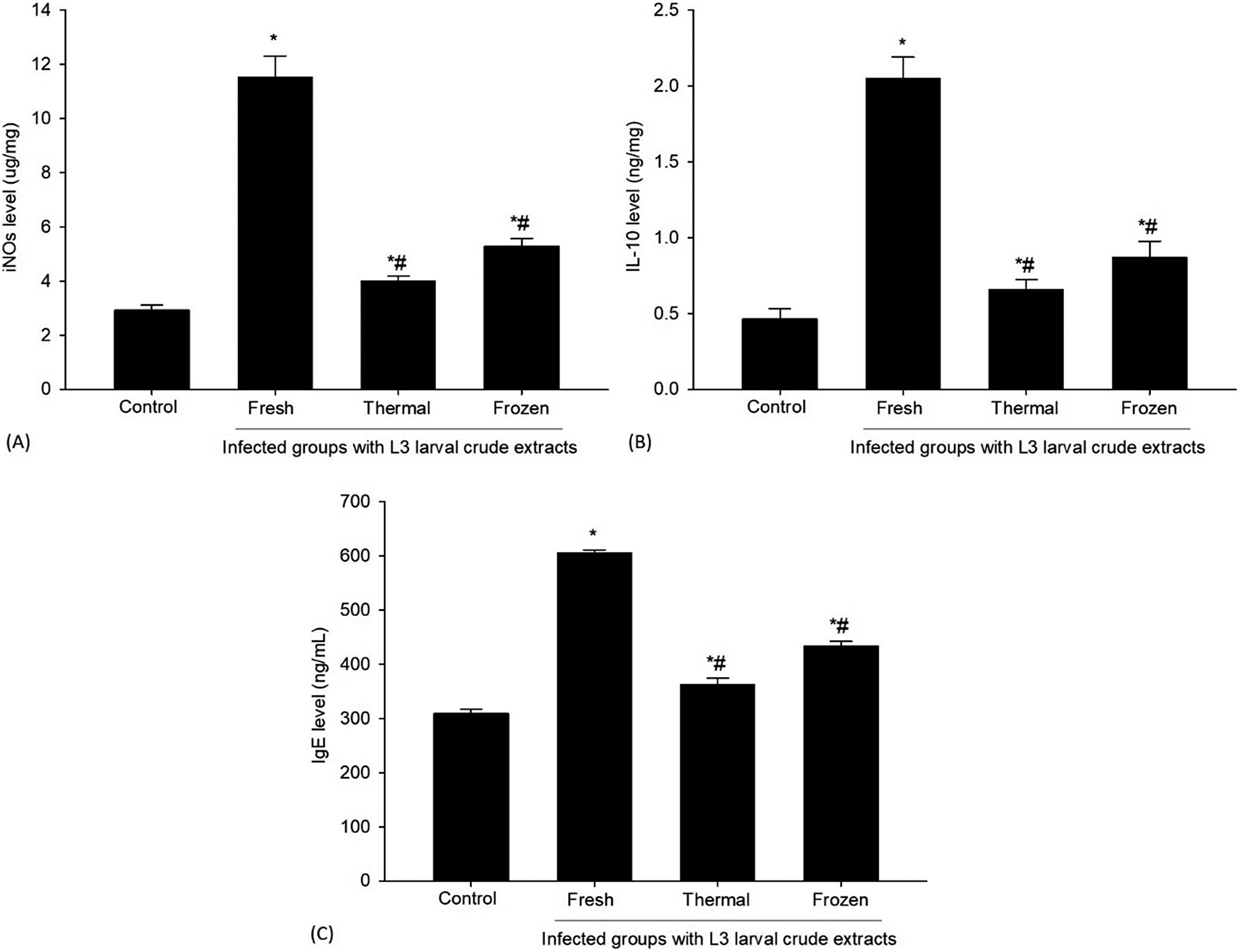
Hepatic concentrations of iNOS, IL-10, and IgE in the experimental mouse groups are presented in Figure 8. Inoculation with fresh L3 larval crude extract caused a marked increase in iNOS expression compared with controls (p ≤0.05, Figure 8A), reflecting heightened inflammatory activity in the liver. In contrast, mice receiving thermally or frozen-treated larval crude extract showed significantly lower iNOS levels than the inoculated group with fresh L3 larval crude extract (p ≤0.05), indicating suppression of NO-mediated inflammation. IL-10 levels were significantly elevated in the inoculated group with fresh L3 larval crude extract relative to controls (p ≤0.05, Figure 8B), suggesting a compensatory anti-inflammatory response. Notably, IL-10 levels were further increased in the treated group (p ≤0.05), implying enhanced immunoregulatory activity that may protect the liver from parasitic damage. Similarly, IgE levels rose significantly in the inoculated group with fresh L3 larval crude extract compared with controls (p ≤0.05, Figure 8C), consistent with activation of a Th2-type immune response. Conversely, the thermally or frozen-treated larval crude extract exhibited a significant reduction in IgE (p ≤0.05), indicating decreased allergic-type immune stimulation and parasite burden. Collectively, these results demonstrate that fresh L3 larval crude extract induces pronounced inflammatory and immune responses in the liver. In contrast, thermally or frozen-treated larval crude extract modulates these responses, promoting immune balance and hepatic protection.
Levels of (A) iNOS, (B) IL-10, and (C) IgE in the liver tissue from the different experimental mouse groups. *p ≤0.05, significant change with respect to control, #p ≤0.05, significant change with respect to inoculated group with fresh L3 larval crude extract.

Discussion
Anisakid nematodes are prevalent marine parasites that could infect a diverse array of fish species and, on rare occasions, mammals. These infections can lead to substantial pathological and metabolic disruptions within the host organism (Fuentes et al. Reference Fuentes, Madrid, Cuesta, Gimeno, Baquedano-Rodríguez, Soriano-Sánchez, Bolívar, Sáez-Durán, Trelis and Debenedetti2022). Among these parasites, H. thalassini has garnered increasing attention due to its potential zoonotic implications and its effects on the physiology of infected hosts. To better comprehend the complex interactions between this parasite and its host, experimental inoculations in mice have been utilised as effective models to investigate the developmental behaviours of H. thalassini and the corresponding immune responses elicited by the host (Greischar et al. Reference Greischar, Reece and Mideo2016). This study specifically aims to evaluate the effects of inoculation using purified crude larvae extracts of H. thalassini (L3) that have been subjected to various treatments, including fresh, thermally treated, and frozen conditions. The research focuses on understanding how these differing larval states impact the host’s growth, liver metabolic processes, and overall immune responses.
The notable decrease in body weight observed in mice inoculated with purified crude larvae extracts of H. thalassini aligns with previous research that highlights how inoculation with anisakid nematodes can lead to both growth suppression and significant loss of body mass. For instance, experimental infections involving Anisakis simplex and Contracaecum species have shown substantial reductions in weight gain, primarily due to factors like impaired nutrient absorption, increased energy expenditure, and the host’s inflammatory responses (Audicana and Kennedy Reference Audicana and Kennedy2008; Mehrdana and Buchmann Reference Mehrdana and Buchmann2017). In the current study, the pronounced weight loss seen in mice inoculated with fresh L3 larval crude extract contrasts with that of mice exposed to larval crude extract that were thermally treated or frozen, emphasising the critical role of larval viability and their metabolic activity in determining the severity of the disease. Fresh larval crude extracts are known to secrete a range of proteolytic and immunomodulatory molecules, which can significantly disrupt the integrity of the intestinal lining. This disruption leads to a decrease in nutrient absorption and triggers systemic inflammation and oxidative stress within the host, ultimately elevating the host’s catabolic metabolism (Caballero and Moneo Reference Caballero and Moneo2004; Pramardika et al. Reference Pramardika, Satrija, Sulistiono, Tiuria and Nugraha2025). These metabolic shifts, combined with a reduction in feeding behaviour and an increase in cytokine production—such as tumour necrosis factor-alpha (TNF-α) and interleukin-1 beta (IL-1β)—further contribute to the breakdown of fats and muscle proteins, exacerbating body weight loss (Kochanowski et al. Reference Kochanowski, Dąbrowska, Różycki, Sroka, Karamon, Bełcik, Korpysa-Dzirba and Cencek2022; Polak et al. Reference Polak, Stryiński, Majewska and Łopieńska-Biernat2023). Thus, the substantial weight reduction observed in this study is likely a result of the intricate interplay between nutrient competition, tissue inflammation, and the metabolic reprogramming instigated by purified crude larvae extracts of H. thalassini.
Furthermore, the observed elevation of liver enzymes, specifically ALT, AST, and ALP, along with increased bilirubin levels in mice inoculated with purified crude larvae extracts of H. thalassini, aligns with earlier studies indicating hepatic dysfunction associated with experimental anisakid inoculations. Similar biochemical changes have been documented in rodent models inoculated with Anisakis simplex and Contracaecum species, which showcase significant liver damage and bile duct obstruction. These issues stem from the migratory behaviour of the larvae combined with localised inflammatory responses that ensue (Bušelić et al. Reference Bušelić, Trumbić, Hrabar, Vrbatović, Bočina and Mladineo2018; Mehrdana and Buchmann Reference Mehrdana and Buchmann2017). The rise in serum transaminases signals membrane damage due to hepatocyte necrosis, exacerbated by oxidative stress. Concurrently, elevated levels of ALP and bilirubin indicate cholestasis and hindered hepatic clearance processes (Contreras-Zentella and Hernández-Muñoz Reference Contreras-Zentella and Hernández-Muñoz2016). Notably, the relatively modest increase in these biochemical markers in mice inoculated with either thermally treated or frozen larval crude extract suggests that the viability of the larvae plays a pivotal role in the extent of hepatic injury observed. This finding is consistent with previous research, which demonstrates that living anisakid larvae actively secrete proteolytic enzymes and metabolic antigens. These elements incite tissue inflammation, the formation of eosinophilic granulomas, and ultimately, the degeneration of hepatocytes (Palomba et al. Reference Palomba, Rughetti, Castrignanò, Napoletano, Pinna, Liberati, Canestrelli and Mattiucci2025; Zamora et al. Reference Zamora, Andreu-Ballester, Rodero and Cuéllar2021). Consequently, our findings highlight that inoculation with purified crude larvae extracts of H. thalassini predominantly induces liver dysfunction, primarily due to the effects of fresh L3 larval crude extract, along with the associated inflammatory and oxidative responses triggered by the host immune system.
The current research findings highlight, also, that inoculation with fresh L3 larval crude extract led to significant and detrimental changes in the hepatic pathology, characterised by hepatocellular degeneration, necrosis, and a pronounced inflammatory response. These histopathological alterations are indicative of severe liver injury. Similar effects have been noted in previous studies involving helminthic infections, such as those caused by anisakid nematodes, where the presence of active larvae triggers substantial inflammatory and degenerative responses in the hepatic tissue (Shamsi and Butcher Reference Shamsi and Butcher2011; Ying et al. Reference Ying, Fang, Wang, Yang, Xu, Liu and Yin2022). In contrast, when the larval crude extract has undergone thermal treatment or freezing, there is a remarkable reduction in tissue damage. This observation suggests that these temperature treatments are highly effective in reducing both the viability and antigenicity of the larval crude extract, which in turn lessens the host’s inflammatory reactions (Shamsi and Butcher Reference Shamsi and Butcher2011). These findings align with earlier research that indicates various temperature treatments—such as heating or freezing—can effectively render helminth larvae non-infectious. This includes species like Anisakis simplex, Pseudoterranova krabbei, and P. decipiens (Wharton and Aalders Reference Wharton and Aalders2002; Podolska et al. Reference Podolska, Pawlikowski, Nadolna-Ałtyn, Pawlak, Komar-Szymczak and Szostakowska2019). The current study further corroborates these findings, emphasising that it is the fresh anisakid larval crude extract—which remains capable of inducing significant hepatic pathology—that is responsible for these harmful effects, while the larvae that have been inactivated by temperature are not associated with such pronounced liver damage. This reinforces the notion of a temperature-dependent reduction in larval pathogenicity, underlining the importance of effective treatment methods in mitigating the risks associated with these parasitic inoculations.
Moreover, the current research findings revealed that inoculation with freshly introduced purified crude larvae extracts of H. thalassini led to a significant elevation in plasma glucose levels. This increase suggests a hyperglycaemic state that is closely linked to the effects of an active parasitic inoculation. Such changes in glucose metabolism are consistent with earlier studies focusing on helminth infections. These studies indicated that the stress associated with inoculation, alongside the release of inflammatory cytokines, triggers the liver to increase gluconeogenesis, which in turn diminishes the uptake of glucose by peripheral tissues (Khudhair et al. Reference Khudhair, Alhallaf, Eichenberger, Whan, Kupz, Field, Krause, Wilson, Daly, Giacomin, Sotillo and Loukas2021; Rennie et al. Reference Rennie, Fernandez, Donnelly and McGrath2021). In contrast, mice that were inoculated with thermally treated or frozen larval crude extract showed a marked decrease in their plasma glucose levels. This decline indicates that the lack of fresh larvae alleviates some of the metabolic stress experienced by the host organism. Previous research has documented similar reductions in hyperglycaemia related to inoculations in models exposed to inactivated or attenuated parasites (Podolska et al. Reference Podolska, Pawlikowski, Nadolna-Ałtyn, Pawlak, Komar-Szymczak and Szostakowska2019). These findings reinforce the notion that the viability of larval crude extract and its antigenic activity play crucial roles in causing disturbances in the host’s glucose regulation.
A significant decline in total plasma protein levels observed in mice inoculated with fresh L3 larval crude extract points to significant impairment in liver function and disruptions in protein metabolism. This observation aligns with previous studies on nematode and trematode infections, which established a connection between hypoproteinaemia and damage to hepatic tissues, as well as an increase in protein catabolism during inoculation (Starr et al. Reference Starr, Odiere, Koski and Scott2014; Zaparina et al. Reference Zaparina, Kapushchak, Lishai, Hong, Sripa and Pakharukova2024). Conversely, the notable recovery of plasma protein levels in mice inoculated with larval crude extract that had undergone thermal treatment or freezing suggests that the loss of larval viability induced by temperature fluctuations serves as a protective factor for liver function, aiding in the preservation of protein synthesis. These findings are consistent with earlier research indicating that larval crude extract inactivated by heat or freezing demonstrates significantly reduced pathogenicity and leads to more favourable biochemical outcomes. This emphasises how temperature treatments can mitigate the metabolic disruptions caused by parasites (Wharton and Aalders Reference Wharton and Aalders2002).
The current research findings reveal that the inoculation of mice with purified crude larvae extracts of H. thalassini leads to significant changes in the activities of hepatic antioxidant enzymes, as well as alterations in markers of oxidative stress. Specifically, the notable decrease in the activities of CAT and SOD in mice inoculated with fresh L3 larval crude extract suggests that the liver’s antioxidant defence mechanisms are compromised. Concurrently, the observed increase in the levels of NO and H₂O₂ indicates a heightened state of oxidative stress and liver damage linked to active parasitic inoculation. These outcomes align with previous studies on helminthic infections, which have demonstrated that both the invasion of parasites and the corresponding immune response of the host can generate excessive reactive oxygen species (ROS) and reactive nitrogen species (RNS). This overproduction can lead to oxidative damage, resulting in the depletion of endogenous antioxidant enzymes (Chiumiento and Bruschi Reference Chiumiento and Bruschi2009; Pawłowska et al. Reference Pawłowska, Mila-Kierzenkowska, Szczegielniak and Woźniak2023). In contrast, mice that were inoculated with larval crude extract that had been treated through thermal or freezing processes displayed a marked improvement in their antioxidant status. This improvement was characterised by increased activities of CAT and SOD, alongside decreased levels of NO and H₂O₂. Such trends have been documented previously, in which reports indicate that a reduction in larval viability or metabolic activity significantly lessens the parasite’s ability to induce oxidative stress. Consequently, this reduction helps to mitigate tissue damage and restore a balanced redox state (Matoo et al. Reference Matoo, Ivanina, Ullstad, Beniash and Sokolova2013). In summary, these findings suggest that the inactivation of L3 larval crude extract through temperature treatment effectively reduces the oxidative stress associated with inoculation and helps to maintain a healthier balance of hepatic antioxidants.
The findings of the present study revealed that inoculation with purified crude larvae extracts of H. thalassini has a profound impact on hepatic gene expression that is closely tied to inflammation and the regulation of immune responses. Mice that were inoculated with fresh L3 larval crude extract exhibited a remarkable increase in the expression of transcripts for iNOS, TNF-α, and IL-10. This pronounced upregulation indicates a robust pro-inflammatory response and an enhanced production of NO, both of which are associated with tissue damage and oxidative stress induced by the inoculation. These observations align with previous research, which has demonstrated that helminth infections can trigger host immune reactions through the activation of inflammatory cytokines and iNOS. This process is instrumental not only in the elimination of the parasite but also in causing collateral damage to host tissues (Klotz et al. Reference Klotz, Ziegler, Figueiredo, Rausch, Hepworth and Obsivac2011; Rolot and Dewals Reference Rolot and Dewals2017; Mabbott Reference Mabbott2018). Also, the concurrent increase in IL-10 expression appears to serve as a protective, compensatory mechanism. This response likely aims to regulate the excessive inflammatory processes and limit the resulting hepatic injury, a phenomenon that has been documented in earlier studies involving experimental helminthic and protozoan infections (Redpath et al. Reference Redpath, Fonseca and Perona-Wright2014; Stijlemans et al. Reference Stijlemans, Korf, De Baetselier, Brys, Van Ginderachter, Magez and De Trez2020). In contrast, the inoculation with L3 larval crude extract that had been subjected to thermal or freezing treatments resulted in significantly lower levels of expression for iNOS, TNF-α, and IL-10. This finding suggests that the temperature treatments may have compromised larval viability or their metabolic activity. This observation is consistent with studies indicating that a decrease in parasite infectivity correlates with a reduction in cytokine expression, leading to a shift towards immune balance and tissue repair mechanisms (Allen and Sutherland Reference Allen and Sutherland2014; Boccardo et al. Reference Boccardo, Rodriguez, Gimenez, Araujo Furlan, Abrate and Almada2025; Bosurgi and Rothlin Reference Bosurgi and Rothlin2021). Taken together, these results underscore the critical role that larval viability plays in governing the intensity and nature of host inflammatory and regulatory immune responses during inoculation.
Moreover, the present study reveals compelling evidence that inoculation with fresh L3 larval crude extract triggers pronounced inflammatory and immune responses in the liver. This is evidenced by significantly elevated levels of key biomarkers, including iNOS, IL-10, and IgE. The marked increase in iNOS expression in mice inoculated with fresh L3 larval crude extract suggests a heightened production of NO, which is closely associated with inflammation. This enhanced inflammation likely contributes to oxidative damage and subsequent liver injury. These observations resonate with earlier studies on helminthic infections, wherein the activation of iNOS has been correlated with tissue inflammation and the initiation of immune defence mechanisms (Kim and Lee Reference Kim and Lee2025; Zamora et al. Reference Zamora, Vodovotz and Billiar2000). Furthermore, the elevated levels of IL-10 detected in the inoculated group imply a compensatory regulatory mechanism that strives to counterbalance excessive inflammation and mitigate damage to host tissues. This finding aligns with previous research indicating that IL-10 plays a pivotal role in immunoregulation during helminth infections, promoting a delicate equilibrium between pro-inflammatory and anti-inflammatory responses. This balance is crucial for fostering host tolerance while minimising tissue pathology (Al-Qahtani et al. Reference Al-Qahtani, Alhamlan and Al-Qahtani2024; Iyer and Cheng Reference Iyer and Cheng2012; Redford and Murray Reference Redford and Murray2011; Saraiva et al. Reference Saraiva, Vieira and O’Garra2020). In addition, the significant increase in IgE concentrations among mice inoculated with fresh L3 larval crude extract underscores a characteristic Th2-type immune polarisation that is typically observed in helminthic infections (Cho et al. Reference Cho, Park, Kang, Caballero, Perez-Pinar, Rodriguez-Perez, Ock, Cha, Hong and Yu2014; Santiago et al. Reference Santiago, Ribeiro-Gomes, Bennuru and Nutman2014; Hidano et al. Reference Hidano, Mizukami, Shirakami, Ito, Matsumura, Dewayani, Ozaka, Saechue, Ariki, Ozaki, Noguchi, Kagoshima, Ekronarongchai, Okamoto, Kamiyama, Sachi, Sekiya, Takaki, Murakami and Kobayashi2025; Watanabe Reference Watanabe2023). The elevation of IgE levels signifies enhanced activation of mast cells and eosinophils, immune cells that play critical roles in the expulsion of parasites and the modulation of immune responses. Conversely, inoculation with thermally or frozen-treated larval crude extract led to reduced levels of iNOS, IL-10, and IgE. This pattern suggests a shift in the immune response towards greater regulation and hepatic protection. Comparable findings have been documented in studies where attenuated or metabolically inactivated larvae elicited diminished pro-inflammatory responses and fostered an anti-inflammatory environment, which is conducive to tissue repair and the maintenance of homeostasis (Boccardo et al. Reference Boccardo, Rodriguez, Gimenez, Araujo Furlan, Abrate and Almada2025; Bosurgi and Rothlin Reference Bosurgi and Rothlin2021). In summary, these findings collectively underscore the critical role of larval viability in shaping not only the intensity of inflammatory responses but also the regulatory immune responses in the liver.
Conclusion
This study reveals that inoculation with fresh L3 larval crude extract causes marked liver dysfunction, oxidative stress, and both inflammatory and Th2-type immune responses in mice, as indicated by disrupted liver enzyme levels, hyperglycaemia, hypoproteinaemia, histopathological liver damage, weakened antioxidant defences, and increased iNOS, IL-10, and IgE expression. Treatments involving heat or freezing of the larval crude extract significantly alleviated these effects, minimising liver injury, restoring antioxidant activity, and enhancing immunoregulatory responses. These results underscore the importance of larval viability in determining pathogenicity and suggest that simple physical treatments can effectively limit larval-induced hepatic damage. Future research should investigate the molecular mechanisms driving host-parasite interactions and explore additional intervention strategies to further reduce larval pathogenicity and protect liver function.
Availability of data and materials
All the datasets generated or analysed during this study are included in this published article.
Acknowledgements
This study was supported by the Ongoing Research Funding Program (ORF-2026-25), King Saud University, Riyadh, Saudi Arabia.
Financial support
No funding was received for this research.
Competing interest
The author(s) declare that they have no conflict of interest regarding the content of this article.
Ethical approval
This research was approved by the Research Ethics Committee (REC) at King Saud University (approval number KSU-SE-25-28).
Consent to participate
All authors agreed to participate in this study.
Consent to publish
All authors agreed to publish the data in this study.