INTRODUCTION
Trigeminal ganglia function to relay sensory information from peripheral tissues in the face, oral cavity, nasal cavity and dura mater to the central nervous system via trigeminal nerves. Trigeminal ganglion neurons innervate primarily mechanoreceptors, thermoreceptors and nociceptors (Dubner, Reference Dubner1978; Davies, Reference Davies1988; Lazarov, Reference Lazarov2002). Central projecting nerve processes terminate on several groups of neurons in the brainstem and upper spinal cord and relay sensory and nociceptive information via the thalamus to the somatosensory cortex. Pain in the head and face, which can be very severe and debilitating, often involves activation of the fifth cranial nerve or trigeminal ganglion nerve, which is the largest and most complex of the 12 cranial nerves. In response to trigeminal nerve activation, craniofacial symptoms can manifest as transient painful conditions as reported with toothaches and headaches, or can transform into more chronic pain conditions such as migraine, rhinosinusitis, temporomandibular joint (TMJ) disorder or trigeminal neuralgia. Importantly, increased neuron–glia interactions are thought to play an important role in the development of persistent pain (Watkins and Maier, Reference Watkins and Maier2002; Ji and Suter, Reference Ji and Suter2007; Cheng and Ji, Reference Cheng and Ji2008; Takeda et al., Reference Takeda, Takahashi and Matsumoto2009).
The trigeminal ganglion is comprised primarily of sensory neurons and their fibers as well as two types of glial cells, satellite glial cells and Schwann cells (for a review see Hanani, Reference Hanani2005). The cell bodies of two main types of primary afferent neurons reside in trigeminal ganglia, which are referred to as C fibers and A fibers (Lawson, Reference Lawson and Scott1992). Trigeminal neurons have a unique morphology and are classified as psuedounipolar since a single axon, which arises from the cell body, divides into two branches with one branch projecting to peripheral target tissue and the other branch projecting to the central nervous system. In adult mammals, the neuronal cell bodies are completely enveloped by satellite glial cells, which together form distinct, functional units (Pannese et al., Reference Pannese, Bianchi, Calligaris, Ventura and Weibel1972; Pannese, Reference Pannese1981). Satellite glial cells facilitate direct signaling between neurons and satellite glial cells via exchanging ions and small molecules (Pannese, Reference Pannese1981; Hanani, Reference Hanani2005). There is increasing evidence to support a significant role of glial cells in pathological states by directly modulating the threshold of activation and excitability state of neurons, and thus their function (Watkins and Maier, Reference Watkins and Maier2002; Hanani, Reference Hanani2005; Takeda et al., Reference Takeda, Tanimoto, Kadoi, Nasu, Takahashi and Kitagawa2007). Furthermore, increased neuron–glia interactions are thought to play an important role in the induction and maintenance of peripheral sensitization of trigeminal nociceptors (Cheng and Ji, Reference Cheng and Ji2008; Takeda et al., Reference Takeda, Takahashi and Matsumoto2009).
During mammalian development, neural crest cells in the embryo migrate away from the dorsal neuronal tube and aggregate to form the sensory and autonomic ganglia of the peripheral nervous system (Pannese, Reference Pannese1974; Kalcheim and Le Douarin, Reference Kalcheim and Le Douarin1986; Le Douarin and Kalcheim, Reference Le Douarin and Kalcheim1999; Lazarov, Reference Lazarov2002). Trigeminal ganglia have a dual origin since they are formed by cranial neural crest cells and trigeminal ectodermal placodes (Hamburger, Reference Hamburger1961; Altman and Bayer, Reference Altman and Bayer1982; D'Amico-Martel and Noden, Reference D'Amico-Martel and Noden1983). In the mouse, trigeminal ganglia first become discernable by the ninth embryonic day with the earliest nerve fibers emerging around day 9.5 and the last fibers exiting at day 13. These fibers do not reach their peripheral target fields until day 10.5 in the mandibular process, day 11 in the maxillary and just after day 15 in the other regions (Davies, Reference Davies1988). Mammalian ganglia are comprised of neurons and glia that differentiate from precursor neural crest cells. Complex processes regulate ganglia development whereby instructive extracellular signals such as bone morphogenic proteins and sonic hedgehog initially promote neuronal differentiation and are essential for specification of trigeminal sensory neurons (Ota and Ito, Reference Ota and Ito2003). Recently, it was shown that although expression of the proneural transcription factor neurogenin 1 is a key regulator of sensory neuron development, blocking its function promotes differentiation of glial cells (McGraw et al., Reference McGraw, Nechiporuk and Raible2008). Since blocking development of trigeminal neurons prevents formation of the ganglia, the differentiation of glial cells appears to be regulated by the neurons they become associated with during development. However, severe degeneration of sensory and motor neurons results in differentiation and proliferation of peripheral glial cells and is prevented by blocking expression of the transcription factor Sox 10 (Britsch et al., Reference Britsch, Goerich, Riethmacher, Peirano, Rossner and Nave2001). Regulatory factors such as Neuregulin (Nrg1, glial growth factor) and Notch ligands are reported to promote glial differentiation at a later time in development (Shah et al., Reference Shah, Marchionni, Isaacs, Stroobant and Anderson1994, Reference Shah, Groves and Anderson1996). It has been proposed that Notch ligands promote the differentiation of satellite glial cells that envelop neuronal cell bodies whereas Neuregulin likely functions to facilitate proliferation and differentiation of glial cells associated with myelinated nerves such as Schwann cells (Morrison et al., Reference Morrison, Perez, Qiao, Verdi, Hicks and Weinmaster2000). Taken together, these findings are suggestive that bidirectional neuron–glia interactions are essential for normal development of trigeminal neurons as well as satellite glial cells and Schwann cells.
Although much research has focused on identifying the signaling molecules required for the differentiation of neuronal and glial cells within sensory ganglia during embryonic development, less attention has been directed at the cellular changes that occur after birth that coincide with formation of functional units. It is known that interactions between a neuron and associated satellite glial cells that comprise a functional unit are required for regulating the excitability of trigeminal nociceptors under normal conditions (Hanani, Reference Hanani2005; Vit et al., Reference Vit, Jasmin, Bhargava and Ohara2006, Reference Vit, Ohara, Bhargava, Kelley and Jasmin2008; Ohara et al., Reference Ohara, Vit, Bhargava and Jasmin2008). Findings from recent studies have also provided evidence of increased and sustained interactions between neuronal cell bodies and satellite glial cells within the trigeminal ganglion in response to inflammatory or nociceptive agents (Vit et al., Reference Vit, Jasmin, Bhargava and Ohara2006, Reference Vit, Ohara, Bhargava, Kelley and Jasmin2008; Thalakoti et al., Reference Thalakoti, Patil, Damodaram, Vause, Langford and Freeman2007; Ohara et al., Reference Ohara, Vit, Bhargava and Jasmin2008). Given the emerging importance of satellite glial cells in the development of pathological pain (Takeda et al., Reference Takeda, Takahashi and Matsumoto2009), the goal of this study was to correlate the expression of proteins known to be involved in facilitating neuron–glia interactions with the formation of functional units during postnatal development of trigeminal ganglia. Specifically, the spatial and temporal expression of the inward rectifying K+ channel Kir4.1 in satellite glial cells and the expression of the vesicle docking protein synaptosomal-associated protein-25 (SNAP-25) as well as the neuropeptide calcitonin gene-related peptide (CGRP) in trigeminal neurons was investigated during postnatal development of rat trigeminal ganglia. Kir4.1 functions to remove extracellular K+ ions from around neurons and thus modulates the excitability state of neurons. SNAP-25 is a protein that facilitates stimulated release of excitatory neurotransmitters such as glutamate and CGRP from sensory neurons. CGRP is implicated in the development of peripheral and central sensitization. Based on data from our study, the expression of each of these proteins known to facilitate neuron–glia interactions and control the excitability state of neurons coincides with the formation of mature functional units during postnatal development of rat trigeminal ganglia.
OBJECTIVE
During postnatal development, neuronal cell bodies become completely surrounded by satellite glia cells and together form a functional unit that facilitates interactions between neurons and glia and hence, the excitability state of trigeminal nociceptors. Based on recent findings, increased neuron–satellite glia interactions are thought to play an important role in peripheral sensitization and the development of chronic pain. The goals of this study were to investigate the postnatal temporal and spatial morphological changes in trigeminal ganglia that lead to formation of functional units and correlate their formation with expression of the inwardly rectifying K+ channel Kir4.1, the vesicle docking protein SNAP-25 and the neuropeptide CGRP.
METHODS
Animal subjects
The animal studies were approved by the Institutional Animal Care and Use Committee at Missouri State University in accordance with the guidelines established in the Animal Welfare Act and National Institutes of Health. Every effort was made to minimize animal suffering and reduce the number of animals used in our study. Sprague–Dawley rat were sacrificed at five different time points postnatal: days 1, 5, 9, 13, 21, as well as young adult males (day 56). Animals were housed in structurally sound, clean plastic cages on a 12-h light/dark cycle and with unrestricted access to food and water for the duration of the experiment.
Immunohistochemistry
Trigeminal ganglia were extracted and placed in 4% paraformaldehyde overnight for fixation, then placed in 15% sucrose for 1 h, followed by 30% sucrose to prevent freeze fracturing. Tissues were next placed in optimal cutting temperature (OCT; Sakura Finetex, Torrance, CA, USA) mounting media and dorsal to ventral 14 µm serial longitudinal sections of the entire ganglion were prepared using a cryostat (Microm HM 525, Richard Allan Scientific). Typically six individual ganglion sections, which included one at each developmental stage, were mounted on Superfrost Plus microscope slides (Fischer Scientific, Pittsburg, PA). The tissue was blocked and permeabilized with 0.1% triton X-100 in PBS containing 5% donkey serum for 20 min. Tissue sections were incubated for 3 h at room temperature with mouse monoclonal antibodies directed against Neurofilament 200 (1:1000, Millipore, Billerica, MA, USA), or rabbit polyclonal antibodies directed against Kir4.1 (1:500, Alomone Labs, Jerusalem, Israel), SNAP-25 (1:1000, Sigma-Aldrich, St. Louis, MO, USA) and CGRP (1:1000, Sigma-Aldrich, St. Louis, MO, USA). After washing several times in PBS, sections were incubated with alexa-fluor 594 (Kir4.1 and SNAP-25) or 488 (NF200 and CGRP) conjugated antibodies (1:500, Invitrogen, Eugene, OR, USA) for 60 min at room temperature. Sections were mounted using Vectashield medium (H-1200) containing 4′,6-diamidino-2-phenylindole (DAPI; Vector Laboratories, Burlingame, CA, USA) and viewed at wavelength 350 nm to identify neuronal and glial cell nuclei. Immunoreactive proteins were visualized by fluorescent microscopy using the Zeiss Imager Z1.
Cell counts
Three representative images (400×) of tissues stained with the nuclear dye DAPI were used to determine the ratio of glial cells to neurons at each stage of trigeminal ganglion development. On average, 20 neurons from images of day 1, 5, 9 and 13 or 15 neurons from images on day 21 and 56 were assigned a number and two investigators each counted the number of glial cells in close proximity to an individual neuronal cell body. At the younger ages, care was taken to make sure that individual glial cells were only counted once. The data are reported as the average number (±standard error of the mean) of glial cells in juxtaposition to an individual neuronal cell body.
RESULTS
Cellular organization of neuronal cell bodies and glia in adult trigeminal ganglia
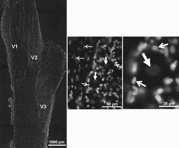
Trigeminal ganglia from naive adult rats were isolated and longitudinal sections of the ganglia were stained with the nuclear dye DAPI to show the distribution of the nuclei of all neurons and glial cells in the ganglia. Multiple image alignment was used to obtain images of the entire ganglion (at 40× magnification) so that the unique arrangement of neurons and glial cells in the V1 (anteromedial), V2 (anterolateral) and V3 (posterolateral) regions was clearly visible (Fig. 1). Within a given region, neuronal cell bodies and their associated satellite glial cells are organized in discreet bands that are aligned in an anterior–posterior orientation. Also, trigeminal nerve fibers and associated Schwann cells are observed between the bands of neuronal cell bodies and satellite glial cells. As seen at higher magnification, functional units comprised of a single neuronal cell body completely surrounded by satellite glial cells are clearly discernible. In adult ganglia, the nuclei of neuronal cells are typically round and >20 µm whereas glial cells show more elliptical nuclei that are >10 µm in diameter.
Cellular organization of neuronal cell bodies and glial cells in adult rat trigeminal ganglia. The fluorescent dye DAPI was used to stain the nuclei of all neuronal and glial cells in the ganglion. A longitudinal section of the entire ganglion from a naïve animal is shown at 40× magnification in the left panel with relative regions indicated. The middle panel shows a representative image of the distribution of neurons (large arrows), satellite glial cells (short arrows) and Schwann cells (thin arrows) in the V2 region of the ganglion. Shown in the right panel at higher magnification is a functional unit that is comprised of a single neuronal cell (large arrow) and satellite glial cells (small arrows).

Spatial organization of neuronal cell bodies and glia in developing trigeminal ganglia
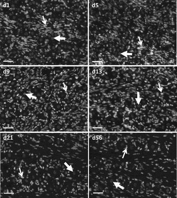
Trigeminal ganglia obtained from rats at days 1, 5, 9, 13, 21 and 56 after birth were stained with the nuclear dye, DAPI, to study the spatial and temporal development of functional units. The ganglia were visualized at 200× magnification in the V1/V2 region of the trigeminal ganglia. In the first week of postnatal development, neuronal and glial cells are found in distinct tight clusters or bands with very little association between the different cell types (Fig. 2; d1, d5). The overall structure of the V1/V2 region displays a high density of cells with very little organization when compared to the adult ganglion. During the second week of development, there is still a high density of cells in the V1/V2 region of the ganglion (Fig. 2; d9, d13). However, within that region of the ganglion, some glial cells are now beginning to rearrange such that more glial cells are found in association with individual neuronal cell bodies to form functional units. At day 9, although some functional units are visible, there remain separate clusters of neurons consisting of three to five cells as well as dense regions of glial cells. By day 13, the number of neuronal only clusters is markedly decreased and readily identifiable functional units are more abundant. In addition at day 13, glial cells are becoming aligned with nerve fibers and are observed between bands of neuronal cell bodies and glial cells as seen in adult tissue. Three weeks after birth, the density of cells has decreased greatly and fiber tracts are clearly seen between the bands of functional units that are aligned primarily in an anterior–posterior orientation (Fig. 2; d21). At this stage of development, the overall cellular organization in the V1/V2 region is similar to that seen in the adult tissue (Fig. 2; d56).
Spatial organization of neurons and glia in the developing trigeminal ganglion. Sections were stained with the nuclear dye DAPI and viewed at 200× magnification. Neurons (large arrows) are identified by their large round nuclei whereas glia (small arrows) are identified by their elongated nuclei. In the first week after birth (d1, d5), the neurons and glial cells are localized in separate clusters. During the second week (d9, d13), glial cells begin to associate with neurons and the early formation of functional units is seen. After three weeks of postnatal development (d21), functional units are abundant and discrete neuronal bands are observed and the overall morphology is similar to that seen in young adult tissue (d56). Scale bars = 50 µm.

Ratio of glial cells to neuronal cell bodies increases throughout development
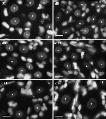
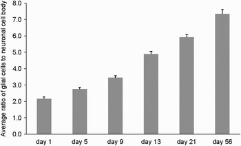
To determine the ratio of glial cells that are associated with a single neuronal cell body during development, the number of glial cells found in juxtaposition (10 μm) to a neuronal cell body was counted at each stage of development. As seen in Fig. 3, the number of glial cells associated with a single neuronal cell body increases steadily from the first day after birth into the young adult. During the first week of postnatal development, the neuronal cells are seen in dense clusters and the average ratio of glial cells to neurons is less than three (Fig. 2; d1, d5; Fig. 4). During the second week, the number of neurons localized in clusters is lower and there are more glial cells associated with each neuronal cell body. At this stage of development, some functional units are observed and there is a corresponding increase in the ratio of glial cells to neurons from 3.45 ± 0.12 at day 9 to 4.89 ± 0.16 at day 13. By postnatal day 21, many of the neuronal cell bodies are almost entirely surrounded by glial cells and the ratio of glial cells to neuronal cell bodies increased to 5.92 ± 0.16. In young adult ganglia, functional units are clearly defined and there are even a greater number of glial cells associated with each neuronal cell body (7.35 ± 0.27). However, it should be noted that the number of glial cells associated with a single neuronal cell body is likely underrepresented because only a single plane (14 µm) of tissue was used for the cell counts. Therefore, glial cells above or below the plane would not be counted. Nonetheless, there is a steady increase in the number of glial cells associated with a neuronal cell body during postnatal development and into young adults that correlates with the formation of functional units.
Ratio of satellite glia to neurons increases in the trigeminal ganglion during postnatal development. Sections were stained with the nuclear dye DAPI and viewed at 400× magnification. Neurons are identified by their large round nuclei (asterisks) whereas glial cells are identified by their elongated nuclei. In the first week of development (d1, d5), the neurons are found clustered in small groups or bands with very little glial cell association. In the second week (d9, d13), the ratio of glial cells to neurons increases and some discrete functional units are discernible. By three weeks (d21), most neuronal cell bodies are almost completely surrounded by glial cells and some functional units are clearly visible as they appear abundantly in adult tissue (d56). Scale bars = 10 µm.

Ratio of glial cells to neurons increases during postnatal development. Using representative images (400×) of tissues stained with the nuclear dye, DAPI, the average ± SEM ratio of glial cells to a single neuronal cell body was determined at multiple stages of postnatal development.

The expression of Kir4.1 in satellite glial cells temporally correlates with formation of functional units during postnatal development
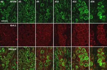
As an alternative method to studying when functional units form during development of trigeminal ganglia, tissues were stained with antibodies directed against Neurofilament 200, a structural protein found in neurons, and Kir4.1, a K+ channel found in satellite glial cells. As seen in Fig. 5, the diameter of the neuronal cell bodies has greatly increased by day 9 when compared to days 1 and 5 after birth. However, the size of the neuronal cell body does not appear to change appreciably after day 9 of postnatal development. Interestingly, day 9 is also the stage at which Kir4.1 expression is increased in satellite glial cells and functional units are beginning to form. Although the general pattern of Kir4.1 expression in satellite glial cells seen at day 13 is also observed at days 21 and 56, the level of expression is increased at the later postnatal ages. Taken together, the level of Kir4.1 expression spatially and temporally correlates with an increase in the size of the neuronal cell bodies, number of glial cells associated with neuronal cell bodies and the appearance of distinct functional units.
Temporal and spatial expression of Neurofilament 200 and Kir4.1 in trigeminal ganglia during postnatal development. The cellular organization of trigeminal ganglia at days 1, 5, 9, 13, 21 and young adult was studied using immunohistochemistry and antibodies directed against Neurofilament 200 (NF200), a structural protein found in neurons, and Kir4.1, a satellite glia cell marker. A merged image of both proteins is shown in the lower panel. During the first week of development (d1, d5), NF200 is expressed in small neuronal cell bodies and Kir4.1 is diffusely expressed in glial cells. During the second week (d9, d13), the neurons have increased in size and Kir4.1 staining is increased in satellite glia. At three weeks (d21), the neurons have further increased in size to a diameter similar to adult neurons and Kir4.1 staining levels in satellite glia is similar to that seen in the adult tissue (d56). Scale bar = 50 µm.

SNAP-25 expression in neuronal cell bodies during postnatal development
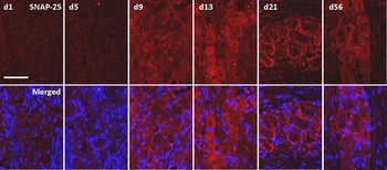
SNAP-25 staining was barely noticeable in trigeminal ganglia neurons at days 1 and 5 after birth (Fig. 6). However, by day 9, the expression of SNAP-25 was greatly increased in the cytosol and cell membrane of neuronal cell bodies. The level of SNAP-25 was further increased at day 13 and showed a similar pattern of cellular expression. By day 21, SNAP-25 was primarily localized to the cell membrane of neuronal cell bodies but some staining was detected in nerve fibers at this stage of development. Both the intensity and cellular distribution of SNAP-25 in neuronal cell bodies and nerve fibers was similar at days 21 and 56.
Increased expression of SNAP-25 in the trigeminal ganglion neurons temporally correlates with formation of functional units. The expression of SNAP-25, a vesicle docking protein, was studied using immunohistochemistry at days 1, 5, 9, 13, 21 and adult (d56). In the first week (d1, d5) SNAP-25 levels are barely detectable, but beginning at day 9 SNAP-25 levels were markedly increased through development with elevated levels maintained into adulthood (d56). Scale bar = 50 µm.

Neuronal levels of CGRP are developmentally regulated
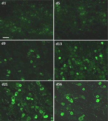
During the first week of postnatal development, low levels of CGRP were detected in the cytosol of many neuronal cell bodies (Fig. 7; d1, d5). The intensity of CGRP staining was greatly increased in a subset of neurons during the second postnatal week (d9, d13). By day 21, when there is a noticeable increase in the diameter of neuronal cell bodies, CGRP is detectable at a level above background in most neuronal cells with around 30–40% expressing higher levels of CGRP, an example of differential gene expression. This level of CGRP expression is maintained in young adult (d56). In addition, the number of detectable CGRP positive nerve fibers steadily increases throughout development reaching the greatest number in young adults.
Expression of CGRP in trigeminal ganglion neurons coincides with development of functional units. Expression of CGRP, a neuropeptide that mediates inflammatory and nociceptive responses, was studied using immunohistochemistry at days 1, 5, 9, 13, 21 and adult (d56). During the first week (d1, d5) CGRP levels are barely detectable in trigeminal neurons. During the second week (d9, d13), CGRP is more readily detected in neurons. By three weeks (d21), CGRP is abundantly expressed in neurons at levels similar to that observed in young adult tissue (d56). CGRP was also readily observed in nerve fibers at day 21 and in adult ganglia. Scale bars = 50 µm.

CONCLUSIONS
• The number of glial cells associated with a single neuronal cell body increases steadily during the first three weeks of development in conjunction with an increase in neuronal cell body volume and formation of functional units.
• Increased expression of the inwardly rectifying K+ channel Kir4.1 in satellite glial cells throughout development temporally correlates with the formation of functional units.
• Neuronal expression of the vesicle docking protein SNAP-25 is increased beginning on day 9 and becomes predominantly localized to the cell membrane by day 21 and this cellular localization pattern is maintained in young adult ganglia.
• Although the neuropeptide CGRP is barely detectable during the first week after birth, its increased expression temporally correlates with an increase in the diameter of neuronal cell bodies, increased SNAP-25 expression and formation of functional units.
• Taken together, it appears that the expression of proteins known to facilitate neuron–glia interactions and thus, function to regulate neuronal excitability, coincide with the formation of mature functional units during postnatal development of rat trigeminal ganglia.
DISCUSSION
Based on embryonic studies on mammals, it is known that both trigeminal neurons and glial cells differentiate from neural crest cells via complex regulatory events involving numerous transcription factors and signaling molecules (Shah et al., Reference Shah, Marchionni, Isaacs, Stroobant and Anderson1994; Britsch et al., Reference Britsch, Goerich, Riethmacher, Peirano, Rossner and Nave2001; Ota and Ito, Reference Ota and Ito2003; McGraw et al., Reference McGraw, Nechiporuk and Raible2008). During postnatal development, satellite glial cells proliferate and migrate to completely envelope a single neuronal cell body to form functional units (Pannese, Reference Pannese1969; Hanani, Reference Hanani2005). In agreement with previous studies, functional units are found in discrete bands or clusters in all three regions in adult trigeminal ganglia (Thalakoti et al., Reference Thalakoti, Patil, Damodaram, Vause, Langford and Freeman2007; Freeman et al., Reference Freeman, Patil and Durham2008; Cady and Durham, Reference Cady and Durham2010). Based on findings from this study, the formation of a few functional units was observed beginning on day 9 after birth and the number of functional units continued to increase during development up to day 56, the latest time point examined in our study. Not surprisingly, we found that the number of glial cells associated with a neuronal cell body increased during development coincident with an increase in the size (diameter) of the neuronal cell body. Our findings are in agreement with the seminal work of Pannese in spinal ganglia in which it was reported that the number of satellite glial cells increased with increasing volume of the neuronal cell body (Pannese, Reference Pannese1960). This result was confirmed in a more recent study involving the analysis of ganglia from five different species, including lizard, gecko, mouse, rat and rabbit (Ledda et al., Reference Ledda, De Palo and Pannese2004). Interestingly, each of the mammals had a higher ratio of satellite glial cells to neuronal cell body when compared to reptiles. Furthermore, in adult mammals, the average number of satellite cells per neuron correlated not only with the volume of the neuronal cell body but also with the overall size of the animal with the mouse < rat < rabbit. In addition, there is evidence that the size of the neuronal cell body positively correlates with the distance between the cell body and the target tissue (Lieberman, Reference Lieberman1976). Based on findings from several morphological studies, it has been proposed that the number of glial cells associated with a neuronal cell body is regulated by the neuron to quantitatively balance the metabolic needs of the neuronal cell (Pannese, Reference Pannese1964; Pannese et al., Reference Pannese, Bianchi, Calligaris, Ventura and Weibel1972; Pannese, Reference Pannese1981; Zimmerman and Braun, Reference Zimmerman and Braun1999). Somewhat surprisingly, findings from a study by Lagares et al. (Reference Lagares, Li, Zhou and Avendano2007) provide evidence that the number of sensory neurons in trigeminal ganglia of adult male rats nearly doubles between the third and eighth months of age. Even at this stage of later development, the increase in number of neurons was accompanied by a corresponding increase in the number of satellite glial cells.
In addition to meeting the metabolic needs of neurons, there is accumulating evidence that satellite glial cells play an important role in modulating the excitability state of sensory neurons by regulating the levels of ions and small molecules in the microenvironment around the neuronal cell body (Hanani, Reference Hanani2005). Importantly, increased neuronal excitability of primary sensory neurons has been shown to contribute to the development of persistent neuropathic pain by causing neurons to become spontaneously active or fire at a lower than normal threshold (Amir and Devor, Reference Amir and Devor2003a,Reference Amir and Devorb; Cherkas et al., Reference Cherkas, Huang, Pannicke, Tal, Reichenbach and Hanani2004; Robinson et al., Reference Robinson, Boissonade, Loescher, Smith, Yates and Elcock2004). One mechanism by which satellite glial cells control neuronal excitability is by regulating the resting membrane potential in neurons. In particular, satellite glial cells are known to express two ion channels, the inwardly rectifying potassium (K+) channel Kir4.l and the small-conductance calcium-activated potassium channel SK3, which function to maintain normal levels of extracellular K+ around neuronal cell bodies (Vit et al., Reference Vit, Jasmin, Bhargava and Ohara2006). In our study, we found that the level of Kir4.1 expression in satellite glial cells temporally correlated with the development of functional units in trigeminal ganglia and the increased size of the neurons during postnatal development. An important role of Kir4.1 in the development of neuropathic pain involving trigeminal ganglion neurons has recently been demonstrated (Vit et al., Reference Vit, Jasmin, Bhargava and Ohara2006). Notably, decreased expression of Kir4.1 channel activity was found to cause spontaneous and evoked pain like behavior in free moving rats that was similar to that caused by chronic constriction injury of the infraorbital nerve. In addition, regulating the levels of K+ ions and expression of Kir4.1 is likely to have significant implications for determining the glutamate levels around neurons since Kir4.1 activity is reported to modulate the ability of GLAST to remove extracellular glutamate (Djukic et al., Reference Djukic, Casper, Philpot, Chin and McCarthy2007; Kucheryavykh et al., Reference Kucheryavykh, Kucheryavykh, Nichols, Maldonado, Baksi and Reichenbach2007; Olsen and Sontheimer, Reference Olsen and Sontheimer2008). Taken together, these data support an important role of Kir4.1 expression in satellite glial cells, which is developmentally regulated to coincide with the formation of functional units within trigeminal ganglia.
We also found that the level of expression of the vesicle docking protein SNAP-25 and the neuropeptide CGRP correlated with the formation of functional units. Both SNAP-25 and CGRP were expressed at low levels in trigeminal ganglia neurons during the first week after birth but their staining levels were greatly increased in neuronal cell bodies beginning at day 9 and in nerve fibers later in development. SNAP-25 in cooperation with syntaxin and synaptobrevin are the soluble N-ethylmaleimide-sensitive factor attachment protein receptor (SNARE) proteins responsible for action potential-dependent, calcium-triggered release of multiple neurotransmitters and neuropeptides (Jahn and Scheller, Reference Jahn and Scheller2006). For example, calcium-dependent stimulation of glutamate release from synatposomal membrane preparations was shown to require SNAP-25 (Mehta et al., Reference Mehta, Battenberg and Wilson1996). Similarly, KCl-stimulated release of the neuropeptide CGRP from trigeminal neurons was shown to be mediated via SNAP-25, since treatment with botulinum toxin type A, which selectively cleaves SNAP-25, blocked CGRP secretion from cultured neuronal cell bodies (Durham et al., Reference Durham, Cady and Cady2004). In agreement with our previous study, CGRP and SNAP-25 were colocalized in the cell bodies of adult neurons as well as neuronal fibers (Thalakoti et al., Reference Thalakoti, Patil, Damodaram, Vause, Langford and Freeman2007). These data provide evidence that the cellular machinery for vesicle docking and calcium-dependent release of CGRP and other neurotransmitters is present in neuronal cell bodies and fibers.
Although most studies to date have focused on understanding the functions of CGRP released from peripheral or central terminals, there is evidence that sensory ganglion cell bodies are transiently depolarized and become more excitable by repetitive action potential activity in neighboring axons in the same ganglion (Amir and Devor, Reference Amir and Devor1996, Reference Amir and Devor2000; Zhang et al., Reference Zhang, Chen, Wang and Huang2007a). Based on results from studies on signaling within trigeminal ganglia (Neubert et al., Reference Neubert, Maidment, Matsuka, Adelson, Kruger and Spigelman2000; Ulrich-Lai et al., Reference Ulrich-Lai, Flores, Harding-Rose, Goodis and Hargreaves2001; Thalakoti et al., Reference Thalakoti, Patil, Damodaram, Vause, Langford and Freeman2007), CGRP released from the cell body of activated neurons would excite other neuronal cells and satellite glial cells via CGRP receptors that are expressed on these cells (Zhang et al., Reference Zhang, Winborn, Marquez de Prado and Russo2007b; Li et al., Reference Li, Vause and Durham2008). Thus, CGRP release from neuronal cell bodies within the ganglion could function as an autocrine signal to increase synthesis and further release of CGRP (Zhang et al., Reference Zhang, Winborn, Marquez de Prado and Russo2007b) or function in a paracrine manner to cause excitation of satellite glial cells and release of inflammatory molecules (Thalakoti et al., Reference Thalakoti, Patil, Damodaram, Vause, Langford and Freeman2007; Li et al., Reference Li, Vause and Durham2008; Vause and Durham, Reference Vause and Durham2010). Thus, release of cytokines, nitric oxide and other inflammatory molecules from satellite glial cells that mediate sensitization and activation of neurons could generate a pathological inflammatory loop within the ganglion that initiates and sustains a hyperexcitable state of the neurons (Rothwell and Hopkins, Reference Rothwell and Hopkins1995; Vitkovic et al., Reference Vitkovic, Bockaert and Jacque2000; Capuano et al., Reference Capuano, De Corato, Lisi, Tringali, Navarra and Dello Russo2009). This type of neuron–glia inflammatory loop has been reported in response to pathological conditions in dorsal root ganglion where cross-depolarization and cross-excitation of neurons contributes to a hyperexcitability state characteristic of injured dorsal root ganglion nerves (Oh and Weinreich, Reference Oh and Weinreich2002). Taken together, these results support a model by which activation of neurons and satellite glia in one region of the trigeminal ganglion initiate an inflammatory cascade involving other neurons and satellite glia, leading to increased intraganglion communication (Thalakoti et al., Reference Thalakoti, Patil, Damodaram, Vause, Langford and Freeman2007; Freeman et al., Reference Freeman, Patil and Durham2008).
This type of nonsynaptic communication, which has also been reported to occur in dorsal root ganglion (Amir and Devor, Reference Amir and Devor1996, Reference Amir and Devor2000; Ulrich-Lai et al., Reference Ulrich-Lai, Flores, Harding-Rose, Goodis and Hargreaves2001), would allow for a coordinated response to a particular inflammatory stimuli or possibly would be involved in sensitization of neurons in other regions of the ganglion. In that way, cross-excitation (propagation of inflammatory signals) within the ganglion would lower the activation threshold of neurons in one region and thus help explain the high level of comorbidity reported in diseases of the head and face that involve trigeminal nerve activation such as migraine and sinus pathology or TMJ disorder and headache (Cady and Schreiber, Reference Cady and Schreiber2002; Schreiber et al., Reference Schreiber, Hutchinson, Webster, Ames, Richardson and Powers2004; Ku et al., Reference Ku, Silverman, Prifti, Ying, Persaud and Schneider2006). In a proof-of-concept study (Damodaram et al., Reference Damodaram, Thalakoti, Freeman, Garrett and Durham2009), tumor necrosis factor alpha (TNF-α), a cytokine whose levels are elevated in nasal secretions during allergic rhinitis and acute sinusitis (Bachert et al., Reference Bachert, Wagenmann and Hauser1995; Bradding et al., Reference Bradding, Mediwake, Feather, Madden, Church and Holgate1995; Bensch et al., Reference Bensch, Nelson and Borish2002; Repka-Ramirez et al., Reference Repka-Ramirez, Naranch, Park, Clauw and Baraniuk2002) was injected in one region of the ganglia (whisker pad, V2) while a subthreshold concentration of the noxious agent capsaicin was injected in a different region (eye brow, V1). Significantly, TNF-α injection in the V2 region of the ganglion caused sensitization of trigeminal nociceptive neurons located the V1 region leading to activation of V1 neurons within the ganglia (Damodaram et al., Reference Damodaram, Thalakoti, Freeman, Garrett and Durham2009). Results from that study may help, at least in part, to explain how pathology directly involving one region of the ganglion can cause sensitization of neurons in a different region and thus, provide a cellular basis for the significant comorbidity associated with diseases involving trigeminal nerves.
In conclusion, we have shown that expression of proteins known to facilitate neuron–glial cells interactions coincides with formation of mature functional units during postnatal development of rat trigeminal ganglia. Recent findings from studies on sensory ganglia support an emerging central role of neuron–glia interactions in the development of peripheral sensitization as well as induction and maintenance of persistent pain states. Thus, a better understanding of the role of functional units within trigeminal ganglia under normal and pathological conditions has considerable health implications. Furthermore, although much progress has been made in identifying processes required for the differentiation of sensory neurons and glial cells during embryonic development, further studies focused on understanding the cellular and molecular mechanisms that regulate the formation of functional units is warranted.
ACKNOWLEDGEMENTS
We would like to thank Jordan Hawkins for her technical assistance. This work was supported by a research grant from NIH (DE017805).
Statement of interest
None.