Introduction
Nematodes of the family Mermithidae are endoparasites of arthropods and, occasionally, other invertebrates. The first described genus was Mermis, erected by Dujardin (Reference Dujardin1842) for the species M. nigrescens, which he found on foliage after rain in France. In the same paper Dujardin admitted that many species previously described as Gordius L., 1766 may also belong to the genus Mermis. Subsequently, Mermis was used as a nominal genus for all nematodes that we would now characterize as mermithids, and very few Mermis spp. in the literature would nowadays fit within the genus as we know it. Indeed, over 100 Mermis species have been named since 1842, of which more than 80% were published prior to 1950. As far as we can ascertain, approximately 20 of these species have been re-assigned to new genera, but many of the species names have not appeared in print since their original description, and their generic designation may depend upon re-examination of the original Type material. There now appear to be 11 species that are accepted in the literature as belonging to the genus Mermis: M. athysanota Steiner, 1921; M. changodudus Poinar, Remillet & Van Waerebeke, 1978; M. gigantea Artyukovsky & Lisikova, 1977; M. kenyensis Baylis, Reference Baylis1944; M. mirabilis von Linstow, 1903; M. nigrescens Dujardin, Reference Dujardin1842; M. papillus Gafurov, 1982; M. paranigrescens Rubstov, 1976; M. quirindiensis Baker & Poinar, 1986; M. savaiiensis Orton Williams, 1984; M. xianensis Xu & Bao, 1995 (Poinar et al., Reference Poinar, Rémillet and van Waerebeke1978; Baker & Poinar, 1986, 1988; Baker & Capinera, 1997).
The life cycle of M. nigrescens is well documented (e.g. Christie, Reference Christie1937; Baylis, Reference Baylis1947; Poinar, Reference Poinar and Poinar1983; Baker & Capinera, 1997) and larvae of the species have been reported from a large number of insect hosts, mainly of the order Orthoptera (Baylis, Reference Baylis1944; Baker & Capinera, 1997). The species also occurs in the European earwig in Tasmania (Crowcroft, Reference Crowcroft1947), Canada (Wilson, Reference Wilson1971) and Britain (Baylis, Reference Baylis1944). The life cycle of Mermis species is unusual in that they infect the insect host via passive infection – the ingestion of embryonated eggs which are deposited on foliage – as opposed to that of most other mermithid genera, which hatch from the eggs in water and actively penetrate the host (Christie, Reference Christie1937; Baylis, Reference Baylis1944, Reference Baylis1947; Poinar, Reference Poinar and Poinar1983).
Mermis nigrescens has a wide distribution, occurring in Europe, Asia, and North and South America, and it has become established in Tasmania since the introduction of European earwigs (Crowcroft, Reference Crowcroft1947; Baker & Capinera, Reference Baker and Capinera1997). The species has been reported from New Zealand only in passing, with MacFarlane (Reference MacFarlane and Gordon2011) and Thomas (Reference Thomas, Cameron, Hill, Bain and Thomas1989) each offering personal observations that M. subnigrescens is common in earwigs in Canterbury; M. subnigrescens was synonymized with M. nigrescens by Nickle (Reference Nickle1972). Other reports of mermithid genera from New Zealand include unidentified mermithid species in Ephemeroptera, Coleoptera, Phasmatodea and Orthoptera (Barker et al., Reference Barker, Pottinger and Addison1989; Ward, Reference Ward1998; Williams et al., Reference Williams, Townsend and Poulin2001; Bell et al., Reference Bell, Davis, Sarathchandra, Barratt, Ferguson and Townsend2005; Yeates & Buckley, 2009), Agamomermis sp. in midges (Craig, Reference Craig1963), Aranimermis giganteus Poinar & Early, Reference Poinar and Early1990 in mygalomorph spiders (Poinar & Early, 1990), Thaumamermis zealandica Poinar, Latham & Poulin, Reference Poinar, Latham and Poulin2002 in supralittoral amphipods (Poinar et al., Reference Poinar, Latham and Poulin2002), Psammomermis canterburiensis Poinar & Jackson, Reference Poinar and Jackson1992 in scarabid beetle larvae (Hoy, Reference Hoy1952; Poinar & Jackson, 1992), and Austromermis namis Poinar, Reference Poinar1990 and Blepharomermis craigi Poinar, Reference Poinar1990 in blackfly and midge larvae (Poinar, Reference Poinar1990).
The European earwig, Forficula auricularia L. (Dermaptera: Forficulidae), was first reported in New Zealand at the end of the 19th century (Smith, Reference Smith1898). Specimens were found in Canterbury, brought in with plants from London, UK, importations that had been going on for some years prior to Smith’s report. It therefore seems likely that the species arrived in New Zealand some time in the latter half of the 19th century. Early after establishment the species became abundant in central Otago and is now widely distributed in open lowland regions of South Island and in parts of North Island (Scott, Reference Scott and Scott1984). An omnivore, the earwig causes feeding damage to vegetables, flowers, pollen and fruits, and has been reported as a serious pest of stonefruit in central Otago (Thomas, Reference Thomas, Cameron, Hill, Bain and Thomas1989). Offsetting this nuisance value is their predation of a wide range of deleterious insect and mite species (Buxton & Madge, 1976; Carroll & Hoyt, 1984; Mueller et al., Reference Mueller, Blommers and Mois1988). Here, we provide a morphological description and a genetic characterization of M. nigrescens from European earwigs in Dunedin, South Island, New Zealand.
Materials and methods
Nematode isolation and vouchering
A total of 198 adult earwigs (Forficula auricularia) was collected between 21 March and 11 April 2012 from two sites in Dunedin, New Zealand. Specimens were housed in sex-segregated 850-ml airtight, clear plastic containers (ClickClack®, Innova Products Ltd, Auckland, New Zealand). Earwigs were maintained in a room receiving natural light and with the temperature maintained between 15 and 20°C. Infected earwigs were found dead at different times during a period of approximately 6 weeks between April and May 2012. Emerged nematodes were separated into two groups: one group (n= 30) which was directly preserved in 96% ethanol, and a second group where the nematodes (13 females and 8 males), which were still alive, were allowed to mature, moult and mate in larger, round Petri dishes (7.5 cm in diameter) containing a 0.5 cm depth of sterilized soil and water. Cultures were incubated at room temperature for 2–8 weeks. No gravid females were obtained by this method. In consideration of this, adult gravid females were sought the following austral summer (December 2012 and January 2013) from one of the previous earwig collection sites, on mornings and evenings after rain. Worms were observed on several different plants (Hypericum sp., Agapanthus sp., Leucanthemum sp., Lilium sp., Iris sp., Camellia sp.) at 580–1460 mm above soil level, and a total of 14 gravid females, as well as leaves with recently deposited eggs, were collected. Nematode fixation and preparation for observation were performed following Johnson & Kleve (Reference Johnson and Kleve2010). Fixed specimens were mounted on temporary slides with a glycerol:water (1:1) solution and observed at different magnifications using a light microscope (Olympus CX41, Olympus Australia Pty Ltd, Notting Hill Victoria, Australia). All specimens were photographed using a digital Olympus DP25 camera and measured with the DP2-BSW software (Olympus®).
DNA extraction and sequencing
Extraction of genomic DNA was performed on a tissue fragment of one post-parasitic juvenile and one mature female (collected in austral autumn and summer, respectively), using the PureLink® Genomic DNA Kit from Invitrogen according to the manufacturer's protocol (Invitrogen New Zealand Ltd, Auckland, New Zealand). For phylogenetic inference, a partial fragment of the 18S small subunit of ribosomal DNA (18S) was amplified, using the primers 1.2 F (5′-TGCTTGTCTCAAAGATTAAGC-3′) and 9 R (5′-GATCCTTCCGCAGGTTCACCTAC-3′) designed by Whiting (Reference Whiting2002). Two additional nuclear markers were also amplified, the 28S large subunit of ribosomal DNA (28S) and the ribosomal DNA first internal transcribed spacer (ITS1) region. The 28S was amplified using the primers 28S rD1.2a (5′-CCCSSGTAATTTAAGCATATTA-3′) and 28S B (5′-TCGGAAGGAACCAGCTAC-3′) described by Whiting (Reference Whiting2002). The ITS1 was amplified with the primers rDNA2 (5′-TTGATTACGTCCCTGCCCTTT-3′; Vrain et al., Reference Vrain, Wakarchuk, Levesque and Hamilton1992) and rDNA1.58 s (5′-ACGAGCCGAGTGATCCACCG-3′; Cherry et al., Reference Cherry, Szalanski, Todd and Powers1997). Polymerase chain reactions (PCR) were performed in a total volume of 20 μl, comprising 4 μl of MyTaqTM Red reaction buffer (Bioline (Aust) Pty Ltd, Alexandria, New South Wales, Australia), primers at 0.5 mm each, MyTaqTM Red DNA Polymerase (Bioline) at 0.025 units/ml and 10–20 ng of DNA template. The PCR reactions consisted of 35 iterations of the following cycle: 30 s at 95°C, 30 s at 54–58°C (depending on the primers used) and 1 min at 72°C, beginning with an additional denaturation step of 3 min at 95°C, and ending with a final extension at 72°C for 10 min. PCR amplicons were purified prior to sequencing using exonuclease I and shrimp alkaline phosphatase enzymes (Werle et al., Reference Werle, Schneider, Renner, Völker and Fiehn1994). The purified PCR products were sequenced using Big Dye Terminator technology (BigDye v. 3.1) on a 3730XL DNA Analyser (Applied Biosystems, Foster City, California, USA). The 18S amplified fragments were sequenced for both strands with the same primers as used in the amplification process. The 28S and the ITS1 fragments were sequenced only for the forward strand with the 28S rD1.2a and rDNA2 primers, respectively.
Phylogenetic analysis
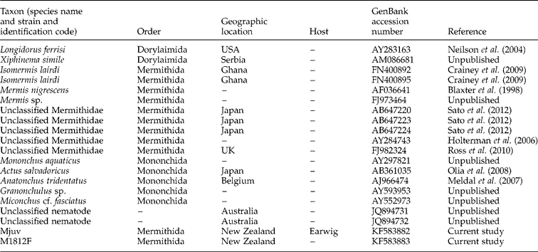
The sequences obtained were imported into the software Geneious 6.1.2 (Drummond et al., Reference Drummond, Ashton, Buxton, Cheung, Cooper, Duran, Field, Heled, Kearse and Markowitz2011) and the posterior end regions were trimmed using the trim option with the default parameters (error probability limit = 0.05). For each 18S sequence pair, a contiguous sequence was assembled. All sequences were uploaded into BLAST (Zhang et al., Reference Zhang, Schwartz, Wagner and Miller2000) to confirm the specimen's identity as a mermithid. However, for the 28S no other mermithid sequences were available (presenting a high of 85% maximum identity with Mononchidae sequences), while for the ITS1 only a maximum of 12% query cover was obtained with other available mermithid sequences. As result, phylogenetic analysis was only performed for the 18S dataset. Nematode 18S rRNA gene sequences with highest identity from BLAST (more than 92% maximum identity) comprising two different genera (Mermis and Isomermis), plus several unclassified mermithids and additional sequences from the sister group, order Mononchida (Ross et al., Reference Ross, Ivanova, Spiridonov, Waeyenberge, Moens, Nicol and Wilson2010), were downloaded to Geneious 6.1.2 (Drummond et al., Reference Drummond, Ashton, Buxton, Cheung, Cooper, Duran, Field, Heled, Kearse and Markowitz2011), providing a total of 16 sequences plus two outgroup sequences (order Dorylaimida) for analysis (table 1). Sequences were aligned with CLUSTALW implemented in Geneious using the default parameters (Cost matrix = ClustalW; gap open cost = 15; gap extended cost = 6.66). In order to determine the best fitting nucleotide model for the dataset, the software jModelTest 2 (Darriba et al., Reference Darriba, Taboada, Doallo and Posada2012) was used, selection based on the Akaike Information Criterion (AIC). The estimated model of nucleotide substitution that best fit the 18S dataset was TIM2+I+ G. Phylogenetic analyses were performed using Bayesian inference (BI) and maximum likelihood (ML) methods, implementing the most appropriate parameters according to the estimated model. Bayesian analyses were performed in MrBayes 3.2.1 (Huelsenbeck & Ronquist, 2001) and ran for 10 × 106 generations with random starting trees, sampling every 100 generations. The first 25,000 trees were discarded as ‘burn-in’, after verifying that a stationary distribution was reached by plotting log-likelihood values against generation time. A 50% majority rule consensus tree was used to summarize the trees sampled with the post-burn-in trees. Maximum likelihood analysis was performed using PhyML 3.0 (Guindon & Gascuel, 2003). Branch support was estimated by bootstrap analysis (Felsenstein, Reference Felsenstein1985) with 1000 replicates. Estimate of the pairwise uncorrected differences among the sequences obtained in this study for the 18S, 28S and ITS1 fragments were calculated in MEGA 5 (Tamura et al., Reference Tamura, Peterson, Peterson, Stecher, Nei and Kumar2011), on a total of 1676, 1020 and 919 bp, respectively. Genetic divergence (p-distance) for the 18S was also calculated on a total of 1641 bp between these sequences and M. nigrescens (GenBank accession number AF036641). New sequences generated in this study were submitted to GenBank under Accession Numbers KF583882–KF583883 for 18S, KF886018–KF886019 for 28S and KF886020–KF886021 for the ITS1.
Accession numbers of 18S rRNA gene sequences incorporated in the phylogenetic analyses.

Results
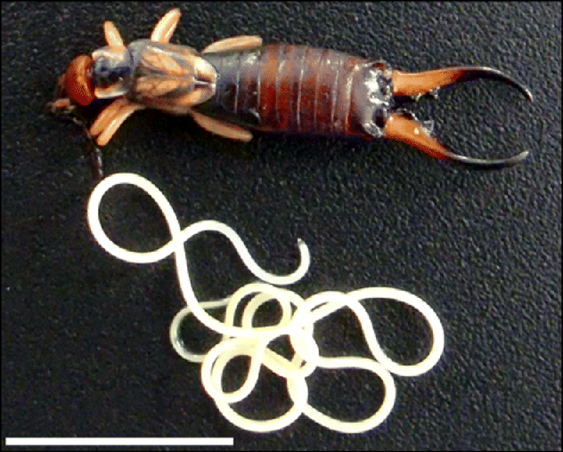
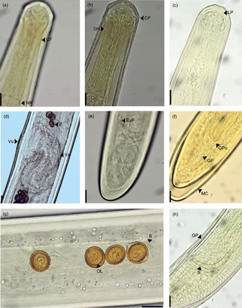
From the 198 earwigs collected, 19 were infected (prevalence 9.6%) with 51 nematodes in total, and all were found dead after the nematodes had emerged (fig. 1). The number of nematode parasites per earwig host ranged from 1 to 7 (mean intensity 2.68 ± 1.83). All post-parasitic worms that emerged were 4th stage juveniles. After allowing them to mature (maximum incubated time of 8 weeks) the majority of the nematodes had not completed the final moult (fig. 2f). However, it was still possible to recognize diagnostic characteristics that allowed us to identify the specimens as mermithids, belonging to the genus Mermis Dujardin, Reference Dujardin1842. These characters included: the presence of cuticle with criss-cross fibres (fig. 2a), females with S-shaped vagina without vulval flap (fig. 2d), and males with equal paired spicules (fig. 2 h). Mature females collected in summer were found actively climbing to the upper parts of plants (see supplementary video V1, available online). These mature females presented the same morphological characteristics and additionally the presence of a yellowish pigment near the apical end above the nerve ring (ocelli) (fig. 2b), and thick, smooth brown eggs with polar knobs and branched byssi (fig. 2 g). On the strength of these morphological characters the nematodes found infecting the earwigs were assigned to the species Mermis nigrescens. Below we present a morphological description of New Zealand M. nigrescens.
Newly emerged Mermis nigrescens and European earwig host. Scale bar = 1 cm.

Mermis nigrescens Dujardin 1842: (a) female, anterior end showing criss-cross fibres; (b) female, anterior end showing ocellar haemoglobin; (c) male, anterior end; (d) female, genital region, lateral view; (e) female, posterior end; (f) moulting male, posterior end, ventral view; (g) eggs in utero; (h) male, posterior end, lateral view. CF, cross fibres; NR, nerve ring; CP, cephalic papilla; E, egg; GP, genital papilla; GPo, genital pore; LP, labial papilla; MC, moulting cuticle; B, byssi; OH, ocellar haemoglobin; OL, ovic larva; S, spicule; Vu, vulva; Va, vagina; Exp, vestigial excretory pore. Scale bars = 50 μm.

Morphological description
Mermithidae Braun, 1883; Mermis Dujardin, Reference Dujardin1842 (emended by Poinar, Rémillet & van Waerebeke, 1978); Mermis nigrescens
Synonyms. M. subnigrescens Cobb, Reference Cobb1926, M. meisseneri Cobb, Reference Cobb1926 , M. kirgisica Kiryanova, Karavaeva & Romanenko, 1959.
Measurements. All measurements are in μm unless otherwise stated. Measurements are given as mean ± standard deviation (range).
Adult females (n= 14): length = 87 ± 21 mm (55–121); mid-body width = 338 ± 55 (231–391); head width (at level of cephalic papillae) = 122 ± 10 (104–142); cuticle width (at mid-body) = 20 ± 3 (14–24); hypodermis width (mid-body) = 30 ± 8 (22–48); distance of nerve ring from mouth = 397 ± 38 (306–441); position of vulva = 49.1 ± 7% body length (38.9–69.4); length of vagina (from vulva to junction with uterus) = 593 ± 73 (460–694); distance of vestigial excretory pore to tail = 512 ± 298 (302–991); eggs in utero= 50 ± 4 (33–58) long × 44 ± 5 (35–53) wide.
Post-parasitic males (n= 8): length = 36 ± 2 mm (32–39); mid-body width = 212 ± 10 (205–233); head width (at level of cephalic papillae) = 117 ± 5 (109–123); distance of nerve ring from mouth = 320 ± 12 (307–340); spicule length = 220 ± 9 (205–232); tail length = 292 ± 18 (267–317); total number of genital papillae = 64 ± 7 (55–75) (medial pre-genital = 18 ± 3 (15–24), medial post-genital = 8 ± 2 (5–10), left lateral = 10 ± 2 (8–13), right lateral = 9 ± 2 (7–14)).
Post-parasitic females (n= 9): length = 79 ± 16 mm (58–100); mid-body width = 431 (n= 1); head width (at level of cephalic papillae) = 146 ± 12 (132–162); distance of nerve ring from mouth = 430 (n= 1); position of vulva = 52.7 ± 2% body length (51.1–54.3).
Description.Long nematodes, post-parasitic females twice the length of post-parasitic males. Cuticle with criss-cross fibres intersecting at angles of 75° and 105°. Head rounded. Mouth apical, surrounded by slight swelling approximately 15 μm in diameter and 5 μm deep. Paired lateral lip papillae (15 μm high); four cephalic papillae; paired amphids posterior to level of cephalic papillae. Amphids very small.
Females: vulva a longitudinal oval, 46–74 μm long; muscular vagina, S-shaped twisted in two planes, opening to a wide vulval cone. Adult females generally white, with eggs in uterus showing as a core of golden brown, except at the region of the vagina. Ocellar haemoglobin (see Burr et al., Reference Burr, Hunt, Wagar, Dewilde, Blaxter, Vanfleteren and Moens2000) near the anterior tip region, yellowish and diffuse. Tail conoid, slightly flattened ventrally, convex dorsally. Vestigial excretory pore present.
Eggs: embryonated in uterus; with small polar knobs and paired, branched byssi on rope-like stems; golden brown colour and smooth; slightly ovoid with short axis between polar knobs; contain fully developed stage two juveniles when deposited on foliage; ovic juveniles with distinctive larval stylet (see Cobb, Reference Cobb1926).
Post-parasitic males: tail conoid and slightly flattened ventrally. Spicules paired, separate, strongly curved; tips curving towards each other; head of spicule not wider than shaft; spicule tip rounded, plain; genital papillae arranged in three distinct single rows, except for medial row in which 3–6 papillae are alternately offset immediately anterior, and occasionally posterior, to genital opening; medial row usually longer than lateral rows.
Sequence data and phylogenetic analysis
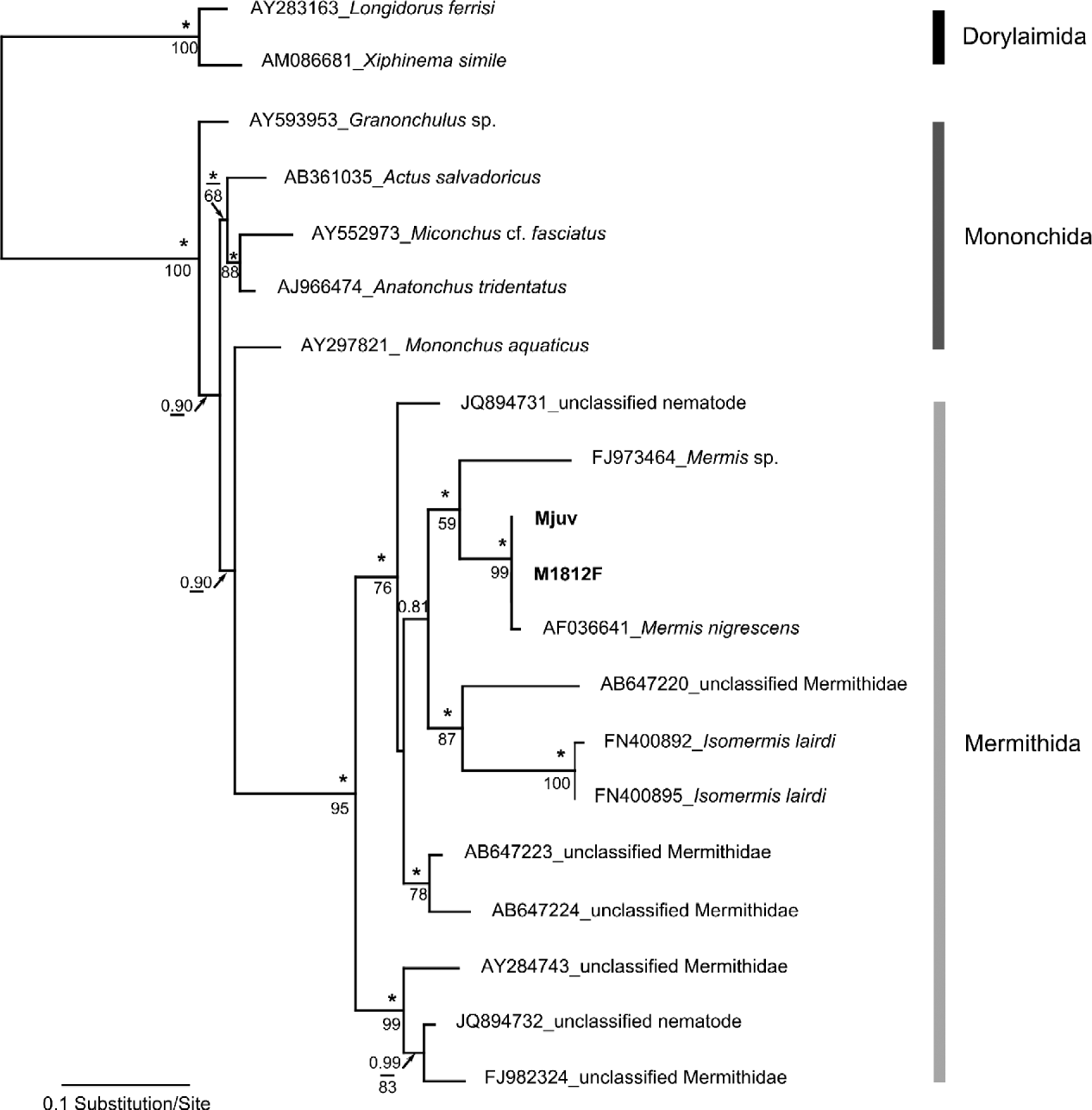
Sequences from the post-parasitic nematode and from the mature female collected in different seasons were identical (100% pairwise identity) for the 18S, 28S and ITS1 amplified fragments. For the 20 sequences used in the phylogenetic analyses, an alignment was obtained for a total of 798 bp. Trees inferred from both Bayesian inference and maximum likelihood presented similar topologies. The phylogenetic analyses supported the morphological identification, placing the sequence obtained in this study within the Mermithidae, and most closely related to M. nigrescens (1.00 posterior probability and 99% bootstrap support; fig. 3). A genetic divergence of 0.7% (uncorrected p-distance) was found between the sequences obtained in this study and that of M. nigrescens (AF036641) from Canada. Unfortunately, most other mermithid sequences in GenBank are unclassified to genus, making it impossible to infer relationships between the genus Mermis and other members of the family Mermithidae at this stage.
Maximum likelihood inference tree derived from 18S rRNA gene sequences. Values above branches represent Bayesian posterior probabilities and those below represent ML bootstrap support values (bootstrap values < 50 not reported). *Corresponds to a posterior probability value of 1. Specimens sequenced in this study are in bold.

Discussion
No other species of Mermis has been reported from New Zealand, either in the earwig, or in the more usual orthopteran hosts. The European earwig and the mermithid described here were probably introduced together in the 19th century and were able to establish easily in the temperate climate, in the same way as happened in Tasmania (Crowcroft, Reference Crowcroft1947) probably at around the same time. The prevalence in earwigs in our localities was less than 10%, lower than that found in other studies, including 50% in Tasmania (Crowcroft, Reference Crowcroft1947) and 11–63% in Ontario, Canada (Wilson, Reference Wilson1971). However, prevalence is strongly influenced by temperature and rainfall, both geographically and temporally (Baker & Capinera, 1997), and levels of infection most likely change from year to year depending upon summer rainfall levels.
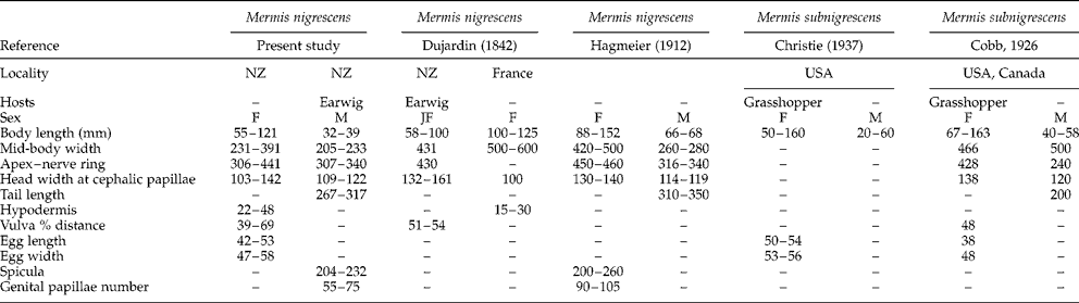
The absence of eggs in post-parasitic females that emerged from the earwigs did not allow us to perform a full morphological identification. This was rectified by the mermithid females collected in the summer, which were all mature with eggs. The nematodes were identified as belonging to the species M. nigrescens, according to the presence of the main diagnostic features: cuticle with criss-cross fibres, females with S-shaped vagina, thick brown eggs with polar knobs and branched byssi, and males with two equal spicules. Despite its many records in the literature, M. nigrescens and its junior synonym M. subnigrescens have rarely been described using detailed measurements of morphological features (but see Dujardin, Reference Dujardin1842; Hagmeier, Reference Hagmeier1912; Cobb, Reference Cobb1926; Christie, Reference Christie1937). Table 2 compares the morphometric data that are available from Europe and North American specimens. Overall size is not a reliable point of comparison, as size varies greatly due to duration of the parasitic stage and the number of nematodes per host (Christie, Reference Christie1937; Baker & Capinera, Reference Baker and Capinera1997). Variation in most features is within, or extends, the range found for the New Zealand specimens, although the mid-body width of our female specimens appears to be considerably smaller. Hagmeier (Reference Hagmeier1912) described his male specimens as having ‘anal papillae in three simple rows, of which the medial row near to the anus is doubled for a short distance’. He also states ‘the number [of papillae] in each row is 30–35’. This description closely resembles that of our specimens from New Zealand, except for the number of papillae in each row. As can be seen from the paucity of data in table 2, in few instances have the same features been characterized in different studies, therefore comparison of previously described material with the New Zealand specimens is incomplete. Certainly, more detailed morphometrics are required for the European and North American specimens, to allow a definitive comparison.
Morphometrics (in μm unless stated) for M. nigrescens and M. subnigrescens from the literature, compared with those from the present study.

F, Female worms; JF, juvenile females; M, males.
Because of a lack of diagnostic taxonomic characters, morphological identification of closely related nematode species is often difficult. There is abundant evidence that species described by morphology alone may represent complexes of cryptic species, revealed when molecular evidence is examined (e.g. Anderson et al., Reference Anderson, Blouin and Beech1998; Hoberg et al., Reference Hoberg, Monsen, Kutz and Blouin1999; Hung et al., Reference Hung, Chilton, Beveridge, Zhu, Lichtenfels and Gasser1999; Powers, Reference Powers2004; St-Onge et al., Reference St-Onge, LaRue and Charpentier2008). The 18S sequences exhibited a 0.7% distance (uncorrected p-distance) from the only other sequence for M. nigrescens present in GenBank, which, for this highly conserved gene, is probably sufficient in nematodes for them to be considered separate species. For instance, Wang et al. (Reference Wang, Xu, Liu and Wang2007) found a distance of 0.4% between species of Romanomermis. The sequence from GenBank was from a specimen collected in Canada (J. Burr & M. Blaxter, pers. comm.). However, we do not yet know how the sequences from Canada and New Zealand compare to M. nigrescens in Europe, which is assumed to be the geographic origin of this species. Two alternative hypotheses are possible, which could be resolved when molecular data from Europe are available. First, the Canadian, New Zealand and European isolates may be different from one another, in which case they may be considered a complex within the concept of M. nigrescens. Second, the New Zealand and European isolates may be the same but differ from the Canadian isolate, in which case the latter could represent M. subnigrescens.
Mermis nigrescens has been reported from many places worldwide (Baker & Capinera, Reference Baker and Capinera1997). Given enough specimens of M. nigrescens from a wide range of localities throughout its currently known distribution, molecular data could shed light on the biogeographic history of this species. Without molecular evidence for specimens from Europe we refrain from erecting a new species for the New Zealand specimens, notwithstanding the genetic difference. It seems likely that M. nigrescens could provide an interesting model of cryptic species in a parasitic nematode that has been introduced more-or-less worldwide from its presumed origin in Europe. This apparent species complex would benefit from an integrative taxonomic approach, combining genetic and multivariate morphometric analysis, and typological taxonomy (Fonseca et al., Reference Fonseca, Derycke and Moens2008).
As molecular identification of morphologically similar species becomes the norm, no doubt the pattern of geographic and taxonomic distribution for many groups of parasites, including the important Mermithidae, will become clearer.
Supplementary material
Video V1. Mermis nigrescens female climbing to the tip of fern frond in the summer after rainfall. Note the anterior tip region exhibiting the yellowish colour characteristic of the female (ocellar haemoglobin), and the brownish eggs visible in the posterior body.
To view this supplementary material, please visit http://dx.doi.org/10.1017/S0022149X14000017.
Acknowledgements
We would like to thank Professors J. Burr and M. Blaxter for helpful information. Special thanks to I. Blasco-Costa and H.S. Randhawa for their helpful comments.
Financial support
F.J. was funded through a doctoral grant (SFRH/BD/77332/2011). This work has been supported indirectly by the Marsden Fund (Royal Society of New Zealand).
Conflict of interest
None.