Introduction
Primary cholesteatoma of the middle ear is, in general, treated surgically by a combined approach or using a canal wall up tympanoplasty technique. Patients require follow up after surgical treatment, as residual or recurrent cholesteatoma can develop. ‘Second-look’ surgery is considered the ‘gold standard’ for follow up, and is performed 6–12 months after primary surgery. A considerable amount of patients undergo unnecessary second-look surgery; this increases the risk of intra-operative complications, amplifies hospital costs and is a burden for patients.
A reliable imaging technique for detecting residual and recurrent middle-ear cholesteatoma after surgery could decrease the number of unnecessary second-look surgical procedures performed. High-resolution computed tomography (CT) has been shown to be an unreliable technique for this purpose, with a sensitivity of 43 per cent and a specificity of 48 per cent.Reference Tierney, Pracy, Blaney and Bowdler1 In the last decade, several studies have suggested a role for magnetic resonance imaging (MRI) in the detection of primary and residual or recurrent cholesteatoma. A major advantage of MRI compared to CT is that it does not use potentially harmful ionising radiation. Differentiation between inflammation and cholesteatoma is possible using gadolinium-enhanced T1-weighted images, as cholesteatoma is avascular tissue and is not enhanced in contrast to inflammation.Reference Martin, Sterkers and Nahum2 The differentiation between cholesteatoma and inflammation can be improved further with echo-planar, diffusion-weighted MRI. However, this technique has some major drawbacks, including low resolution, relatively thick slices, and susceptibility artefacts at the interface of the temporal lobe and temporal bone, which limit the ability to detect smaller cholesteatomas.Reference Vercruysse, De Foer, Pouillon, Somers, Casselman and Offeciers3
Recent studies have found non echo planar, diffusion-weighted sequences to be superior in detecting cholesteatoma tissue. These sequences mostly concern single shot or multi shot based, turbo spin-echo, diffusion-weighted sequences. Advantages of this technique include thinner slices, slightly higher resolution and minimal artefacts compared with echo-planar, diffusion-weighted MRI. Non echo planar, diffusion-weighted MRI therefore seems to be more reliable in detecting cholesteatoma, and might be more suitable for identifying patients without residual or recurrent cholesteatoma. The relatively high negative predictive value of non echo planar, diffusion-weighted MRI may help to prevent unnecessary second-look surgery.Reference Jindal, Riskalla, Jiang, Connor and O'Connor4
This study aimed to determine the value of non echo planar, diffusion-weighted MRI used during follow up for the detection of residual and recurrent middle-ear cholesteatoma after primary combined-approach tympanoplasty.
Materials and methods
A retrospective study was conducted at the Diakonessenhuis Hospital Utrecht, a large general hospital in the centre of The Netherlands.
Patients
Those patients who underwent non echo planar, diffusion-weighted MRI of the petrous bone for post-operative follow up of cholesteatoma between January 2010 and August 2012 were included in the study. The MRI findings were retrospectively correlated with the peri-operative findings at second-look surgery. In those patients who had not yet undergone second-look surgery, the findings of non echo planar, diffusion-weighted MRI were compared to the clinical follow-up results. If the non echo planar, diffusion-weighted MRI investigation revealed primary cholesteatoma, these results were excluded for the assessment of sensitivity and specificity. Furthermore, in cases where multiple non echo planar, diffusion-weighted MRI studies were performed during follow up, only the latest study was included in the analyses for follow-up time and assessment of sensitivity and specificity. All patients underwent regular clinical follow-up examinations; otoscopy, audiometry and non echo planar, diffusion-weighted MRI were conducted at least once a year.
Imaging technique
Magnetic resonance imaging was performed using a 1.5 Tesla MRI scanner (Magnetom Avanto; Siemens, Erlangen, Germany). The following MRI protocols were carried out in all patients 45 minutes after intravenous administration of gadolinium-based contrast: (1) coronal, spin-echo, T1-weighted, fat saturation sequences: repetition time = 623 ms, echo time = 9.4 ms, slice thickness = 2 mm, field of view = 250 mm, number of excitations = 2.00 and shooting time = 292 seconds; (2) transverse, spin-echo, T1-weighted, fat saturation sequences: repetition time = 531 ms, echo time = 9.4 ms, slice thickness = 2 mm, field of view = 250 mm, number of excitations = 2.00 and shooting time = 249 seconds; (3) coronal, half-Fourier single-shot turbo spin-echo (‘HASTE’) diffusion, T2-weighted sequences: repetition time = 1250 ms, echo time = 109 ms, slice thickness = 3 mm, field of view = 220 mm, number of excitations = 12.00 and shooting time = 227 seconds; (4) coronal, turbo spin-echo, T2-weighted sequences: repetition time = 3500 ms, echo time = 125 ms, slice thickness = 2 mm, field of view = 230 mm, number of excitations = 4.00 and shooting time = 201 seconds; and (5) transverse, turbo spin-echo, T2-weighted sequences: repetition time = 3500 ms, echo time = 125 ms, slice thickness = 2 mm, field of view = 230 mm, number of excitations = 4.00 and shooting time = 201 seconds.
Radiological interpretation
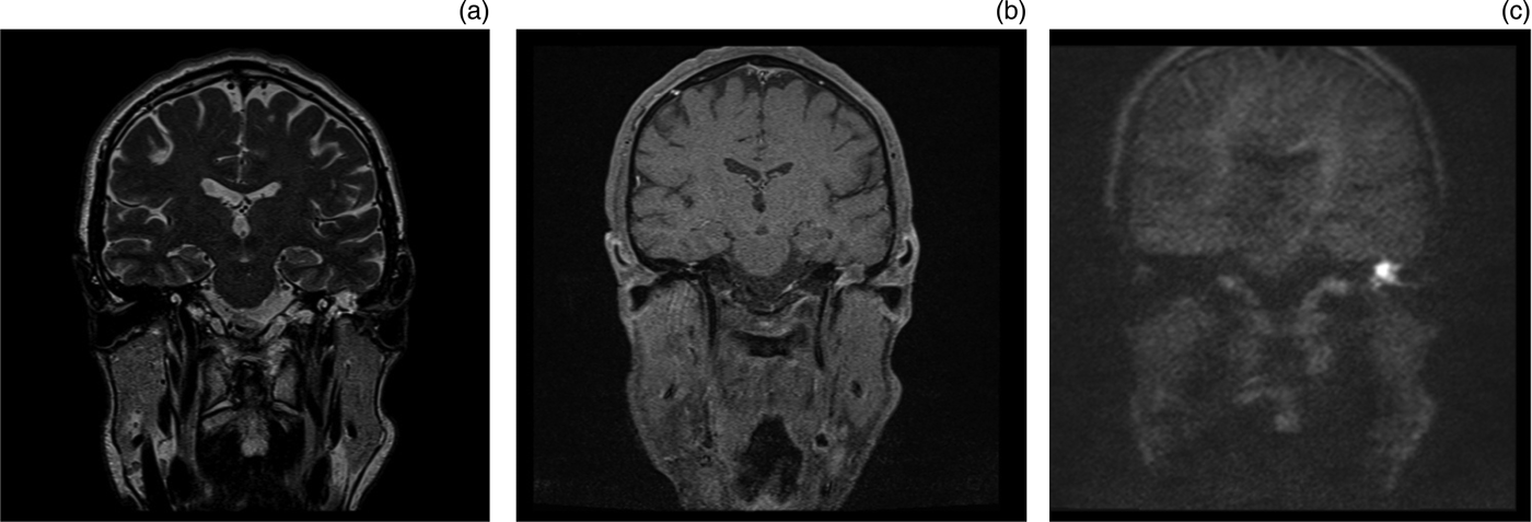
All non echo planar, diffusion-weighted MRI studies were evaluated by two experienced head and neck radiologists, who were blinded to the results of second-look surgery. The diagnosis of cholesteatoma was based on increased diffusion-weighted signal intensity on non echo planar, diffusion-weighted MRI, as compared with brain tissue in the same region in the T2-weighted MRI scan. An example is given in Figure 1. The initial diagnosis from the radiologist's original report was used as the primary measure. The mean maximum diameter of the cholesteatoma was also recorded.
Coronal magnetic resonance imaging scans of recurrent cholesteatoma in the left middle ear. The turbo spin-echo, T2-weighted image (a) shows a hyperintense lesion, which appears as a lesion of low signal intensity on the spin-echo, T1-weighted image (b). At the same location, increased diffusion-weighted signal intensity is shown on the non echo planar, half-Fourier single-shot turbo spin-echo (‘HASTE’), diffusion-weighted imaging sequence (c), indicative of cholesteatoma.

Statistical analysis
The sensitivity and specificity values for non echo planar, diffusion-weighted MRI were calculated on the basis of the findings at second-look surgery and those at clinical follow up. The diagnostic usefulness of non echo planar, diffusion-weighted MRI for the detection of residual and/or recurrent cholesteatoma after cholesteatoma surgery was described in terms of the means of positive and negative predictive values, and sensitivity and specificity.
Results
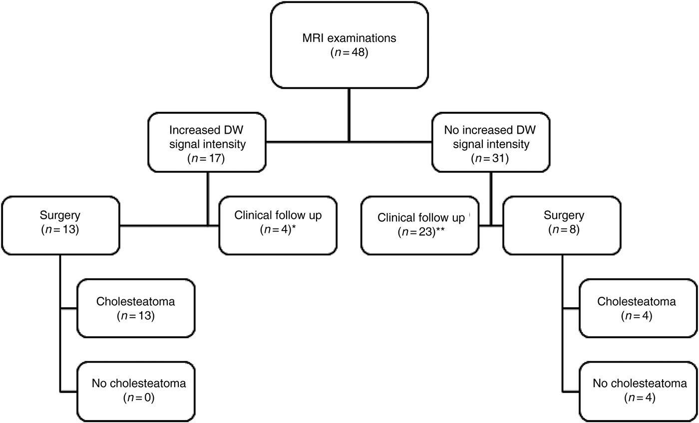
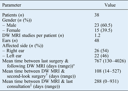
A schematic overview of the results is provided in Figure 2. A total of 48 non echo planar, diffusion-weighted MRI studies were performed in 38 patients. Second-look surgery was performed 21 times in 18 patients. The remaining patients were followed up at the out-patient clinic. Patients' demographics are depicted in Table I.
Schematic overview of the results. *Clinically proven cholesteatoma; **clinically non-suspicious for cholesteatoma. MRI = magnetic resonance imaging; DW = diffusion-weighted
Patient demographics
*n = 48 (note: the four ears not included in the cells below were positive on diffusion-weighted magnetic resonance imaging (MRI) and confirmed by otoscopy, and will be strictly monitored at the out-patient clinic). †Positive and negative diffusion-weighted MRI studies, followed by ‘second-look’ surgery; n = 21. ‡Negative diffusion-weighted MRI studies, not verified by second-look surgery; n = 23. DW = diffusion-weighted
Non echo planar, diffusion-weighted MRI showed increased diffusion-weighted signal intensity in 17 ears. Second-look surgery was performed in 13 of those 17 ears. Cholesteatoma was found during all 13 surgical procedures. Four ears with increased diffusion-weighted signal intensity belonged to patients who refused to undergo surgery, or in whom it was considered safe to monitor the cholesteatoma with close clinical follow up because the patients had no complaints and/or minimal hearing loss.
There was no increase in diffusion-weighted signal intensity in 31 ears. Of these, eight underwent second-look surgery and cholesteatoma was found in four ears during the surgical procedure. The remaining 23 ears were followed up at the out-patient clinic. During the follow up (which ranged from 0 to 931 days, with a mean follow-up period of 288 days), there was no clinical suspicion of residual or recurrent cholesteatoma.
There were no false-positive findings for the non echo planar, diffusion-weighted MRI studies; however, there were four cases of false-negative findings. This resulted in a sensitivity of 0.76, a specificity of 1.00, a positive predictive value of 1.00 and a negative predictive value of 0.50.
When the patients who did not undergo surgery for confirmation of the non echo planar, diffusion-weighted MRI results were included in the analysis as well, the sensitivity increased to 0.81, the specificity and positive predictive values remained at 1.00, and the negative predictive value increased to 0.87.
The mean maximum diameter of residual or recurrent cholesteatoma, as measured on diffusion-weighted MRI, was 11.7 mm (range, 4.4–25.3 mm).
Discussion
Recent studies investigating the accuracy of primary and recurrent cholesteatoma detection with diffusion-weighted MRI have shown promising results. These new MRI techniques may inform decisions regarding surgery and help to reduce the number of unnecessary second-look procedures performed. To date, several types of diffusion-weighted sequences have been evaluated for the detection of primary and residual or recurrent cholesteatoma. Initial studies described single-shot, spin echo planar imaging.Reference Fitzek, Mewes, Fitzek, Mentzel, Hunsche and Stoeter5–Reference Evlice, Tarkan, Kiroglu, Bicakci, Ozdemir and Tuncer7 More recent studies have evaluated the use of non echo planar imaging to detect cholesteatoma.Reference De Foer, Vercruysse, Bernaerts, Maes, Deckers and Michiels8–Reference Dubrulle, Souillard, Chechin, Vaneecloo, Desaulty and Vincent10
The current study aimed to evaluate the use of non echo planar, diffusion-weighted MRI for the detection of post-operative residual or recurrent cholesteatoma, and determine whether diffusion-weighted MRI could replace routine second-look surgery. There were no false-positive findings, resulting in specificity and positive predictive values of 100 per cent.
These results are in contrast to those of Dremmen et al.Reference Dremmen, Hofman, Hof, Stokroos and Postma11 These authors retrospectively evaluated the diagnostic accuracy of non echo planar, diffusion-weighted MRI for detecting residual or recurrent cholesteatoma. Examination of the correlation between the MRI findings and the peri-operative second-look surgery findings revealed false-positive MRI findings for 2 out of 27 patients. In one of the false-positive cases, the increased diffusion-weighted signal intensity was caused by empyema. The other false-positive case remained unexplained.
Dhepnorrarat et al. performed a similar study in a smaller population of cholesteatoma patients.Reference Dhepnorrarat, Wood and Rajan12 In seven cases, diffusion-weighted MRI results indicated cholesteatoma; these findings were confirmed by intra-operative evidence of cholesteatoma in all seven cases. Of a total of 16 cases shown to have negative diffusion-weighted MRI findings, all were confirmed to be disease-free on second-look surgery. The sensitivity, specificity, positive predictive and negative predictive values were all 100 per cent.
In contrast to our study, neither of the two studies mentioned above reported any false-negative findings. In our study, four ears with cholesteatoma-negative diffusion-weighted MRI findings appeared to contain cholesteatoma during second-look surgery. In order to explain these false-negative findings, the radiologists were first asked to independently and blindly re-evaluate these four cases, along with four other random cases. In this manner, we tried to exclude any inter-individual variability in the radiologist's interpretation. The radiologists had no access to the original diffusion-weighted MRI reports or the patients' files. There were no differences in results between the re-evaluation and the first assessment. We therefore considered a learning curve as the cause of the false-negative findings unlikely.
As described in other studies, false-negative findings for non echo planar, diffusion-weighted MRI are most likely due to a failure of the technique to demonstrate lesions smaller than 2–3 mm.Reference Vercruysse, De Foer, Pouillon, Somers, Casselman and Offeciers3, Reference De Foer, Vercruysse, Bernaerts, Maes, Deckers and Michiels8, Reference Dhepnorrarat, Wood and Rajan12–Reference Kasbekar, Scoffings, Kenway, Cross, Donnelly and Lloyd14 We reviewed the original surgery reports in an attempt to determine the cholesteatoma size in the four cases with false-negative findings. Only two of the four reports accurately described the size of the cholesteatoma; the cholesteatoma appeared to be smaller than 5 mm in both cases. We considered the small size of the cholesteatomas to be a plausible explanation for the false-negative findings.
Another factor that might influence false-negative diffusion-weighted MRI findings is a longer period between the diffusion-weighted MRI and the following second-look procedure, which allows time for cholesteatoma to develop. This can be avoided by conducting several MRI studies at regular time intervals after primary surgery for cholesteatoma. Although there was no significant difference in the time interval between the false-negative diffusion-weighted MRI studies and the other diffusion-weighted MRI studies to second-look surgery, we think that this time interval is important for an accurate assessment of diffusion-weighted MRI sensitivity.
In general, non echo planar, diffusion-weighted MRI is the recommended technique for those who want to implement diffusion-weighted MRI for clinical follow up of patients who have undergone primary surgery for cholesteatoma.Reference Aarts, Rovers, van der Veen, Schilder, van der Heijden and Grolman15 This technique is superior in identifying patients without recurrent or residual disease, which reduces the need for second-look surgery. False-negative findings for non echo planar, diffusion-weighted MRI can, in most MRI studies, be attributed to the small size of the cholesteatoma (i.e. smaller than 2–4 mm). This was the case in at least two of four cases with false-negative MRI findings in our study. Continued follow up with non echo planar, diffusion-weighted MRI is therefore needed to detect recurrent or growing residual disease. This should be performed on a regular basis, at 6–12-month intervals, for up to 2–3 years after primary surgery. Further studies are needed so that different protocols can be designed that vary depending on whether the cholesteatoma is in children or adults, or on the origin of the cholesteatoma (attic or other location, for example).
• Diffusion-weighted magnetic resonance imaging (MRI) is now used on a regular basis in otology for detecting primary and recurrent cholesteatoma
• Many patients can be spared unnecessary ‘second-look’ surgery with a reliable imaging technique such as non echo planar, diffusion-weighted MRI
• If non echo planar, diffusion-weighted MRI is used instead of second-look surgery, strict clinical follow up and regular MRI studies are mandatory as false-negative results may occur
Although the retrospective study design was a limitation of our study, we think it shows that there is a place for non echo planar, diffusion-weighted MRI in otology practice for follow up after primary surgery for cholesteatoma. Variations in the time intervals between the primary surgery and the diffusion-weighted MRI, and between the diffusion-weighted MRI and the second-look surgery, may have both influenced the outcome of our study, as might the fact that only 21 of 48 diffusion-weighted MRI studies were followed by second-look surgery. We think, however, that non echo planar, diffusion-weighted MRI is a promising technique capable of reducing the number of unnecessary second-look surgical procedures performed. However, repeated MRI studies are mandatory; these should be conducted at least once or twice a year over a two to three year follow-up period after primary surgery.
Conclusion
Recurrent cholesteatoma can be accurately detected by non echo planar, diffusion-weighted MRI. Our study, however, also showed some false-negative results with this technique. Strict out-patient follow up with otoscopy and MRI therefore remains necessary when non echo planar, diffusion-weighted MRI is used as an alternative to second-look surgery for the detection of residual or recurrent cholesteatoma.