Introduction
Honey bees, Apis mellifera, are important emerging livestock in many African countries, and an increasing number of small-scale farmers are adopting apiculture to supplement their livelihoods. Honey bees are essential pollinators of wild and cultivated plants, and are vital to food production and biodiversity. Pollination by honey bees accounts for approximately US$3.2 million of added crop value in Kakamega, western Kenya, which is almost 40% of the annual market value (Kasina et al., Reference Kasina, Mburu, Kraemer and Holm-Mueller2009) and includes fruits (mango, guava, avocado, passion fruit, papaya, and tomato), vegetables (onion, beans, carrot, coriander, and cabbage), nuts (macadamia), and seeds (sunflower and sesame). The added crop value is expressed as the increased yield and quality are achieved. The value of honey bee pollination worldwide exceeds $215 billion (Gallai et al., Reference Gallai, Salles, Settele and Vaissière2009). Klein et al. (Reference Klein, Vaissière, Cane, Steffan-Dewenter, Cunningham, Kremen and Tscharntke2007) estimated that around one-third of the food that humans consume is produced from plant life pollinated by bees.
The diversity of wild honey bees has decreased in many regions of Western Europe, mostly owing to habitat destruction (Biesmeijer et al., Reference Biesmeijer, Roberts, Reemer, Ohlemüller, Edwards, Peeters, Schaffers, Potts, Kleukers, Thomas, Settele and Kunin2006; Fitzpatrick et al., Reference Fitzpatrick, Murray, Paxton, Breen, Cotton, Santorum and Brown2007) and diseases that infect many managed honey bee populations (Graystock et al., Reference Graystock, Blane, McFrederick, Goulson and Hughes2015). Additionally, populations of wild pollinators are declining in several regions (Kluser and Peduzzi, Reference Kluser and Peduzzi2007; Potts et al., Reference Potts, Biesmeijer, Kremen, Neumann, Schweiger and Kunin2010), raising concerns of a potential global pollination crisis that could threaten our food supply (Withgott, Reference Withgott1999; Kremen and Ricketts, Reference Kremen and Ricketts2000; Richards, Reference Richards2001; Westerkamp and Gottsberger, Reference Westerkamp, Gottsberger, Kevan and Imperatriz-Fonseca2002; Steffan-Dewenter et al., Reference Steffan-Dewenter, Potts and Packer2005). Epidemiological data indicate that honey bee pathogens have spread worldwide (OIE, 2012). Colonies infected by pathogens may experience reduced size and sudden losses. In the USA, honey bees have been reported to experience colony collapse, due to a combination mostly of pathogens and parasites and other harmful environmental factors (vanEngelsdorp et al., Reference Van Engelsdorp, Underwood, Caron and Hayes2007). Compared with honey bees in many countries in different regions of the world, so far, there is no evidence that African honey bees have experienced the devastating effects of the ectoparasitic mite Varroa destructor and honey bee pathogens (Strauss et al., Reference Strauss, Human, Gauthier, Crewe, Dietemann and Pirk2013; Muli et al., Reference Muli, Patch, Frazier, Frazier, Torto, Baumgarten, Kilonzo, Kimani, Mumoki, Masiga, Tumlinson and Grozinger2014). Continental data are also limited (Mumoki et al., Reference Mumoki, Fombong, Muli, Muigai and Masiga2014) on the epidemiological status of diseases among populations of African honey bees, due to the near absence of the routine screening for pathogens (Pirk et al., Reference Pirk, Strauss, Yusuf, Démares and Human2016), which can play an invaluable role in the detection of diseases in pre-clinical stages and prevention of losses (Ryba et al., Reference Ryba, Titera, Schodelbauerova-Traxmandlova and Kindlmann2012). Standards set by the World Organisation for Animal Health (OIE) are recognized internationally as a basis for declaring a country or region free from disease and, therefore, any successful surveillance programme should meet OIE criteria, to maintain a disease-free status (OIE, 2012).
Good beekeeping management requires the training, constant inspection for diseases, and monitoring of colonies, which are vital elements in recognizing disease symptoms. Regular disease reporting can be achieved only with the participation of beekeepers themselves in disease management in honey bee populations (OIE, 2012). Apiary inspectors and beekeepers must be able to recognize diseases and parasites and to differentiate the notifiable diseases from those that are of less importance (Shimanuki and Knox, Reference Shimanuki and Knox2000). Early identification is the key to protecting honey bees from diseases, parasites, and other harmful conditions (Frazier et al., Reference Frazier, Caron and vanEngelsdorp2011). Early detection allows prompt remedial action, which can prevent diseases from spreading to other colonies, as spread can potentially lead to a serious disease outbreak that can result in widespread economic losses to beekeeping communities. Disease control strategies depend on the timely and accurate identification of pathogens.
In apiculture, the trade in hive products (Costopoulou et al., Reference Costopoulou, Lambrou and Harizanis2000) and the need for increased yield and quality of pollinated products (Klatt et al., Reference Klatt, Holzschuh, Westphal, Clough, Smit, Pawelzik and Tscharntke2014) are growing in importance. Pathogen surveillance is, therefore, important to support these economic activities. General surveillance is an effective arrangement for detecting circulating disease agents and does not focus on a particular pest or disease. Targeted surveillance, on the other hand, looks for specific disease agents. Surveillance is employed to provide evidence to support the presence or absence of a disease agent, and can demonstrate the prevalence and distribution of circulating pests and diseases.
In 2013, the African Reference Laboratory for Bee Health was established at the International Centre of Insect Physiology and Ecology (icipe) in Nairobi, Kenya, to coordinate efforts in disease surveillance efforts across the continent. This study was, therefore, initiated as part of a larger surveillance effort to identify the determinants of African honey bee health, and, particularly, to identify common pathogens that exist in Kenyan colonies, to gain a better understanding of how factors such as husbandry and disease epidemiology affect them. Data emanating from the umbrella study will go a long way in supporting the subsequent development of appropriate disease diagnostic methods and other control and apiary management tools, to improve the health of African honey bees. The present study was conducted to investigate the prevalence of common bee pathogens at selected apiaries in Kenya. Pathogens were detected using established molecular methods from published literature and their occurrence assessed, to create baseline disease levels that will support future epidemiological studies.
Materials and methods
Selection of study sites
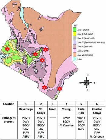
Selection of study sites was guided by agroecological zone. Kenya has seven agro-ecological zones, as per the index developed by Sombroek et al. (Reference Sombroek, Braun and Van der Pouw1982). These are Zone I (humid); Zone II (sub-humid); Zone III (semi-humid); Zone IV (semi-humid to semi-arid); Zone V (semi-arid); Zone VI (arid); and Zone VII (very arid) (Fig. 1). The six selected study sites are representative of five of the seven agroecological zones: Kakamega 0°16′60.00″ N 34°44′59.99″ E (humid); Mt Kenya 0°07′15.60″ N 37°20′7.20″ E (humid); Isiolo 0°52′60.00″ N 38°40′0.12″ E (arid); Mwingi 0°55′59.99″ N 38°03′60.00″ E (semi-arid); Taita Hills −3°22′59.99″ S 38°33′59.99″ E (semi-humid to semi-arid); and coastal Kenya −4°02′20.40″ S 39°40′12.59″ E (semi-humid).
A map of Kenya highlighting the presence of honey bee pathogens at the study sites superimposed on the agroecological map adapted from the Macharia (Reference Macharia2004). The study locations are highlighted with a numbered star shape and the pathogens detected at those sites are listed in the panel beneath the map.

Sampling
The study adopted the stratified sampling design. The apiary was the epidemiological unit and only those apiaries with more than 10 hives were considered for the study. From each study site, apiaries were conveniently selected from locations with established beekeeping activities. For this cross-sectional study, sampling was done over a span of 6 months during the hot dry months from September 2013 up to the end of February 2014. Altogether, 32 apiaries were visited. Five colonies were randomly selected from each apiary, giving a total of 160 colonies. From each colony, 20 individual foragers, nurse bees, worker pupae, and drone pupae were sampled separately. For each colony, foraging bees returning to the hive entrance were collected using vacuum aspiration into 50 mL sterile Falcon tubes. Nurse bees were collected directly from the brood comb into 50 mL sterile Falcon tubes. Worker and drone pupae were picked individually from the brood comb using toothpicks and placed into separate 50 mL sterile Falcon tubes. As part of the sampling process, the sample collectors looked out for any manifested signs or symptoms of disease. The sampled bees were preserved directly on dry ice at the field site and transported to the African Reference Laboratory for Bee Health in Nairobi, Kenya, where they were stored at −80 °C until processing. Repeated sampling was not carried out, since the study was not designed to look at changes in the same colonies over time.
Sample processing (homogenization)
In the laboratory, the bees from each apiary (hives 1–5) were pooled in groups as foragers, nurse bees, worker pupae, and drone pupae. Twenty bees were randomly selected from each pool and homogenized in batches of four bees per 2 mL tube in 500 μL of 1× phosphate buffered saline (PBS) pH 7.4 (137 mM NaCl, 2.7 mM KCl, 10 mM Na2HPO4, 2 mM KH2PO4) with 2 mm zirconia beads with a density of 5.5 g/cc (BioSpec Products, USA) using the TissueLyser II (QIAGEN). Following homogenization, the batch homogenates (of the selected 20 bees from a pool) were mixed by vortexing.
Nucleic acid extraction
Viral RNA/DNA and microbial DNA were extracted from 200 μL of the bee homogenate following magnetic bead-based isolation with the NucleoMag® VET kit (Macherey-Nagel Inc., USA). This process was automated on the epMotion® 5075 (Eppendorf, Germany) liquid handling instrument following the manufacturer's instructions.
PCR amplification of targeted pathogens
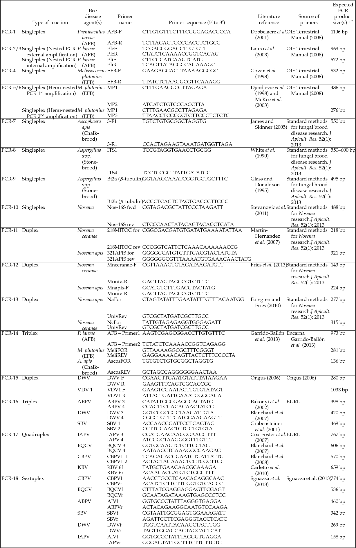
Molecular assays were designed to detect the presence of disease agents that had been previously identified to have the gravest consequences on honey bee health (OIE, 2012; Dietemann et al., Reference Dietemann, Ellis and Neumann2013). Many of the diagnostic methods applied in this study have been documented in the OIE Manual of Diagnostic Tests and Vaccines for Terrestrial Animals 2012 and COLOSS BEEBOOK, Volume II: Standard Methods for Apis mellifera Pest and Pathogen Research (Dietemann et al., Reference Dietemann, Ellis and Neumann2013). Other methods developed later have also been incorporated (Table 1). The target pathogens were two bacterial agents: Paenibacillus larvae (causing American foulbrood - AFB) and Melissococcus plutonius (causing European foulbrood – EFB), three groups of fungal agents: Ascophaera apis (causing chalkbrood), Aspergillus spp. (causing stonebrood), Nosema ceranae, and N. apis (causing nosema dysentery) and eight viral agents: Deformed Wing Virus (DWV), Varroa destructor virus 1 (VDV 1), Acute Bee Paralysis Virus (ABPV), Sacbrood virus (SBV), Israeli Acute Paralysis Virus (IAPV), Black Queen Cell Virus (BQCV), Chronic Bee Paralysis Virus (CBPV), and Kashmir Bee Virus (KBV). Positive controls included a combination of plasmids carrying the targeted segments from the pathogens of interest, complementary DNA (cDNA) of targeted viruses, and genomic DNA of specific pathogens as listed (Table 1). PBS was used as negative control in all reactions in place of the template.
A summary of the 18 PCRs applied for multi-gene targeted analysis to detect the different honey bee disease agents and the sources of PCR primers and reaction parameters employed

1 Positive Controls.
•For the primer sets that were obtained from the European Union Reference Laboratory (EURL) for Bee Health, the positive controls were respective plasmids carrying the targeted segments from seven viruses (DWV, BQCV, KBV, CBPV, SBV, IAPV, ABPV), M. plutonius (EFB), P. larvae (AFB), Nosema (N. apis and N. ceranae).
•In addition, complementary DNA (cDNA) of seven viruses (DWV, BQCV, KBV, CBPV, SBV, IAPV, ABPV) and genomic DNA (gDNA) of P. larvae, M. plutonius, and A. apis were obtained from the Swedish University of Agricultural Sciences, honey bee research group. Specific plasmid controls for the various primer sets were prepared from PCR amplicons cloned into pGEM-T Easy Vector (Promega, USA). These were subsequently used in the respective PCR reactions.
•No control was obtained for the stonebrood agents.
2 Negative controls.
•1× PBS was used as negative control in all reactions in place of the template.
Multi-loci targeting with several primer pairs
Each sample in this study was screened using 18 different PCR reactions with a panel of primers as described (Table 1). Since these primers were not designed specifically to detect the circulating genotypes of African honey bee pathogens, a multi-loci targeted approach was employed to cater for the undefined genetic incompatibility at the primer annealing sites and to maximize the chances of detecting the disease agents. For the detection of RNA viruses, the MyTaq™ One-Step RT-PCR Kit (BIOLINE) was used for amplification. Reverse transcription for first-strand cDNA synthesis was carried out at 45 °C for 20 min according to the manufacturer's instructions. The subsequent PCR cycle parameters were set following the optimized reactions in published literature (Table 1). The PCR products were observed on 1% Tris-acetate-EDTA buffer agarose gel stained with ethidium bromide. For the detection of the bacterial and fungal pathogens, MyTaq™ DNA Polymerase Kit (BIOLINE) was used for the amplification. The reaction set-ups were done following the manufacturer's instructions.
Sequencing of PCR amplicons
All the amplified PCR bands were excised from the agarose gel and purified using the ISOLATE II PCR and Gel Kit (BIOLINE). Purified PCR fragments were individually cloned into pGEM-T Easy vector (Promega, USA) and screened according to the manufacturer's instructions. Recombinant plasmid DNAs were purified using the ISOLATE II Plasmid Mini Kit (BIOLINE) and sequenced from both the forward and reverse directions at Macrogen (Korea) using universal SP6 and T7 primers.
Data analysis
Disease prevalence among apiaries was measured as the proportion of apiaries whose honey bees tested positive for a pathogen using PCR. The sequenced PCR products were compared with the GenBank database using the standard nucleotide BLAST search tool on the National Center for Biotechnology Information (NCBI) site.
Results
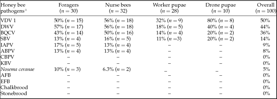
VDV 1 was the most abundantly circulating virus with a prevalence of 50% in the 100 bee pools. It was closely followed by DWV at 44%. The other disease agents detected were BQCV, SBV, IAPV, ABPV, and N. ceranae (Table 2). VDV 1, DWV, BQCV, and SBV were detected in both adults and brood; IAPV, ABPV, and N. ceranae were detected only in adults (foragers and nurse bees).
Prevalence of common honey bee pathogens in Kenya in 2013/2014 in the honey bee pools

+ Varroa destructor virus 1 (VDV 1), Deformed wing virus (DWV), Black queen cell virus (BQCV), Sacbrood virus (SBV), Israeli acute paralysis virus (IAPV), Acute bee paralysis virus (ABPV), Chronic bee paralysis virus (CBPV), Kashmir bee virus (KBV), American foulbrood (AFB), and European foulbrood (EFB).
Kakamega and Coastal Kenya, followed by Mt Kenya Region, were the locations most burdened by disease (Fig. 1). The semi-arid region of Mwingi had reduced disease load and the arid region of Isiolo did not register any pathogens.
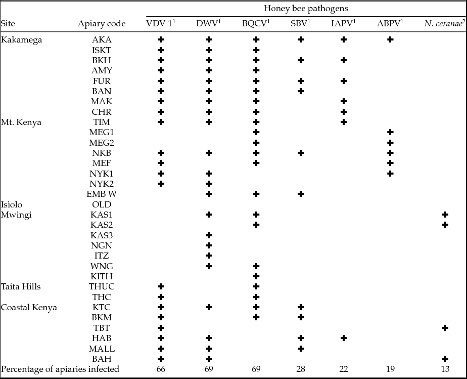
A summary of disease agents detected in each apiary (Table 3) indicated a co-existence of multiple infections within apiaries across the study sites. Co-infections within colonies were, however, not investigated in this study.
Co-existence of honey bee pathogens at the study apiaries in Kenya

1 The presence of a pathogen at an apiary is indicated using the ✚ symbol. Pathogens not listed were not detected in any sample.
2 Varroa destructor virus 1 (VDV 1), Deformed wing virus (DWV), Black queen cell virus (BQCV), Sacbrood virus (SBV), Israeli acute paralysis virus (IAPV), Acute bee paralysis virus (ABPV).
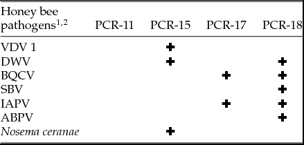
Products in the multi-gene targeted approach were not amplified at all times by all primers. Some primers were more effective than others. Table 4 shows all the PCR reactions targeted at disease agents that resulted in an amplified product, and thus provided all the data presented in this study.
PCR reactions that detected common honey bee pathogens in Kenya

1 The presence of a pathogen at an apiary is indicated using the ✚ symbol. Pathogens not listed were not detected in any sample.
2 Varroa destructor virus 1 (VDV 1), Deformed wing virus (DWV), Black queen cell virus (BQCV), Sacbrood virus (SBV), Israeli acute paralysis virus (IAPV), Acute bee paralysis virus (ABPV).
PCR-11, a duplex PCR, detected only N. ceranae for Nosema. PCR-15 detected both VDV 1 and DWV, effectively differentiating between the two closely related viruses. PCR-17 detected BQCV and IAPV; PCR-18 was the most efficient at detecting multiple pathogens, amplifying five of the six target viruses. Only CBPV was undetected by PCR-18. There was no detectable evidence of the circulation of CBPV, KBV, P. larvae (causing AFB), M. plutonius (causing EFB), A. apis (causing chalkbrood), and Aspergillus spp. (causing stonebrood) in bees collected from the investigated locations.
The nucleic acid sequence reads in this study were far too small and also not necessarily targeted at the optimal segments for reliable phylogenetic analysis to be conducted for the respective pathogens. Similarity searches indicated different mutations (single nucleotide insertions, deletions, and nucleic acid substitutions). Examples are single nucleotide deletions in the N. ceranae (99% identity), single nucleotide polymorphisms as in the case of VDV 1 (99% identity), multiple nucleotide substitutions in both BQCV (81% identity) and SBV (91% identity).
PCR-16, a method designed at the European Union Reference Laboratory (EURL) for Bee Health, was developed to detect ABPV, DWV, and SBV. Even though these viruses were positively detected using other PCR methods in the panel (Table 4), they were not detected by PCR-16 at all. PCR-17 and PCR-18 are both multiplexes that were designed to detect BQCV among other viruses (Table 1). Their results matched in detecting BQCV in 12 samples. However, PCR-17 detected BQCV in eight other samples when PCR-18 did not. On the other hand, PCR-18 detected BQCV in 16 different samples when PCR-17 did not. The samples that tested positive for IAPV by PCR-17 were not the same as those that tested positive for IAPV by PCR-18.
Discussion
The present study identified seven pathogens causing the greatest disease burden to honey bees in Kenya. These are DWV, BQCV, ABPV, VDV 1, SBV, IAPV, and N. ceranae. A preliminary nationwide survey to evaluate the status of pathogens in honey bee populations conducted in 2010 identified four disease agents: DWV, BQCV, ABPV, and N. apis (Muli et al., Reference Muli, Patch, Frazier, Frazier, Torto, Baumgarten, Kilonzo, Kimani, Mumoki, Masiga, Tumlinson and Grozinger2014). This is the only prior honey bee disease surveillance study conducted countrywide and it was somewhat restrictive in sample type and method applied. In addition to the four disease agents identified by Muli et al. (Reference Muli, Patch, Frazier, Frazier, Torto, Baumgarten, Kilonzo, Kimani, Mumoki, Masiga, Tumlinson and Grozinger2014), the present study identified four other pathogens: VDV 1, SBV, IAPV, and N. ceranae. Whereas Muli et al. (Reference Muli, Patch, Frazier, Frazier, Torto, Baumgarten, Kilonzo, Kimani, Mumoki, Masiga, Tumlinson and Grozinger2014) described disease agents solely from the perspective of foragers, in this study, both adults and brood were analysed. All infected bees in the present study were asymptomatic.
This study describes N. ceranae infections in both foragers and nurse bees at the coast (semi-humid) and at another inland location in the semi-arid region of Mwingi. The inland transmission is likely to continue spreading to other beekeeping regions in the near future.
Muli et al. (Reference Muli, Patch, Frazier, Frazier, Torto, Baumgarten, Kilonzo, Kimani, Mumoki, Masiga, Tumlinson and Grozinger2014) described infections by N. apis in foragers at three sites along the coast and in one interior site on the western border of Kenya and Uganda. The disease agents were almost evenly spread across the study sites. VDV 1, DWV, and BQCV were equally spread across apiaries in different agroecological zones. The apiaries in Kakamega (humid), presented the highest positivity rate for all the disease agents detected and would form an ideal location for future focused disease studies. An investigation of the presence of the agents in apiaries revealed the common occurrence of more than one pathogen per apiary. Other studies investigating the prevalence of honey bee viruses in and around the Gauteng region of South Africa (Strauss et al., Reference Strauss, Human, Gauthier, Crewe, Dietemann and Pirk2013) and in the Biobío Region of Chile (Rodríguez et al., Reference Rodríguez, Vargas, Antúnez, Gerding, Castro and Zapata2014) reported similar mixed infections in apiaries. There appears to be a heavy disease burden on the Kenyan bees and it would be important to investigate the mixed infections further, to see if they manifest as co-infections within the colonies. Since the samples were analysed as apiary samples, incidences of co-infection within colonies could not be elucidated. It would also be important to measure pathogen loads to fully understand the extent of the disease burden and lack of clinical symptoms.
PCR has become the standard, reliable, rapid, specific, and accurate method for detecting many pathogens. Based on the PCR results, it is possible to develop molecular diagnostic methods applicable to the circulating bee pathogens for general screening during surveillance activities. Even in the presence of certain pathogens, for example, BQCV and IAPV, the established PCR methods applied were not equally efficient in detecting the pathogens in all the samples where they were present. In some cases, one set of primer pairs would detect the agent and the other would not. This is a strong indication of genetic drift, which is also supported by nucleic acid sequencing and similarity searching on the GenBank database that yielded 81% genetic identity in the BQCV sequences, for example. The observed genetic changes could have occurred also at the primer annealing sites and probably in entire genomes. To support this argument, taking the example of Nosema, Muli et al. (Reference Muli, Patch, Frazier, Frazier, Torto, Baumgarten, Kilonzo, Kimani, Mumoki, Masiga, Tumlinson and Grozinger2014) used the RFLP approach to detect Nosema and found that the restriction enzymes MspI and NdeI followed by nucleic acid sequencing revealed genetic change which removed the NdeI cleavage site in the Nosema isolates circulating in Kenya. Single-nucleotide polymorphisms and other types of mismatches reduce the performance of PCR assays and may interfere with PCR primer annealing efficiency (Lefever et al., Reference Lefever, Pattyn, Hellemans and Vandesompele2013). The genetic segments sequenced in this study indicated mutations sufficient to interfere with PCR. Primer annealing in PCR is a temperature-dependent, sequence-specific step requiring high genetic conservation to support efficient hybridization of the primer to the template, leading to successful amplification (Berg et al., Reference Berg, Tymoczko and Stryer2002). The effect of primer-template mismatches is to reduce PCR product yield and, depending on the extent of the mismatches, amplification will be prevented altogether (Sipos et al., Reference Sipos, Székely, Palatinszky, Révész, Márialigeti and Nikolausz2007). The full extent of the genetic changes can be determined only by a metagenomic analysis (Mokili et al., Reference Mokili, Rohwer and Dutilh2012). Phylotyping gives reliable information about within-species genomic diversity.
The OIE Terrestrial Manual (OIE, 2012) has documented some of the pathogen diagnostic methods that the EURL for Bee Health has adopted, particularly the methods for detecting American foulbrood and European foulbrood. Viral diagnostic methods, on the other hand, are not documented. In this study, PCR-16 and PCR-17 methods sourced from the EURL, were not sensitive enough to detect most of the viruses in the Kenyan samples. Indeed, none of the methods applied in this study were developed in the African honey bee pathogen genotypes. As part of the activities of the African Reference Laboratory for Bee Health, there is the need to conduct further genetic screening to develop better-optimized molecular diagnostic tools for application in the African region. These can then be incorporated to form revised diagnostic methods for surveillance activities to monitor disease agents in colonies in the region.
The data presented in this study will be linked to data from other studies to generate a bigger picture of the health status and epidemiological indices of African honey bees.
Conclusion
This study identified an expanded list of common pathogens in Kenyan bees. This information will support the development of revised diagnostic tools suited for disease surveillance activities in the region. This study also points to genetic change in the circulating pathogens, since some primers in the literature were unsuited for molecular diagnosis, and partial nucleic acid sequencing of the virus genes indicated some changes. Advanced genetic analysis of circulating pathogens will be required to fully describe this phenomenon to support the design of new primers more suited to the circulating genotypes and more optimizations of the revised diagnostic methods. This would improve the chances of identifying pathogens infecting African honey bees with a higher level of reliability. In this study, a great diversity of diagnostic primers targeting multi-loci positions in the pathogens was assessed. The best primer pairs were identified and will support future diagnostic expeditions, pending the full genetic analysis, and revision of the available molecular tools.
Acknowledgements
This study was funded by a European Union Grant, Contract Number DCI-FOOD/2013/313-659 for the project entitled ‘icipe: African Reference Laboratory (With Satellite Stations) for the Management of Pollinator Bee Diseases and Pests for Food Security’, and icipe core funding from UK Aid from the UK Government, Swedish International Development Cooperation Agency (Sida), the Swiss Agency for Development and Cooperation (SDC), and the Kenyan Government. The views expressed herein do not necessarily reflect the official opinion of the donors. We also thank the bee health team for assisting in collecting samples in the field. We thank icipe for providing the facilities to conduct the study. We acknowledge all beekeepers for allowing us to access their apiaries to obtain the samples that were used in this study. The European Union Reference Laboratory (EURL) for Bee Health provided plasmid controls of seven viruses (DWV, BQCV, KBV, CBPV, SBV, IAPV, ABPV), M. plutonius (EFB), P. larvae (AFB), and Nosema (N. apis and N. ceranae). Joachim de Miranda of the Swedish University of Agricultural Sciences (SLU) Honeybee Research Group provided the biological material (cDNA and gDNA) of pathogens.