Introduction
The standard surgical management for neck metastasis has shifted over the last two decades, from the classical radical neck dissection to the modified neck dissection. Accordingly, our department has adopted an organ preservation policy during neck dissections, and in the majority of cases the internal jugular vein is now spared.
However, although rare, we still encounter patients in whom the sacrifice of both internal jugular veins is inevitable. It is well known that simultaneous excision of bilateral internal jugular veins leads to acute reduction of cerebral venous drainage and resultant serious morbidity, including facial oedema (in approximately 30 per cent of cases), blindness (in approximately 1 per cent), inappropriate anti-diuretic hormone hypersecretion, laryngeal oedema, carotid artery rupture, cerebral vessel complications (in approximately 5 per cent) and death (in approximately 10–13 per cent).Reference Frazell and Moore1–Reference Dulguerov, Soulier, Maurice, Faidutti, Allal and Lehmann4 Thus, it is recommended to leave an interval of at least one month before ligating the second internal jugular vein, to promote the formation of collateral drainage. However, even when thus staged, the sacrifice of both internal jugular veins occasionally still leads to grave complications, including stroke (in 1.3 per cent) and death (in 1.6 per cent).Reference Dulguerov, Soulier, Maurice, Faidutti, Allal and Lehmann4 Notably, leaving a longer interval between staged procedures does not guarantee the development of sufficient collateral venous drainage, as reported by Dulguerov et al. Reference Dulguerov, Soulier, Maurice, Faidutti, Allal and Lehmann4 (who described a peri-operative death after a staged bilateral neck dissection with an interval of six months).
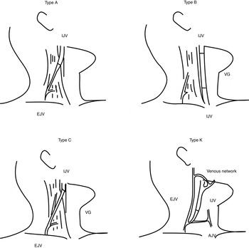
In view of these risks, we opted to undertake unilateral internal jugular vein reconstruction in two patients in whom bilateral internal jugular vein excision was inevitable. In the first case, the internal jugular vein was anastomosed to the external jugular vein using the type A method, according to the classification proposed by Katsuno et al. (Figure 1).Reference Katsuno, Ishiyama, Nezu and Usami5 In the second case, we anastomosed the internal jugular vein to the anterior jugular vein, preserving the venous flow of the external jugular vein. We termed this method type K (Figure 1). In both cases, the post-operative course was uneventful and the reconstructed vessels were functional.
Previously and currently proposed methods of internal jugular vein reconstruction. Type A: end-to-end anastomosis between the internal jugular vein (IJV) and the external jugular vein (EJV). Type B: the internal jugular vein is reconstructed via an interposition, using either an autograft or an artificial vein graft (VG). Type C: either an autograft or an artificial vein graft is interposed between the internal jugular vein and the external jugular vein. Type K: end-to-end anastomosis between the internal jugular vein and the anterior jugular vein (AJV). Methods A, B and C were originally proposed by Katsuno et al. Reference Katsuno, Ishiyama, Nezu and Usami5

We herein report the procedures used in both cases, and we also review the significance of internal jugular vein reconstruction.
Case reports
Case one
A 73-year-old man was diagnosed with supraglottic laryngeal carcinoma (tumour (T) node (N) metastasis (M) staging cT3 N2c M0) following staging investigations. Computed tomography (CT) images with contrast demonstrated multiple cervical lymph node metastases which compressed both internal jugular veins, indicating extra-nodal extension of the tumour to these vessels.
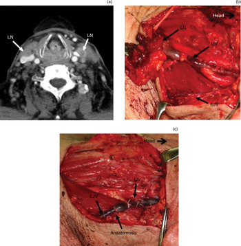
The primary tumour demonstrated a good response to 30 Gy of chemoradiation (consisting of S-1, vitamin A and external radiation).Reference Kumamoto, Masuda, Kuratomi, Toh, Shinokuma and Chujo6 However, the metastatic lymph nodes remained unchanged in size (Figure 2a).
Case one. (a) Axial, contrast-enhanced computed tomography image after 30 Gy chemoradiation treatment, showing metastatic lymph node (LN) densely adherent to both internal jugular veins. (b) Left neck dissection. The internal jugular vein (IJV) was involved by the lymph node (LN). The external jugular vein (EJV) was spared for reconstruction. (c) Internal jugular vein reconstruction using type A method (see Figure 1).

At this point, we decided to perform bilateral neck dissection, as per our treatment protocol, and to preserve the patient's larynx by further administering the same form of chemoradiotherapy up to 70 Gy. Considering the risks associated with bilateral internal jugular vein excision, a staged bilateral neck dissection was planned.
A left neck dissection was carried out first, with the intention of preserving the internal jugular vein. However, the left internal jugular vein was seen to be densely involved by the inferior cervical lymph node (Figure 2b), and so was sacrificed. We reconstructed the internal jugular vein using the type A method (according to Katsuno and colleagues' classification), i.e. end-to-end anastomosis of the internal jugular vein to the external jugular vein (Figure 1).Reference Katsuno, Ishiyama, Nezu and Usami5 Since there was a considerable difference between the diameter of the two veins, we employed a fish-mouth method to achieve anastomosis (Figure 2c).
After confirming the patency of the reconstructed internal jugular vein using Doppler ultrasound, the patient underwent a staged right neck dissection two weeks after the initial, left neck dissection. As expected, preservation of the right internal jugular vein was also difficult. We used a classical radical neck dissection, requiring sacrifice of both the right internal jugular vein and the right external jugular vein.
After the second procedure, there were no signs of serious complications associated with increased intracranial venous pressure. However, facial oedema was initially prominent and lasted a few weeks; this was probably due to reduced facial drainage caused by the sacrifice of both external jugular veins.
Case two
A 77-year-old man with subglottic laryngeal cancer (staged as cT2 N1 M0) was treated with 30 Gy of chemoradiotherapy as above. Since both the primary tumour and the left metastatic lymph nodes displayed a poor response, we decided to proceed to total laryngectomy plus type III left modified radical neck dissection, preserving the left internal jugular vein, the spinal accessory nerve and the sternocleidomastoid muscle.Reference Shah, Patel, Shah and Patel7
However, during operation, the metastatic lymph node were found to be tightly adherent to the left internal jugular vein, necessitating excision of this vessel. Histopathological examination revealed considerable amounts of residual viable tumour cells in both the primary site and the lymph node.
Considering the risk of tumour cell dissemination, we recommended the patient undergo post-operative chemoradiation; however, he refused.
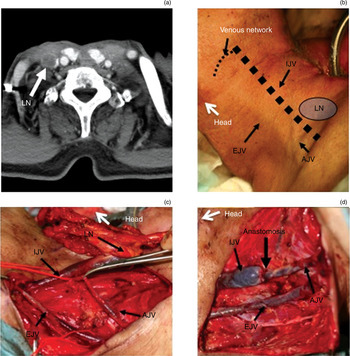
Two months after the initial operation, a rapidly growing mass appeared in the patient's right lower neck. Contrast-enhanced CT images showed a metastatic lymph node strongly compressing the right internal jugular vein (Figure 3a). Hence, right-sided neck dissection was urgent, and preservation of the right internal jugular vein seemed likely to be difficult. Since the metastatic lymph node (located at lower level IV) and the patient's anterior jugular vein were prominent, we planned to anastomose the internal jugular vein to the anterior jugular vein, preserving the flow of the right external jugular vein, in order to maintain facial drainage (Figure 3b). Adopting this method, we also anticipated that the venous networks between the external jugular vein and the upper internal jugular vein (including vessels such as the facial vein) would act as a safeguard should the reconstructed internal jugular vein occlude for any reason.
Case two. (a) Axial, contrast-enhanced computed tomography image, showing the right internal jugular vein strongly compressed by a metastatic lymph node (LN). (b) Pre-operative planning for internal jugular vein (IJV) reconstruction. Since the metastatic lymph node (located in the lower neck) and the anterior jugular vein (AJV) was both prominent, we planned to anastomose the IJV to the AJV, preserving the venous flow of the external jugular vein (EJV) (termed method K). (c) Right neck dissection. (d) End-to-end anastomosis between the IJV and the AJV.

At operation, the metastatic lymph node was tightly adherent to the internal jugular vein, as expected, and intra-luminal invasion was suspected (Figure 3c). The internal jugular vein was sacrificed, and reconstructed using end-to-end anastomosis of the internal jugular vein to the anterior jugular vein, as planned. A fish-mouth method was employed to overcome the discrepancy in diameter between these two veins (Figure 3d).
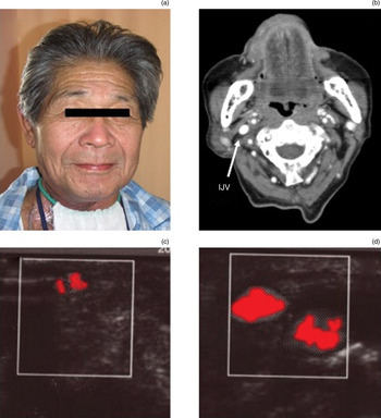
This patient's post-operative results were more favourable than those of our first patient. There was no facial oedema (Figure 4a), nor any more serious complications. The patency of the reconstructed internal jugular vein was confirmed by Doppler ultrasound and contrast-enhanced CT (Figures 4b, 4c and 4d) conducted approximately one month after surgery.
Case two. (a) The patient on post-operative day one. (b) Axial, contrast-enhanced computed tomography scan, and (c) & (d) colour Doppler ultrasonography images, all taken approximately one month after internal jugular vein (IJV) reconstruction.

Discussion
To our knowledge, 25 cases of internal jugular vein reconstruction have previously been reported in the English language literature.Reference Eibling, Snyderman, Weber, Johnson and Steed2, Reference Dulguerov, Soulier, Maurice, Faidutti, Allal and Lehmann4, Reference Katsuno, Ishiyama, Nezu and Usami5, Reference Comerota, Harwick and White8 Katsuno et al. have classified reconstruction methods into three types – methods A, B and C (Figure 1) – and have reported the use of these methods in four cases.Reference Katsuno, Ishiyama, Nezu and Usami5 Another three reports have described the use of the type B method, using either autologous or artificial vein graft.Reference Eibling, Snyderman, Weber, Johnson and Steed2, Reference Dulguerov, Soulier, Maurice, Faidutti, Allal and Lehmann4, Reference Comerota, Harwick and White8
We applied the type A method in our first patient, and satisfactorily reconstructed a functional internal jugular vein. However, this patient suffered from prominent facial oedema which lasted a few weeks. This was probably because his left external jugular vein had been utilised as a counterpart vessel to reconstruct the internal jugular vein, and his right external jugular vein had been sacrificed during neck dissection, resulting in marked reduction of facial drainage bilaterally.
Katsuno et al. did not comment on facial oedema in their original paper.Reference Katsuno, Ishiyama, Nezu and Usami5 However, none of their three methods maintains the facial drainage system of the reconstructed side (as indicated in Figure 1). Thus, when the external jugular vein on the non-reconstructed side is sacrificed during neck dissection, consequent facial oedema would seem to be inevitable, as occurred in our first patient.
• Even when staged, the sacrifice of bilateral internal jugular veins occasionally causes fatal complications
• A new method of internal jugular vein reconstruction (termed method K) is presented, separate from Katsuno and colleagues' previously categorised methods
• Method K has at least two theoretical advantages over previously described reconstruction techniques: preservation of facial drainage; and provision of an alternative venous drainage route (via preserved venous collaterals between the internal and anterior jugular veins) should the reconstructed internal jugular vein occlude
• Reconstruction of at least one internal jugular vein is highly recommended in patients requiring bilateral internal jugular vein excision
For our second patient, we employed a new method (termed type K) for the reconstruction of the internal jugular vein, undescribed by Katsuno et al. (Figure 4).Reference Katsuno, Ishiyama, Nezu and Usami5 The internal jugular vein was anastomosed to the anterior jugular vein, preserving the venous flow of the external jugular vein. We anticipated two benefits from this method. First, the preserved venous flow of the external jugular vein would help reduce facial oedema. Second, the preserved external jugular vein would act as a safeguard if the reconstructed internal jugular vein occluded for any reason (this required preservation of the venous networks between the internal jugular vein and the external jugular vein, during neck dissection). Thus, in our second case we carefully preserved the facial and retromandibular veins, in addition to the external jugular vein, during neck dissection. Presumably as a consequence, this patient did not suffer from post-operative facial oedema (although it is possible that collateral venous drainage, apart from the external jugular vein, may have developed during the two-month interval between the first and second procedures).
We believe that our type K method enables preservation of the venous networks between the internal and external jugular veins, which act as a safeguard in the event of internal jugular occlusion; however, we cannot confirm the functionality of this alternative venous pathway, since our second patient's reconstructed internal jugular vein was patent and functional.
Nevertheless, we believe that our type K method has at least two theoretical advantages compared with the conventional type A, B and C methods, as described above. Thus, we would encourage further validation of the utility and safety of our type K method, for selected patients in whom this method is feasible.
Comerota et al. Reference Comerota, Harwick and White8 measured the pre-bypass stump pressure of the internal jugular vein before reconstruction, in a series of 11 cases, and found that reconstructed internal jugular veins with a venous pressure of less than 30 mmHg had a tendency to collapse and thrombose. In our two cases, we were able to satisfactorily reconstruct the internal jugular vein despite the considerable discrepancy in diameter between the internal jugular vein and the counterpart vessel. This was probably because the reconstructed internal jugular veins had high venous pressures of more than 30 mmHg; however, we did not measure these pressures.
As described above, even when staged, the sacrifice of both internal jugular veins occasionally leads to grave complications (including stroke and death). Moreover, a longer staging interval does not guarantee the development of sufficient collateral venous drainage.Reference Dulguerov, Soulier, Maurice, Faidutti, Allal and Lehmann4 Including the current two patients, 27 cases of internal jugular vein reconstruction have now been reported in the English language literature. No grave complications have occurred in any of these cases, including stroke or death.Reference Eibling, Snyderman, Weber, Johnson and Steed2, Reference Dulguerov, Soulier, Maurice, Faidutti, Allal and Lehmann4, Reference Katsuno, Ishiyama, Nezu and Usami5, Reference Comerota, Harwick and White8 These findings strongly indicate that the reconstruction of at least one internal jugular vein is highly recommended for patients requiring excision of both internal jugular veins, in order to obviate the risk of critical complications.