I. INTRODUCTION
Calcium ruthenates such as CaRuO3, Ca2RuO4, and Ca3Ru2O7 have attracted a lot of attention, as they exhibit antiferromagnetic, metal-to-non-metal, Mott insulator, and non-Fermi-liquid behaviour (Cao et al., Reference Cao, McCall, Crow and Guertin1997, Reference Cao, Lin, Balicas, Chikara, Crow and Schlottmann2004; Nakatsuji et al., Reference Nakatsuji, Ikeda and Maeno1997a, Reference Nakatsuji, Ikeda and Maeno1997b; Braden et al., Reference Braden, André, Nakatsuji and Maeno1998; Yoshida et al., Reference Yoshida, Ikeda, Matsuhata, Shirakawa, Lee and Katano2005; Tripathi et al., Reference Tripathi, Rana, Kumar, Pandey, Singh and Rana2014). Although the synthesis of another calcium ruthenate with chemical formula Ca4RuO6 was already reported by Komer and Machin (Reference Komer and Machin1978), almost nothing is known about the physical properties of this compound. In their publication, Komer and Machin reported that the crystal structure is isostructural to the K4CdCl6 type (Bergerhoff and Schmitz-Dumont, Reference Bergerhoff and Schmitz-Dumont1954); nevertheless they did not carry out a detailed structural analysis.
Using a different synthesis route than Komer and Machin with the flux growth method, we were able to synthesize a non-stoichiometric Ca4−x RuO6−x compound and its crystal structure was determined by the X-ray powder diffraction (XRD).
II. EXPERIMENTAL
A. Sample synthesis
A non-stoichiometric sample of Ca4−x RuO6−x was prepared using a self-flux growth method. An off-stoichiometric mixture of RuO2, CaCO3, and CaCl2 was put into a platinum crucible and heated in an oven, whereas the CaCl2 acts solely as a flux. In the oven, the off-stoichiometric mixture was ramped for 5 h from room temperature to 1300 °C, followed by another ramping for 50 h to 900 °C. After this treatment the oven was turned off and the product was environmentally cooled.
B. XRD data collection
The obtained Ca4−x RuO6−x sample was ground in an agate mortar with an agate pestle to a fine powder and subsequently filled in a glass capillary of 0.3 mm diameter. Room temperature XRD measurements of the Ca4−x RuO6−x specimen were carried out using a Stoe Stadi P diffractometer in Debye–Scherrer geometry. The diffractometer was equipped with a sealed tube Mo X-ray source (operated at 50 kV and 40 mA) and a Ge(111) primary beam monochromator to obtain pure MoKα 1 radiation at a wavelength of λ = 0.7093 Å. Detection of diffracted X-rays was done using a Dectris Mythen 1 K detector. The diffractogram was collected from 2° to 59.9°2θ with a resolution of 0.012°2θ and a total integration time of 12 h.
C. Data treatment
Crystal structure determination by the charge-flipping method and subsequent Rietveld refinements of the measured powder diffraction pattern of Ca4−x RuO6−x were performed using the TOPAS 4.2 software from Bruker AXS Inc. The background of the pattern was modelled by Chebyshev polynomials, whereas the phase peaks where modelled by the fundamental parameter approach.
III. RESULTS AND DISCUSSION
Owing to the off-stoichiometric synthesis, possible impurities of a cubic CaRuO3 perovskite phase were expected. Therefore, for the indexing, only peaks which do certainly not belong to the CaRuO3 phase were accounted. The indexing of the remaining peaks resulted in a suggestion of a hexagonal unit cell with lattice parameters a = 9.2881(1) and c = 11.1634(2) Å and a probable space group R
 $\bar 3$
c [figure-of-merit M
N = 37.57 (M
N after de Wolff, Reference de Wolff1968)]. For the crystal structure determination by the charge-flipping method (Coelho, Reference Coelho2007), integrated intensities were obtained by whole powder pattern decomposition (Pawley, Reference Pawley1981) refinement of the hexagonal unit cell in the suggested space group combined with a simultaneous Rietveld refinement of the CaRuO3 impurity phase. Different solutions obtained by the charge-flipping method indicated, that the unknown crystal structure must be isostructural to the crystal structure type of K4CdCl6 (Bergerhoff and Schmitz-Dumont, Reference Bergerhoff and Schmitz-Dumont1954). For this reason, a Rietveld refinement with two phases, namely CaRuO3 and Ca4RuO6 were carried out. With these two phases, each Bragg reflection in the powder diffraction pattern is accounted by a certain phase-dependent hkl value and there is no indication for further impurities. However, the intensities of the Ca4RuO6 phase were obviously misfitted (R
wp = 6.298%); therefore the occupancies of the two Ca positions and the O position were refined with a constraint between the total amount of Ca atoms and the O atom, resulting in a necessary rewriting of the chemical formula of the phase to Ca4−x
RuO6−x
. Following this procedure, the misfit and R
wp could be remarkably lowered to the final agreement factors of R
p = 3.241, R
wp = 4.431, R
B(Ca4−x
RuO6−x
) = 1.112%, R
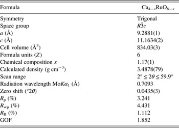
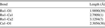
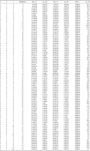
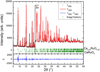
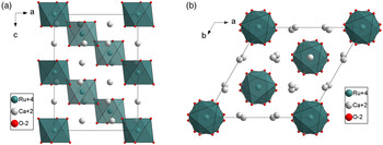
B(CaRuO3) = 1.031%, and GOF = 1.852 (all agreement factors as defined in TOPAS 4.2). The value x of the chemical composition determined by the Rietveld refinement is x = 1.17(1), which results in a final chemical composition of Ca2.83RuO4.83. The calculated weight percentage of the Ca2.83RuO4.83 phase is wt% = 87.7(1), whereas that of the CaRuO3 impurity could be determined to wt% = 12.3(1). The results of the final Rietveld refinement of the Ca2.83RuO4.83 phase (lattice parameters, agreement factors, etc.) are summarized in Table I, whereas atomic positions, occupancies, and anisotropic displacement parameters of this phase are listed in Table II. Selected bond lengths are listed in Table III and the powder diffraction data are given in Table IV. A Rietveld plot of the final refinement is shown in Figure 1 and the crystal structure of Ca4(−x)RuO6(−x) is presented in Figure 2.
Crystal structure data obtained from Rietveld refinement for Ca2.83RuO4.83.

Refined atomic coordinates, occupancy, and atomic anisotropic displacement parameters for Ca2.83RuO4.83.

Selected bond lengths for Ca2.83RuO4.83.

Powder XRD data for Ca2.83RuO4.83.

(colour online) Rietveld plot of the measured powder XRD data. Two phases, Ca2.83RuO4.83 [wt% = 87.7(1)] and CaRuO3 [wt% = 12.3(1)] were refined.

(colour online) Trigonal crystal structure of Ca4(−x)RuO6(−x). (a) View along the crystallographic b-direction, (b) view along the crystallographic c-direction.

IV. CONCLUSION
A non-stoichiometric Ca4−x RuO6−x phase was synthesized and the crystal structure was successfully determined by the charge-flipping method from the XRD data. A Rietveld refinement revealed that the crystal structure of the non-stoichiometric Ca2.83RuO4.83 phase is trigonal and isostructural to the K4CdCl6 type as it was suggested for the stoichiometric Ca4RuO6 phase.
SUPPLEMENTARY MATERIAL
“To view supplementary material for this article, please visit http://dx.doi.org/10.1017/S0885715615000883”
ACKNOWLEDGEMENTS
The authors would like to acknowledge Christine Stefani, Max Planck Institute for Solid State Research for carrying out the powder X-ray diffraction experiments.