Introduction
The genus Galactosomum Looss, 1899 is a group of trematodes belonging to the family of Heterophyidae, parasites in the intestine of fish-eating birds and mammals and, as metacercaria, encysted in fish (Prudhoe, Reference Prudhoe1949; Yamaguti, Reference Yamaguti1971; Pearson, Reference Pearson1973; Dailey et al., Reference Dailey, Demaree and Critchfield2002).
Galactosomum lacteum metacercaria was first described by Jägerskiöld (Reference Jägerskiöld1896) as Monostomum lacteum, in the brain of Cottus scorpius L., from the west coast of Sweden. As mentioned by Prudhoe (Reference Prudhoe1949), the same author in 1908 observed the adult in the intestine of cormorants Phalacrocorax carbo L. Afterwards, this parasite, as reported by Pearson (Reference Pearson1973), was found by different authors as metacercaria encysted in the brain, muscle and peritoneum of Cottus bubalis Euphrasen, 1786, C. scorpius, Smaris chriselis (Valenciennes, 1830), Onos tricirrhata (Bloch, 1785), Blennius sp., Gadus merlangus L., Acipenser sturio L., Bothus maeoticus Pallas, 1814, Trachinus draco L., Trachurus trachurus L., Uranoscops scaber L., Scorpaena porcus L. and Spicara smaris L. from Scotland, Sweden and the former USSR (Black Sea).
During a survey of parasitic infections of economically important teleosts from a fishing area off the south-western coasts of Sardinia (Gulf of Cagliari), from April 2002 to November 2005, 77 Galactosomum lacteum Jägerskiöld, Reference Jägerskiöld1896 (Heterophyidae) naturally encysted metacercariae were recorded for the first time in the Mediterranean Sea in nine species of fish: Diplodus annularis L., D. sargus L., Serranus cabrilla L., Spicara smaris, S. flexuosa Rafinesque, 1810, S. maena L., Symphodus tinca L., S. mediterraneus L. and Gobius cruentatus Gmelin, 1789 (Figus et al., Reference Figus, D'Amico, Loddo, Siddu and Canestri Trotti2005, Reference Figus, D'Amico, Loddo, Siddu and Canestri Trotti2006; Culurgioni et al., Reference Culurgioni, D'Amico, Canestri Trotti and Figus2006; D'Amico et al., Reference D'Amico, Canestri Trotti, Culurgioni and Figus2006).
The aim of this paper was to report the finding of G. lacteum metacercariae in teleosts from the Gulf of Cagliari, its parasitic infection levels and to discuss the morphological differences from G. timondavidi. This Haplorchinae congeneric species, in fact, was previously found naturally encysted in the brain of Diplodus annularis (Pearson & Prévot, Reference Pearson and Prévot1971) from the south of Marseille, and, as reported by Prévot (Reference Prévot1973), in Mugil aurata (Risso, 1810), Syngnathus abaster Risso, 1827, Gobius microps Krøyer, 1838 and Blennius pavo Risso, 1810 from Mediterranean waters, southern France. The authors put this species ‘closest to Knipowischetrema echinatum J. Timon-David, 1955’, found encysted in the brain of Diplodus annularis from the south of Marseille (Timon-David & Timon-David, Reference Timon-David and Timon-David1970).
Materials and methods
From April 2002 to November 2005, 957 teleosts caught in the Gulf of Cagliari, between 38°54′ and 39°10′ N and 8°55′ and 9°32′ E, belonging to eight different families were examined for metazoan parasites: 105 Atherinidae, 180 Centracanthidae, 97 Gobiidae, 55 Labridae, 116 Mullidae, 106 Scorpaenidae, 102 Serranidae and 196 Sparidae.
The metacercarial cysts recovered were measured in a drop of saline and excysted with the aid of two needles. The larvae were fixed under a coverslip in AFA (ethyl alcohol, formalin, acetic acid), stained in acetocarmine, dehydrated in ethyl alcohol and mounted in Canada balsam.
Drawings were made with the aid of a drawing tube mounted on an Jenaval photomicroscope and supplemented with free-hand details from living and/or fixed and photographed specimens. Measurements are given in micrometres. Infection prevalence (P%) and its confidence interval (CI), mean intensity (MI) ± SE and mean abundance (A) ± SE were calculated according to Bush et al. (Reference Bush, Lafferty, Lotz and Shostak1997).
Results
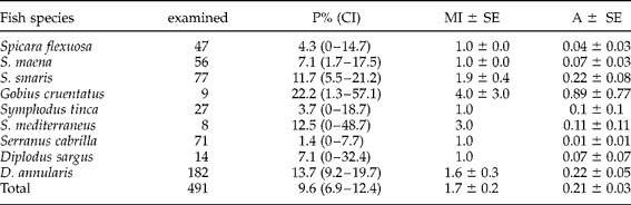
Seventy-seven encapsuled metacercariae of Galactosomum lacteum (Jägerskiöld, Reference Jägerskiöld1896) Looss, 1899 were collected from Spicara maena, S. flexuosa, S. smaris (Centracanthidae), Gobius cruentatus (Gobiidae), Symphodus tinca, S. mediterraneus (Labridae), Serranus cabrilla (Serranidae), Diplodus sargus and D. annularis (Sparidae). Relative to the above-cited species, as reported in table 1, the total prevalence of the infection was 9.6%, with mean intensity of 1.7 and mean abundance of 0.21. A higher prevalence (22.2%) and intensity (4.0) were recorded in G. cruentatus, while the lowest values were in S. cabrilla, 1.4% and 1.0, respectively.
Galactosomum lacteum in teleosts from Gulf of Cagliari (South Sardinia, Italy).
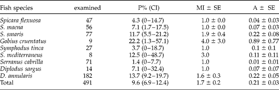
P%(CI), prevalence (confidence interval); MI, mean intensity; A, abundance; SE, standard error.
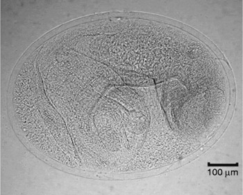
The metacercariae were identified according to Yamaguti (Reference Yamaguti1971) and to Pearson (Reference Pearson1973) by some distinctive morphological features: expanded forebody (clearly visible even in fixed specimens), tubular excretory bladder extending to the posterior border of the ovary, two-chambered seminal vesicle, asymmetrical and parenchymatous ventral sucker with lines of spines within its cavity and unarmed gonotyle. The metacercariae were 673 (565–822) long and 441 (371–584) wide, yellowish brown and elliptical, with a transparent, ~20 μm cyst wall (figs 1234).
Galactosomum lacteum encapsuled metacercaria.

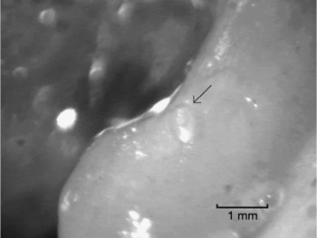
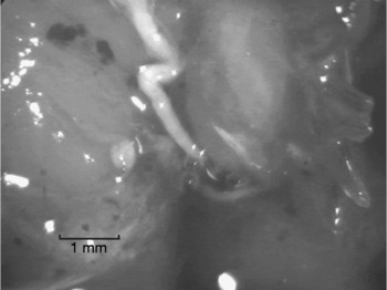
Galactosomum lacteum metacercaria encysted on the brain of Diplodus annularis (Sparidae).

Galactosomum lacteum metacercaria encysted on the optyc nerve of Diplodus annularis (Sparidae).

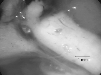
Galactosomum lacteum metacercaria encysted on the connective tissue external to the pharynx of Spicara maena (Centracanthidae).

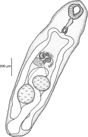
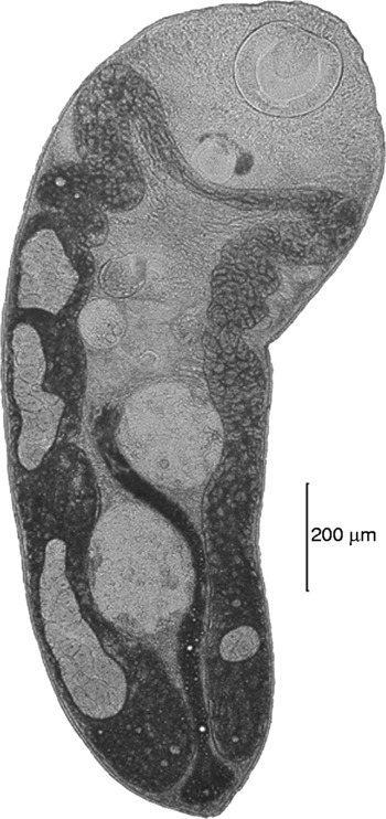
After excystation, the morphometric measures were based on 11 specimens: body elongate, greatest width at level of caecal bifurcation, forebody slightly flattened and hindbody cylindrical; 1380 (925–1970) long and 428 (281–676) wide; tegument covered with scales in the anterior third of the body, then thinly spined. Oral sucker round, subterminal, 180 (126–295) long and 185 (141–285) wide. Prepharynx 79 (28–115) long. Pharynx 76 (41–120) by 75 (34–120). Oesophagus very short, practically not measurable. Caeca simple, straight, not reaching the posterior end of the body (figs 5, 6).
Drawing of Galactosomum lacteum excysted metacercaria (ventral view).

Galactosomum lacteum excysted metacercaria (ventral view).

Ventrogenital sac in the anterior midbody, well behind the gut bifurcation, distant 290 (249–321). Ventral sucker dextrally asymmetric, rounded, 97 (65–112) by 84 (52–100). Gonotyl 47 (40–51) by 52 (47–60).
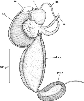
Two testes, entire, rounded to oval, diagonal, both posterior to the midbody. Left testis anterior, 119 (60–229) by 124 (80–175); right testis, posterior, 139 (78–250) by 128 (90–192). Seminal vesicle double, proximal portion smaller, measuring 59 (45–76) by 33 (25–41), and distal portion 100 (82–140) by 54 (42–75). Ovary 55 (30–86) by 62 (44–110), globular or transversely oval, dextral, anterior to testes, behind the posterior end of the seminal vesicle. Excretory bladder tubular, crossing between the testes and reaching the posterior end of the ovary (fig. 7).
Galactosomum lacteum metacercaria, drawing of ventro-genital complex: v.s., ventral sucker; m., mouth; g., gonotyl; l.p., lateral pocket; d.s.v., distal seminal vesicle; p.s.v., proximal seminal vesicle; u., uterus.

Representative specimens of metacercariae are deposited in the Museo di Zoologia del Dipartimento di Biologia Animale ed Ecologia (University of Cagliari).
Discussion and conclusions
Previously, in the Mediterranean area, natural and experimental infections by metacercariae of Galactosomatidae Knipowitschetrema echinatum J. Timon-David, 1955 and G. timondavidi Pearson & Prévot, Reference Pearson and Prévot1971, were observed in D. annularis from the Gulf of Lyon. Cysts of Galactosomatidae found in the same host from the same area (south of Marseille) were always located on the encephalon and on the optic nerve (Timon-David & Timon-David, Reference Timon-David and Timon-David1970; Pearson & Prévot, Reference Pearson and Prévot1971; Prévot, Reference Prévot1973). In the concerned fishing area, on the other hand, metacercariae of G. lacteum were found encysted on the brain (fig. 2), on the optic nerve (fig. 3) and/or on the muscular and connective tissue external to the pharynx and oesophagus (fig. 4), as in D. annularis (D'Amico et al., Reference D'Amico, Canestri Trotti, Culurgioni and Figus2006) and S. maena, while in S. flexuosa, S. smaris (Figus et al., Reference Figus, D'Amico, Loddo, Siddu and Canestri Trotti2006), S. cabrilla (Figus et al., Reference Figus, D'Amico, Loddo, Siddu and Canestri Trotti2005), D. sargus, G. cruentatus, S. tinca and S. mediterraneus (Culurgioni et al., Reference Culurgioni, D'Amico, Canestri Trotti and Figus2006) they were observed exclusively in the pharynx.
The site of encystment in the fish hosts, not limited to nervous tissue, is similar to those of G. lacteum found by Vlasenko (Reference Vlasenko1931) in S. chriselis as reported by Pearson (Reference Pearson1973), who, on the other hand, considers it ‘unlikely that a single species would localise on the brain in one genus of host, but in the muscles (Chulkova, Reference Chulkova1939) or on peritoneum (Vlasenko, Reference Vlasenko1931) in other genera’. In any case, this ‘unlikelihood’ disagrees with the cyst localizations observed in this work. It seems evident that this parasite is not strictly organ specific since the metacercariae have been found in the same specimen, as in D. annularis and in S. maena, both in the pharynx and in the brain. Furthermore, morphological features such as the diagonal testes, the double-chambered seminal vesicle, the structure of the ventro-genital complex and the excretory bladder reaching the posterior end of the ovary, allowed us to distinguish clearly the metacercariae examined here from those of G. timondavidi described by Pearson & Prévot (Reference Pearson and Prévot1971).
The data obtained provided further information about the life cycle of G. lacteum, widening the number of its intermediate host species, i.e. fish, and outlining new geographical records, i.e. Mediterranean waters.
To date, there are no reports concerning both the definitive hosts and the first intermediate hosts in the Gulf of Cagliari. Among fish-eating birds, potential definitive hosts are permanent and/or migratory species, such as Ardea cinerea L., Larus genei Breme, 1839, L. michahellis Naumann, 1840, L. ridibundus L., P. carbo, Podiceps cristatus L. and Sterna L. At the same time, monitoring of the prosobranch gastropods belonging to the genus Cerithium Bruguiere, 1789, its first intermediate hosts, should be helpful in the description of the full life history of G. lacteum in this area.
Acknowledgements
We are grateful to Professor Giorgio Canestri Trotti for helping in the identification of the parasite and to Dr Federica Usai for the drawing of the specimens.