

Introduction
For over a century there have been records of transient global amnesia (TGA) in the literature, with transient amnesic states suggestive of TGA first described in 1882 by RibotReference Ribot1 and in 1909 by Benon.Reference Benon2 In 1956, further cases suggestive of TGA were described by BenderReference Bender3 and by Guyotat and Courjon,Reference Guyotat and Courjon4 and in 1964 Fisher and AdamsReference Fisher and Adams5 published a large case series and introduced the term ‘transient global amnesia’. Although there are well-described clinical features of the syndrome, the cause of TGA remains unclear, despite advances in imaging.
Search strategy
Medline and EMBASE databases from 1956 to February 2012 were systematically searched with the following terms: transient global amnesia; transient global amnesic states and stroke; transient global amnesia and neuroimaging. 612 articles were retrieved and the abstracts reviewed. If the abstract was not available the title was considered. If the article was not in English it was not reviewed as the great majority were published in the English language and we considered bias from excluding other languages was likely to be minimal. We identified 70 papers to read and their references were reviewed for secondary references. We included randomized control trials, case series, case reports and review articles.
Epidemiology
The incidence is reported as 3–8 per 100,000 people.Reference Hodges and Warlow6 A third of these episodes occur in the age group of 50–70 years. The annual recurrence rate is between 6 and 10%.Reference Hodges and Warlow6 There has been conflicting evidence on gender distribution, with Hodges and Warlow claiming that TGA was more common in men,Reference Hodges and Warlow6 whilst other studies have shown the opposite.Reference Melo, Ferro and Ferro7 There is emerging evidence that TGA can be hereditary.Reference Vyhnalek, Bojar, Jerabek and Hort8–Reference Dupuis, Evrard, Jacquerye, Picard, Goossens and Ghysens10
Clinical features
TGA is characterized by an episode of anterograde and retrograde amnesia lasting less than 24 hours. During this episode patients are conscious and able to interact normally. They are typically distressed and noted to repeat questions without remembering explanations. Although often described, a link between preceding emotional or physical stress and TGA has not been proven. Although these clinical features resolve within 24 hours, mild subclinical neurophysiological deficits with vegetative symptoms can last for days to months.Reference Sander and Sander11 However, a recent meta-analysis of 25 studies showed no difference in long-term cognitive performance between patients and the healthy population.Reference Jager, Bazner, Kliegel, Szabo and Hennerici12
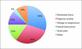
Common triggers are shown in Figure 1 and include emotional stress (30%), vigorous activity (30%), change in temperature (15%) and sexual intercourse (12%).Reference Velasco, Al Hussayni and Bermejo13 Quinette et al.Reference Quinette, Guillery-Girard, Dayan, de La Sayette, Marquis and Viader14 found an increased frequency of patients with emotional or psychological instability, while Pantoni et al.Reference Pantoni, Lamassa and Inzitari15 described an increased incidence of personal and family history of psychiatric or phobic disorders.
Triggers for transient global amnesia (TGA)

Diagnosis is clinical and based on the criteria of Caplan and Hodges,Reference Hodges and Warlow6, Reference Caplan16 shown in Table 1.
Diagnostic criteria for transient global amnesia by Caplan & HodgesReference Hodges and Warlow6, Reference Caplan16

During the acute episode a temporally graded retrograde amnesia can occur, although this is not part of the current diagnostic criteria. Nausea, headache and dizziness may be present. Other features supporting the diagnosis of TGA are a history of excessive physical activity or strong emotions preceding the onset of symptoms and repetition of the same questions. Patients are co-operative, follow instructions and are able to name objects. Features against the diagnosis are a history of a recent change in medications, evidence of recent hypoglycaemia, memory of the acute episode, retrograde amnesia only, or more than three episodes in one year. Further diagnostic assessment is necessary if there was no witness, the presentation was not typical, or there are vascular risk factors present.
Pathophysiology
The underlying pathophysiology of TGA is unclear. The current hypothesis is that it is associated with lesions within the mediobasal temporal region, hippocampus and the para-hippocampus.Reference Forster, Griebe, Gass, Kern, Hennerici and Szabo17 Magnetic resonance imaging (MRI) has demonstrated lesions in the hippocampal formation and memory circuits in the mesiotemporal regionReference Alberici, Pichiecchio, Caverzasi, Farina, Persico and Cavallini18, Reference Badve, Henderson, Jhamb and Read19 and analysis of the functional anatomy of these lesions has shown selective distribution within the CA1 subfield of the hippocampal cornu ammonis.Reference Bartsch, Alfke, Stingele, Rohr, Freitag-Wolf and Jansen20, Reference Bartsch, Alfke, Deuschl and Jansen21 Some images have demonstrated that cellular mechanisms may play a role in developing the lesions, suggesting that a cascade initiated by the selective vulnerability of CA1 neurons to metabolic stress causes a temporary disturbance to the memory pathways. The exact cause of this disturbance in memory is not known, but has been attributed to impaired venous flow, ischaemia, migraine, psychological factors or epilepsy.
Venous flow
Using 3D time-of-flight magnetic resonance angiography (MRA), Chung et al.Reference Chung, Hsu, Chao, Sheng, Soong and Hu22 showed that compression of the left brachiocephalic vein led to retrograde intracranial venous flow in half of the TGA patients studied and none of the age- and gender-matched controls. In addition, right-sided internal jugular vein incompetence was found by Agosti et al.Reference Agosti, Borroni, Akkawi and Padovani23 However, neither is well defined or reproducible.Reference Chung, Hsu, Chao, Sheng, Soong and Hu22, Reference Agosti, Borroni, Akkawi and Padovani23 Lewis has suggested that venous congestion leading to cerebral venous thrombosis and venous ischaemia could cause TGA.Reference Lewis24 Duplex ultrasonography in 114 TGA cases found a high frequency of insufficient jugular vein valves during Valsalva manoeuvre compared with controls. Together, these results suggest that TGA patients may have an underlying impairment of cerebral venous outflow and vulnerability to venous congestion, leading to TGA.
Ischaemia
The speed of onset and predominant age of patients affected by TGA would support transient ischaemia as a cause.Reference Agrawal, Chandak and Zaretskaya25 However, this aetiology is unlikely as there is always prompt resolution of the symptoms and, unlike following a transient ischaemic attack (TIA), patients are less likely to suffer a future stroke.Reference Zorzon, Antonutti, Mase, Biasutti, Vitrani and Cazzato26 There are also no relevant abnormalities found on MRA during an acute episode.Reference Ahn, Kim, Lee, Kim, Lee and Oh27, Reference Auyeung, Tsoi, Cheung, Fong, Li and Chan28 Interestingly, Winbeck et al. did find that carotid atherosclerosis identified by ultrasound was more common among patients with TGA, especially those with diffusion-weighted imaging (DWI) changes, compared with controls. In this subset of patients they concluded that ischaemia may cause the symptoms, although the association was not strong and, compared with TIA patients, other vascular risk factors were not common.Reference Winbeck, Etgen, von Einsiedel, Rottinger and Sander29 The fact that microvascular ischaemic changes on MRI are not increased in TGA patients would also be against an arterial cause.
Migraine
Because migraine is characterized by paroxysmal neuronal dysfunction and a comparable time course, it has been suggested as a cause of TGA. Some investigators have found a strong association,Reference Schmidtke and Ehmsen30 whilst others disagree,Reference Pantoni, Lamassa and Inzitari15 but interpretation is difficult because of the many other disorders associated with migraine, e.g. epilepsy, stroke, patent foramen ovale, high antiphospholipid titers, depression, arterial dissection, fibromyalgia and dementia.Reference Davies and Panayiotopoulos31–Reference Sas, Pardutz, Toldi and Vecsei39
The pathogenesis of migraine with aura is generally accepted to be caused by cortical spreading depression (CSD) of electrical activity characterized by transient neuronal depolarization and hyperperfusion, and subsequent hyperpolarization, hypoperfusion and neuronal dysfunction. This spreads at a rate of 3–5 mm per minute across the cortex and in animal models and humans has been seen to involve the hippocampus, at times leading to hypoxia in the CA1 field.Reference Melo, Ferro and Ferro7, Reference Bartsch, Alfke, Deuschl and Jansen21, Reference Bartsch, Dohring, Rohr, Jansen and Deuschl40 Furthermore, induction of CSD in animals produces amnesic behaviour that resolves spontaneously. Given this background and the high prevalence of migraine in some TGA patients who also had headache as an accompanying symptom or precipitating factor, CSD was proposed by Olesen and Jorgensen in 1986 as a possible mechanism underlying TGA.Reference Olesen and Jorgensen41 They also noted high hippocampal glutamate concentration, the fact that emotional events could trigger release of glutamate and proposed that TGA tends to follow such events. Initiating a stress response can induce hippocampal dysfunction.Reference Cho and Kim42, Reference Hunter43
DWI abnormalities are not described in migraine, unless associated with stroke, but there have been reports of abnormalities in the apparent diffusion coefficient maps in patients who have had prolonged aura.Reference Sander and Sander11, Reference Cho and Kim42, Reference Felix, Castro, Maia and da Rocha44 This would, perhaps, support CSD as a cause of TGA, but migraine aura is more common in young people, whereas TGA is not. This could be explained by compensatory mechanisms in younger patients preventing CSD in the hippocampal region. However, migraine episodes are generally not associated temporally with TGA so an association with migraine remains possible, but unlikely.
Epilepsy
In 1889, Hughlings-Jackson first suggested that epileptic activity could cause transient amnesia.Reference Hughlings-Jackson45 Quinette et al. performed electroencephalograpy (EEG) studies in 106 patients during or after acute TGA.Reference Quinette, Guillery-Girard, Dayan, de La Sayette, Marquis and Viader14 They were unremarkable in 80%, and the remainder showed minor non-epileptiform abnormalities only, which suggests that epilepsy is not a cause of TGA. There is also a low possibility of developing epilepsy after TGA. More recently, however, there have been reports of transient epileptic amnesia (TEA) when classic TGA is associated with clear electrographic seizures.Reference Pearce and Bogousslavsky46 These episodes last for 1–3 hours, rather than the usual 24-hour duration of TGA. After typical complex partial seizures with unresponsiveness, automatisms or secondary generalized seizures, the subject remains alert but amnesic. Non-convulsive status epilepticus has been found in a few cases of apparent TGA. EEG studies demonstrate wicket spikes, which are a benign variant, and underlying epileptiform activity is suggested to be the cause.
TEA needs to be considered when there is a history of epilepsy, recurrent attacks of TGA, or atypical duration. Since up to 70% of this group may have a normal inter-ictal EEG, continued surveillance is sometimes necessary. Prolonged or ambulatory EEG studies can identify the nature and frequency of TEA episodes.
Neuroimaging
Single photon emission computed tomography (SPECT), positron emission tomography (PET) and DWI are the three main neuroimaging modalities used to investigate TGA, but are still unable to find a cause and explain the pathophysiology of TGA.Reference Sander and Sander11, Reference Forster, Griebe, Gass, Kern, Hennerici and Szabo17, Reference Auyeung, Tsoi, Cheung, Fong, Li and Chan28, Reference Takeuchi, Matsuda, Yoshioka and Yonekura47, Reference Guillery, Desgranges, de La Sayette, Landeau, Eustache and Baron48
Findings from PET and SPECT studies suggest that hypoperfusion and hypometabolism of the hippocampus may be characteristic of TGA,Reference Forster, Griebe, Gass, Kern, Hennerici and Szabo17 but there have also been numerous cases of dysfunction in other locations such as the thalamic, frontal, cerebellar and striatal regions.Reference Ahn, Kim, Lee, Kim, Lee and Oh27, Reference Auyeung, Tsoi, Cheung, Fong, Li and Chan28 Haemodynamic changes in SPECT studies confirmed transient dysfunction of the medial temporal lobe during the acute phase of TGA in 42 out of 43 patients.Reference Takeuchi, Matsuda, Yoshioka and Yonekura47, Reference Kirshner49 PET studies performed after an episode of TGA in 15 patients found abnormalities in the hippocampus of four patients and a high incidence of high and giant cavities.Reference Guillery, Desgranges, de La Sayette, Landeau, Eustache and Baron48, Reference Eustache, Desgranges, Laville, Guillery, Lalevee and Schaeffer50, Reference Eustache, Desgranges, Petit-Taboue, de La Sayette, Piot and Sable51 Resolution of these changes usually occurs, but persistent hypoperfusion was seen as late as one year after an episode of recurrent TGA. Altered perfusion and metabolism are also seen in migraine and epilepsy, so these findings do not help to explain the underlying mechanism of TGA.
DWI is a sensitive technique used to detect early cytotoxic oedema in ischaemia and in the past 20 years it has been reported to show abnormalities suggestive of ischaemic injury in TGA.Reference Felix, Castro, Maia and da Rocha44 However, the evidence is conflicting, with some investigators finding no DWI changes during TGA, leading to the conclusion that if changes representing ischaemic injury are seen, a diagnosis of TGA should not be made. Of 31 patients fulfilling Hodges and Warlow's criteria for TGA,Reference Hodges and Warlow6 abnormalities on DWI during and/or after the episode were found in only 56%.Reference Choi, Kim, Jung and Kim52 During a 7-year study by Ahn and colleagues,Reference Ahn, Kim, Lee, Kim, Lee and Oh27 MRI scans were performed on 203 TGA cases. Only 16 of these had changes in the hippocampus, with no obvious clinical difference from those without such changes. However, the scans demonstrating changes were performed at a mean time to scan of 9 hours, whereas the overall mean time to scan was 6 hours. This suggests that precise timing might help to detect more abnormalities, and current recommendations for the imaging of TGA with DWI include use of a 3-T magnet, acquisition between 24 and 72 hours of onset and 3 mm slice thickness.
Bartsch and colleaguesReference Bartsch, Alfke, Deuschl and Jansen21 studied the evolution of MRI changes in 29 patients. Between 24 and 72 hours post-event, 34 DWI lesions were identified in the CA1 sector of the hippocampus, and these had normalized by 10 days. This is a similar time course to ischaemic lesions, which has led to the suggestion that TGA may have a vascular cause.
MR spectroscopy showed a lactate peak in hippocampal DWI lesions,Reference Bartsch, Alfke, Wolff, Rohr, Jansen and Deuschl53 which suggests local dysfunction of CA1 neurons. Certainly the most consistent finding from neuroimaging studies is the location of abnormalities in the hippocampus, but their origin remains debatable. Whilst DWI is highly sensitive for ischaemia, it is not specific, and other causes for cytotoxic oedema could include hypoglycaemia, cortical spreading depression or epileptic seizures.
Management
The diagnosis of TGA is based on the typical clinical presentation, clear collateral history and absence of any features suggestive of underlying pathology. Careful assessment should avoid the patient being labelled as ‘acutely confused’ and assumed to have delirium. Screening for haematological abnormalities and hypoglycaemia should be done promptly and a stroke or transient ischaemic attack must be ruled out, so neuroimaging is usually appropriate. If available, high resolution MRI with DWI may detect acute hippocampal lesions. EEG may be useful to demonstrate any inter-ictal activity if epilepsy is suspected. Cardiological investigations with electrocardiography, Holter monitoring and echocardiogram may sometimes be required. Adequate investigation without any significant pathology being demonstrated should reassure patients and the excellent prognosis needs to be emphasized. There is no need for any restriction on driving or reporting to the Driving and Vehicle Licensing Agency (DVLA) after diagnosis of a single episode of TGA.
Conclusion
The belief that TGA is related to hippocampal dysfunction is no longer a hypothesis. With advanced imaging and laboratory techniques, clearly there is a metabolic stress localizing to CA1 sector. The precise cause and exact triggers of attacks, however, remains unclear. However, stressors precipitating TGA might be purely coincidental. TGA could be caused by energy exhaustion in a metabolically fragile hippocampus, typical in a demand-supply scenario. Optimal imaging parameters were only recently set and are not uniformly available. This explains why the frequency of DWI changes in TGA patients has not been clearly defined. Uncertainty with regard to treatment may be a result of these studies. It remains unclear whether patients with DWI lesions should be managed as stroke or TIA. While imaging findings of presumed stroke make this less clear, epidemiology on the other hand suggests otherwise. There have been no autopsy studies. Autopsy could help by identifying the cause of the dysfunction, if it was caused by ischaemia or by other causes of energy failure. In recurrent cases or those with very short durations, prolonged or ambulatory EEG should be considered to identify the subgroup of patients with epileptic syndromes. Usually the prognosis of TGA is excellent, and the management depends on recognition and reassurance.
Conflicts of interest
None