Case report
We report the course and management of a 15-month-old child with Williams syndrome, born at 35+2 weeks of gestation and diagnosed with Williams syndrome after birth. An initial echocardiogram showed only mild branch pulmonary arterial stenosis.
When she was 5 months old, she was diagnosed with a large Morgagni hernia, and underwent surgical hernia repair at the age of 7 months. She also developed severe seizures with a diagnosis of infantile spasms – West syndrome. Cognitive and neurological development was significantly delayed, and she had a right-sided hearing impairment. Feeding difficulties including gastro-oesophageal reflux required percutaneous endoscopic gastrostomy tube implantation. CT and echocardiography at that time confirmed mild branch pulmonary arterial narrowing, mild supravalvar aortic stenosis, as well as a slightly small aortic arch. The aortic valve was tricuspid with normal function. Biventricular systolic function was good.
Cardiac assessment at the age of 12 months showed little change with mild-to-moderate (gradient 36 mmHg) supravalvar aortic stenosis, left ventricular hypertrophy, and a few hyperechogenic areas in the left ventricular myocardium. Biventricular function remained normal, and there were no clinical signs of congestive heart failure. The electrocardiogram was within normal limits.
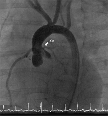
She presented 3 months later with recurrent “blue episodes”. Echocardiography demonstrated moderately impaired left ventricular function (ejection fraction 35%) with dilation, with left ventricular end-diastolic diameter of 27 mm and left ventricular end-systolic diameter of 23 mm, antero- and infero-lateral regional wall motion abnormality, hypertrophy, and endocardial hyperechogenicity. Speckle tracking demonstrated severely impaired global longitudinal strain (average longitudinal strain 7.2%) with antero- and infero-lateral dyskinesia, at the left anterior descending and circumflex coronary artery territory. The degree of supravalvar aortic stenosis was unchanged. The electrocardiogram now showed down-sloping ST segment depression and T wave inversion in the lateral leads, indicating sub-endocardial ischaemia. Troponin I levels were elevated at 231 ng/L. Cardiac catheterisation revealed severe tubular left main coronary arterial stenosis (Fig 1). The supravalvar aortic stenosis was mild with an invasive gradient of 15–20 mmHg. Although it was agreed that high risk surgical intervention was the only treatment option, initial conservative management with medical therapy was begun in the hope that the ischaemia would settle and the ventricular function would improve, and hence lessen the risk of surgery. Unfortunately, a month later, there remained evidence of ongoing ischaemia with raised troponin levels and no improvement in echocardiography parameters, including speckle tracking. After further discussion between congenital cardiac surgeons and paediatric cardiologists, a cardiovascular MRI study was planned to assess myocardial viability and determine the best surgical strategy. The day before the cardiovascular magnetic resonance study, she was admitted to intensive care after an out-of-hospital cardiac arrest, requiring intubation but no inotropic support.
Angiographic fluoroscopy image showing severe stenosis of the left main coronary artery (LCA).

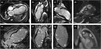
We performed 3-Tesla cardiovascular MRI under general anaesthesia with full extracorporeal membrane oxygenation backup. This demonstrated severe left ventricular dysfunction and dilation (ejection fraction 14%) and a thin-walled akinetic aneurysm (Fig 2a–c). Late gadolinium enhancement imaging revealed widespread left ventricular endocardial hyperenhancement and transmural apical myocardial fibrosis (Fig 2e–g). The entire thoracic aorta was small, and the ascending aorta was thick walled, although there was only mild supravalvar aortic narrowing (Fig 2d and h).
Cardiovascular magnetic resonance cine images showing severe dilatation of the left ventricle ( a – c ), a thick-walled ascending aorta ( d , arrow) and a small aortic arch ( h ). Late gadolinium enhancement images ( e – g ) demonstrate widespread left ventricular endocardial enhancement (arrows) and apical delayed enhancement (arrowhead). Segments affected and late gadolinium transmurality are as follows: basal: anteroseptal, anterior, anterolateral, and inferoseptal <25%; mid: anteroseptal, anterior, anterolateral, and inferolateral <25%; and apical: septal <25%, lateral <25%, and inferior 25–100%.

During cardiovascular magnetic resonance, she had two episodes of mild arterial hypotension, one of which was after gadolinium injection. These resolved with a small dose of vasopressor and a 5 ml/kg fluid bolus.
Unfortunately, back in intensive care the patient had three consecutive episodes of ischaemic cardiac arrest, necessitating cardiopulmonary resuscitation. High doses of vasopressor and inotropic agents were used without associated echocardiographic or clinical improvement. Further discussions between paediatric cardiologists, paediatric intensivists, congenital cardiac surgeons, and her parents were started.
Taking into account the new cardiovascular magnetic resonance findings (severe left ventricular dilatation, global left ventricular dysfunction with ejection fraction 14%, apical left ventricular aneurysm, and extensive left ventricular endocardial and apical transmural fibrosis), the associated increased risks related proceeding to cardiopulmonary bypass, effective myocardial protection, and need for extensive left main coronary arterial plasty in the setting of severe generalised arteriopathy, as well as her ongoing clinical ischaemia and neurological co-morbidities, it was agreed that surgical intervention was not warranted because of being highly risky and likely futile. The patient was not eligible for cardiac transplantation because of her neurological status. She received palliative care, and died a few hours later in the paediatric ICU.
Discussion
Williams syndrome is caused by a deletion on chromosome 7, which involves the elastin gene, leading to vascular abnormalities. Coronary artery involvement has been found in up to 45% of Williams syndrome patients with supravalvar aortic stenosis,Reference Collins 1 , Reference Collins, Kaplan, Somes and Rome 2 usually when severe, and can result in sudden cardiac death.Reference Bird, Billman and Lacro 3 Significant coronary arterial stenosis leading to end-stage ischaemic heart disease or myocardial infarction, however, is rarely seen.Reference Pieles, Ofoe and Morgan 4 – Reference González-López, Pérez-Caballero-Martínez and Granados-Ruiz 6
Although the cardiovascular diagnosis in this child was initially assessed using echocardiography and cardiac catheterisation, cardiovascular magnetic resonance allowed for confirmation of end-stage ischaemic heart disease showing severe regional left ventricular dysfunction and dilation on cine images as well as widespread endomyocardial and transmural apical fibrosis by late gadolinium enhancement imaging.
Late gadolinium enhancement cardiovascular magnetic resonance is widely used to differentiate between viable myocardium and focal myocardial fibrosis including congenital and paediatric cardiovascular disorders.Reference Etesami, Gilkeson and Rajiah 7 – Reference Latus, Gummel and Klingel 9 Obtaining adequate temporal and spatial resolutions in infants and young children, however, can be challenging, and advanced cardiovascular MRI expertise specific to paediatric cardiology patients is essential as technical modifications of the sequences are often necessary.Reference Etesami, Gilkeson and Rajiah 7 , Reference Fratz, Chung and Greil 10 , Reference Stockton, Hughes, Broadhead, Taylor and McEwan 11 In the context of the clinical, echocardiographic, cardiovascular magnetic resonance, and cardiac catheterisation findings, the pattern of myocardial late gadolinium enhancement substantially contributed to the clinical decision.
When using cardiovascular magnetic resonance in small children, the risk of general anaesthesia and related safety aspects should be taken into account. The risks versus benefits of obtaining cardiovascular magnetic resonance under anaesthesia were debated in a joint medical and surgical case conference in this challenging case. Vital to the acquisition is a multispecialty team including an anaesthetist with experience in managing children with heart disease and an infrastructure addressing all patient-specific safety aspects and those of the cardiovascular magnetic resonance environment.Reference Stockton, Hughes, Broadhead, Taylor and McEwan 11 Our case not only demonstrates that patients with Williams syndrome are at risk for coronary heart disease but also highlights an important role of cardiovascular magnetic resonance in the diagnosis and management.
Acknowledgements
S.V.B.-N. is supported by an Intermediate Clinical Research Fellowship from the British Heart Foundation (FS/11/38/28864). This project was supported by the NIHR Cardiovascular Biomedical Research Unit of Royal Brompton and Harefield NHS Foundation Trust and Imperial College London. This report is an independent research by the National Institute for Health Research Biomedical Research Unit Funding Scheme. The views expressed in this publication are those of the author(s) and not necessarily those of the NHS, the National Institute for Health Research, or the Department of Health. The authors thank Dr Mary Lane for facilitating CMR and Dr Sylvia Krupickova and Dr Guido Michielon for their clinical support. Informed consent was obtained from the parents.
Financial Support
This research received no specific grant from any funding agency, commercial, or not-for-profit sectors.
Conflicts of Interest
None.