





Significant outcomes
-
• Long-term (180 min) maternal separation has more severe effects on behaviour and neuropeptides than short-term (15 min).
-
• Long-term maternal separation at postnatal day 8–9 leads to increased anxiety and decreased NPY levels in the hippocampus.
-
• Long-term maternal separation at postnatal day 10–14 leads to decreased anxiety and increased NPY levels in the hippocampus.
Limitations
-
• The study was limited to male rats.
-
• The study does not allow to draw conclusions with respect to the causal relationship between neuropeptide levels and behaviour.
-
• Only one animal model for anxiety was used, limiting the generalisation to other forms of anxiety.
Introduction
Although genes contribute to the aetiology of mental disorders such as schizophrenia and affective disorders, the concordance rates of monozygotic twins consistently falls short of 100%, indicating that non-genetic, environmental factors also play an important role (Ellenbroek & Youn, Reference Ellenbroek and Youn2016). Among these, early adverse life events have been most often studied in animals as well as in humans (Ellenbroek et al., Reference Ellenbroek, van den Kroonenberg and Cools1998; Carr et al., Reference Carr, Martins, Stingel, Lemgruber and Juruena2013; Targum & Nemeroff, Reference Targum and Nemeroff2019; Lippard & Nemeroff, Reference Lippard and Nemeroff2020; Wang et al., Reference Wang, Levine, Avila-Quintero, Bloch and Kaffman2020) and it is now established that individuals exposed to early childhood adversity have an increased risk of developing mental disorders including depression, anxiety and substance abuse (McLaughlin et al., Reference McLaughlin, Greif Green, Gruber, Sampson, Zaslavsky and Kessler2012; Björkenstam et al., Reference Björkenstam, Burström, Vinnerljung and Kosidou2016). For example, it has been well established that prenatal viral or bacterial infection, leading to an activation of the maternal immune system, increase the risk for the offspring to develop autism spectrum disorders (ASDs), schizophrenia, major depressive disorders and other mental health conditions (Brown, Reference Brown2000; Al-Haddad et al., Reference Al-Haddad, Jacobsson, Chabra, Modzelewska, Olson, Bernier, Enquobahrie, Hagberg, Östling, Rajagopal, Adams Waldorf and Sengpiel2019; Croen et al., Reference Croen, Qian, Ashwood, Zerbo, Schendel, Pinto-Martin, Daniele Fallin, Levy, Schieve, Yeargin-Allsopp, Sabourin and Ames2019; Álvarez-Mejía et al., Reference Álvarez-Mejía, Rodas and Leon-Rojas2025; Zhu et al., Reference Zhu, Yang, Feng, Jiang and Zhang2026). Exposure to childhood trauma was significantly associated with obsessive compulsive and generalised anxiety disorders (Park et al., Reference Park, Hong, Bae, Cho, Lee, Lee, Chang, Jeon, Hahm, Lee, Seong and Cho2014), social anxiety disorder (Michail & Birchwood, Reference Michail and Birchwood2014), schizophrenia (van Nierop et al., Reference van Nierop, Lataster, Smeets, Gunther, van Zelst, de Graaf, ten Have, van Dorsselaer, Bak, Myin-Germeys, Viechtbauer, van Os and van Winkel2014) but also migraine (Fuh et al., Reference Fuh, Wang, Juang, Lu, Liao and Chen2010).
These correlative studies in humans are supported by studies in animals where the causal relationship between early stressful life events and later life alterations in behaviour has been shown (McLaughlin et al., Reference McLaughlin, Sheridan and Lambert2014). For instance the offspring of females exposed to restraint and light stressors during pregnancy (from gestational day 11 to date of birth) show a decrease in the time in the open arms of an elevated plus maze (EPM) (Morley-Fletcher et al., Reference Morley-Fletcher, Mairesse, Soumier, Banasr, Fagioli, Gabriel, Mocaer, Daszuta, McEwen, Nicoletti and Maccari2011), indicative of increased anxiety. Likewise, prenatal stress or prenatal immune challenges in rats lead to schizophrenia and autism-like deficits in the adult offspring, including deficits in prepulse inhibition, social behaviour and cognition (Markham & Koenig, Reference Markham and Koenig2011; Patterson, Reference Patterson2011). Early postnatal events such as early handling, maternal separation and maternal deprivation also have long-term (LT) effects on brain and behaviour. Early handling refers to a short-term (ST) (usually 15 minutes) separation of the mother from her litter (Raineki et al., Reference Raineki, Lucion and Weinberg2014). The most well-known LT effect of early handling is a significant reduction in anxiety-like behaviour and a reduction stress response, as measured by the activation of the hypothalamic–pituitary–adrenal (HPA) axis release of ACTH and/or corticosterone (Levine et al., Reference Levine, Haltmeyer, Karas and Denenberg1967; Meaney et al., Reference Meaney, Aitken, Bodnoff, Iny and Sapolsky1985a, Reference Meaney, Aitken, Bodnoff, Iny, Tatarewicz and Sapolsky1985b). Maternal separation refers to repeated separation of the dams from their litter, usually for 180 minutes (Nylander & Roman, Reference Nylander and Roman2013; Vetulani, Reference Vetulani2013). Most results show that maternal separation induces opposite effects in the offspring compared to early handling. Thus, maternal separation leads to reduced exploration of the open arms in an EPM and to an enhanced HPA-axis response to stress (Meaney et al., Reference Meaney, Diorio, Francis, Widdowson, LaPlante, Caldji, Sharma, Seckl and Plotsky1996; Kalinichev et al., Reference Kalinichev, Easterling, Plotsky and Holtzman2002; Gong et al., Reference Gong, Yang, Wang, Liang and Yue2026; Najafabadi et al., Reference Najafabadi, Shamsara, Mirzaie, Sheibani, Ahmadi, Joushi and Basiri2026). Although maternal separation and deprivation are sometimes used synonymously, we prefer to restrict the latter to a single 24 h period of dam removal. Similar to maternal separation, maternal deprivation has been found to induce anxiety in rats and enhanced HPA-axis reactivity (Rots et al., Reference Rots, de Jong, Workel, Levine, Cools and De Kloet1996; Rentesi et al., Reference Rentesi, Antoniou, Marselos, Fotopoulos, Alboycharali and Konstandi2010; Llorente-Berzal et al., Reference Llorente-Berzal, Fuentes, Gagliano, López-Gallardo, Armario, Viveros and Nadal2011). However, these rats also show several other characteristics that are not seen after maternal separation, many of which are reminiscent of schizophrenia (Ellenbroek et al., Reference Ellenbroek, van den Kroonenberg and Cools1998, Reference Ellenbroek, Derks and Park2005; Ellenbroek & Riva, Reference Ellenbroek and Riva2003).
One of the most intriguing aspects related to the LT consequences of early environmental challenges is the influence of timing, although so far the data is still largely circumstantial. For instance, there is a clear season of birth effect in schizophrenia, with children born in late winter/early spring having a greater chance of developing schizophrenia (Davies et al., Reference Davies, Welham, Chant, Torrey and McGrath2003). Likewise exposure to the Dutch famine in 1944/1945 during the 1st trimester of pregnancy enhances the risk for schizophrenia-related disorders and coronary heart disease, while exposure during the 2nd trimester enhances the risk of affective disorders and obstructive airway diseases (Kyle & Pichard, Reference Kyle and Pichard2006).
Such timing effects are by no means limited to the prenatal period, as an extensive study of the Swedish national registries showed that the LT consequences of bereavement depend on whether this occurred preconceptionally, during pregnancy or postnatally (Class et al., Reference Class, Abel, Khashan, Rickert, Dalman, Larsson, Hultman, Långström, Lichtenstein and D’Onofrio2014). Similarly, trauma in early childhood (0–5 years) but not in middle childhood (6–10 years) or adolescence (11–18 years) significantly increased the risk of depression and PTSD (Dunn et al., Reference Dunn, Nishimi, Powers and Bradley2017).
Timing effects have also been described in the animal literature, which typically allow for a more precise delineation of the exact period. Thus we have previously found that prenatal exposure to lipopolysaccharide (to mimic a bacterial infection) led to cognitive deficits in the offspring when injected early (gestational days 10 and 11) but not later (gestational days 15 and 16 or 18 and 19) during pregnancy (Waterhouse et al., Reference Waterhouse, Roper, Brennan and Ellenbroek2016). Likewise, a single period of 24 h of maternal deprivation disrupted prepulse inhibition in adulthood when the deprivation was performed on postnatal day 6 or 9 but not day 3 or day 13 (Ellenbroek & Riva, Reference Ellenbroek and Riva2003). Finally, whereas maternal deprivation on postnatal day 4 significantly disrupted active avoidance, deprivation on postnatal day 9 enhanced it (Lehmann et al., Reference Lehmann, Pryce, Bettschen and Feldon1999).
These data clearly indicate that the consequences of adverse events may critically depend on when in the developmental trajectory they occur. However, systematic studies of early life events at different time points are rare, particularly in the early postnatal period. Therefore, in the present study we exposed rats to an environmental challenge at three different postnatal periods (postnatal days PND 2/7, PND 8/9 or PND 10/14). As mentioned above, ST and LT maternal separation have been found to lead to opposite effects on for instance the HPA axis and behaviour in adulthood. Therefore, we decided to investigate the LT effects of short (15 min) and long (180 min) maternal separation during the three critical developmental periods on anxiety (using the EPM) and biochemical changes in neurokinin A (NKA) and neuropeptide Y (NPY). These neuropeptides are crucially related to normal functioning as well as brain pathology, particularly in depression and anxiety (Thorsell & Mathé, Reference Thorsell and Mathé2017; De Zoysa et al., Reference De Zoysa, Govinnage, Giorlando, Dodd, Narayanaswamy and Berk2026).
Experimental procedures
Animals and early stressors
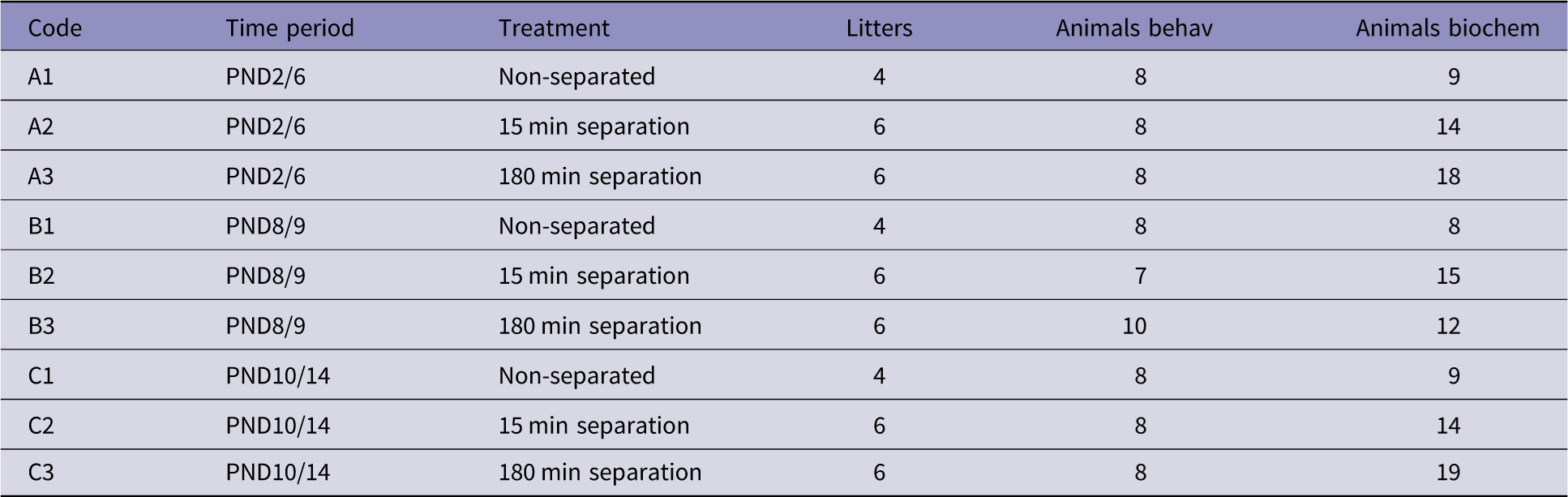
All experiments were performed in accordance with institutional guidelines as well as with EU regulations (EU directive 86/609/EEC). Male and nulliparous female Wistar rats (>4 months old) were mated and, when the females were clearly pregnant, the males were removed from the cage. Female rats were then observed twice daily (08:00 and 17:00 h) for the birth of the litters, which was designated as postnatal day 0. Litters were randomly assigned to one of 9 groups as shown in Table 1. In total 48 litters were used, though animals were also used for other experiments not described here. Control non-deprived litters (A1, B1 and C1) were left undisturbed except the mothers were quickly picked up from the litters and immediately (within 30 s) placed back on the designated days as indicated in Table 1.
Description of the different experimental groups. The number of litters refers to the total number of litters from which the number of animals per experimental group were drawn. PND: postnatal day; Behav: behaviour; Biochem: Biochemistry

Table 1. Long description
A table with 9 rows and 6 columns comparing different experimental groups of animals. The columns are labeled Code, Time period, Treatment, Litters, Animals behav, and Animals biochem. The table details the number of litters and animals used in each experimental group, categorized by different time periods and treatments. Row 1: Code A1, Time period PND2 – 6, Treatment Non-separated, Litters 4, Animals behav 8, Animals biochem 9. Row 2: Code A2, Time period PND2 – 6, Treatment 15 min separation, Litters 6, Animals behav 8, Animals biochem 14. Row 3: Code A3, Time period PND2 – 6, Treatment 180 min separation, Litters 6, Animals behav 8, Animals biochem 18. Row 4: Code B1, Time period PND8 – 9, Treatment Non-separated, Litters 4, Animals behav 8, Animals biochem 8. Row 5: Code B2, Time period PND8 – 9, Treatment 15 min separation, Litters 6, Animals behav 7, Animals biochem 15. Row 6: Code B3, Time period PND8 – 9, Treatment 180 min separation, Litters 6, Animals behav 10, Animals biochem 12. Row 7: Code C1, Time period PND10 – 14, Treatment Non-separated, Litters 4, Animals behav 8, Animals biochem 9. Row 8: Code C2, Time period PND10 – 14, Treatment 15 min separation, Litters 6, Animals behav 8, Animals biochem 14. Row 9: Code C3, Time period PND10 – 14, Treatment 180 min separation, Litters 6, Animals behav 8, Animals biochem 19.
In the ST early stress litters (A2, B2 and C2), the mothers were removed from the litter for 15 minutes on the designated days and placed in a separate cage with clean bedding in the same room as the litters. After 15 minutes the females were placed back.
In the LT early stress litters (A3, B3 and C3), the mothers were removed from the litters for 180 min (on the designated days) and again placed in a separate cage with clean bedding in the same experimental room as the litters. All procedures were started in the morning between 09:00 and 10:00.
After these procedures the litters were left undisturbed except for a weekly change of bedding. On postnatal day 21 ± 2 days the litters were weaned with males and females placed in different cages (in groups of 3 to 4 animals per cage). Only males were used for the present study. All animals were housed in a well-controlled environment (21 ± 1°C, 60 ± 2% humidity) under a normal light/dark regime (lights on from 07:00 to 19:00 h).
Elevated plus maze
On PND70 ± 2 rats were subjected to the EPM. Rats were isolated 24 h before the test. On the day of the test, each rat was placed in the experimental room for at least 30 minutes to adapt to the environment. The procedure was similar to that described before (Olivier et al., Reference Olivier, Van Der Hart, Van Swelm, Dederen, Homberg, Cremers, Deen, Cuppen, Cools and Ellenbroek2008). The EPM, made of black polyvinylchloride, consisted of two open and two closed arms each 50 cm long and 10 cm wide. The closed arms also had 40 cm high walls. The entire maze was positioned 50 cm above the ground in a neutral room without any obvious cues. The room was dimly lit measuring 2.5 Lx in the open and 0.2 Lx in the closed arms. Each rat was placed in the central area between the open and closed arms facing the open arm and allowed to freely explore the maze for 5 minutes. The behaviour was recorded and offline tracked with the video tracking program (developed by Dick Heeren, University of Nijmegen, Holland). Dependent variables were time spent in open and closed arms as well as number of visits to open and closed arms, in addition to total distance moved.
Biochemical analysis
24 hours after the EPM all animals were quickly sacrificed by cervical dislocation and the brain removed and snap-frozen in liquid nitrogen. To ensure that the behaviour had no influence on the brain levels of NPY and NKA, we additionally took brains from animals that had not been subjected to the EPM (see Table 1). NPY and NKA were assessed by radioimmunoassays (RIAs) as previously described in detail (Husum et al., Reference Husum, Vasquez and Mathé2001, Reference Husum, Termeer, Mathé, Bolwig and Ellenbroek2002). In brief, brain tissues were homogenised, ultrasonicated, and extracted twice in 1 M acetic acid and water. Following centrifugation, supernatants were lyophilised and stored at −20°C until analysis. Lyophilised samples were reconstituted and diluted in phosphate buffer before quantification by RIA. NPY-like immunoreactivity (NPY-LI) was measured using an NPY antibody that does not cross-react with other peptides in the pancreatic polypeptide family. NKA-like immunoreactivity (NKA-LI) was measured using a specific NKA antibody. Bolton–Hunter labelled 125I-NPY and 125I-NKA served as tracers. All samples from a given brain region were assayed in the same RIA run to minimise inter-assay variability. Samples were analysed in triplicate, and the mean value was used for statistical analysis.
Statistical analysis
The behavioural and biochemical data were analysed using a one-way ANOVA with factor timing as the independent (between subject) factor, separately for ST and LT separation. When a significant main effect was obtained, LSD post-hoc tests were conducted to identify the source of significance. A p-value of less than 0.05 was considered statistically significant.
Results
Statistical analysis showed that there were no significant differences between the three non-handled groups (A1, B1 and C1 in Table 1) in any of the behavioural or biochemical parameters and hence they were grouped together.
LT separation affects anxiety in a time-dependent manner
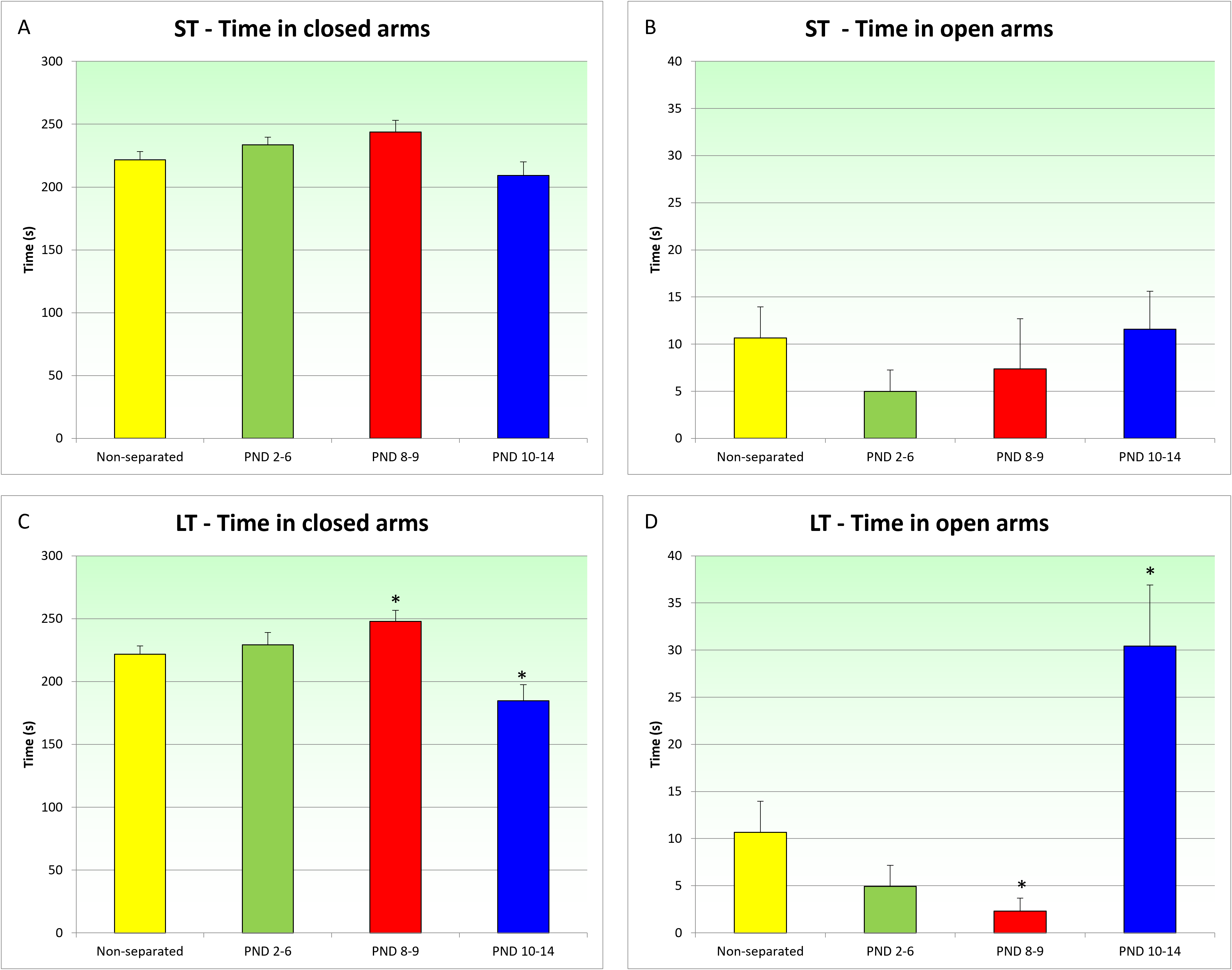
The effects of ST and LT separation on the behaviour in the EPM are displayed in Figure 1. ST separation did not significantly change the time spent in the closed (Figure 1A) or open (Figure 1B) arms (closed arms: F (3,42) = 2.15, p > 0.1; open arms: F (3,42) = 0.46; p > 0.1). Likewise, the total distance travelled was not influenced by early handling (data not shown, F (3,42) = 1.96; p > 0.1).
The effects of maternal separation on behaviour in the elevated plus maze. A: Effects of ST separation on time spent on the closed arms; B: Effects of ST separation on time spent on the open arms; C: Effects of LT separation on time spent on the closed arms; D: Effects of LT separation on time spent on the open arms. Represented are mean + SEM.

Figure 1. Long description
Panel A: A bar graph shows the effects of short-term (ST) separation on time spent in closed arms. The y-axis represents time in seconds, ranging from 0 to 300. The x-axis includes categories: Non-separated, PND 2-6, PND 8-9, and PND 10-14. The Non-separated group shows the highest time, followed by PND 2-6, PND 8-9, and PND 10-14. Panel B: A bar graph shows the effects of ST separation on time spent in open arms. The y-axis represents time in seconds, ranging from 0 to 40. The x-axis includes the same categories. The Non-separated group shows the highest time, followed by PND 10-14, PND 8-9, and PND 2-6. Panel C: A bar graph shows the effects of long-term (LT) separation on time spent in closed arms. The y-axis represents time in seconds, ranging from 0 to 300. The x-axis includes the same categories. The PND 8-9 group shows the highest time, followed by Non-separated, PND 2-6, and PND 10-14. Panel D: A bar graph shows the effects of LT separation on time spent in open arms. The y-axis represents time in seconds, ranging from 0 to 40. The x-axis includes the same categories. The PND 10-14 group shows the highest time, followed by Non-separated, PND 8-9, and PND 2-6.
LT separation, on the other hand, led to a significant effect of timing of separation on time spent in the closed arms (F (3,45) = 3.29; p < 0.03) and a trend towards an effect on time spent in the open arms (F (3,45) = 2.52; p = 0.07). Post-hoc analysis showed significant differences between the control animals and those separated on days 8/9 as well as those separated on days 10/14. Intriguingly, whereas separation on days 8/9 led to an increased time spent on the closed arms and a decreased time spent on the open arms, the reverse was true for separation on days 10/14. In other words, whereas separation on days 8/9 had an anxiogenic effect, separation on days 10/14 had an anxiolytic effect (Figure 1C and 1D).
Maternal separation increases NKA in specific brain regions
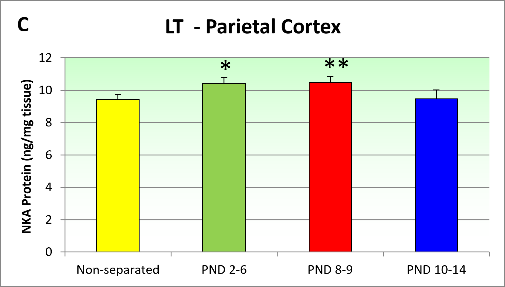
The effects of LT separation on brain levels of NKA are shown in Figure 2. There was a significant effect of LT maternal separation on NKA levels in the neostriatum (F (3,71) = 2.7, p < 0.05), parietal cortex (F (3,71) = 5.5, p < 0.005) and nucleus accumbens (F (3,71) = 3.0, p < 0.05). Post-hoc analysis showed a significant increase in NKA levels in all three areas after separation on days 2/6 and in the parietal cortex and nucleus accumbens also after separation on days 8/9. ST separation only induced a significant change in NKA levels in the hypothalamus (F (3,62) = 3.3, p < 0.05) which was due to an increase in NKA levels after separation on days 8/9 (data not shown).
The effects of LT separation on levels of neurokinin A in the neostriatum (A), nucleus accumbens (B) and parietal cortex (C). Represented are mean + SEM.

Maternal separation has time-dependent effects on NPY brain levels
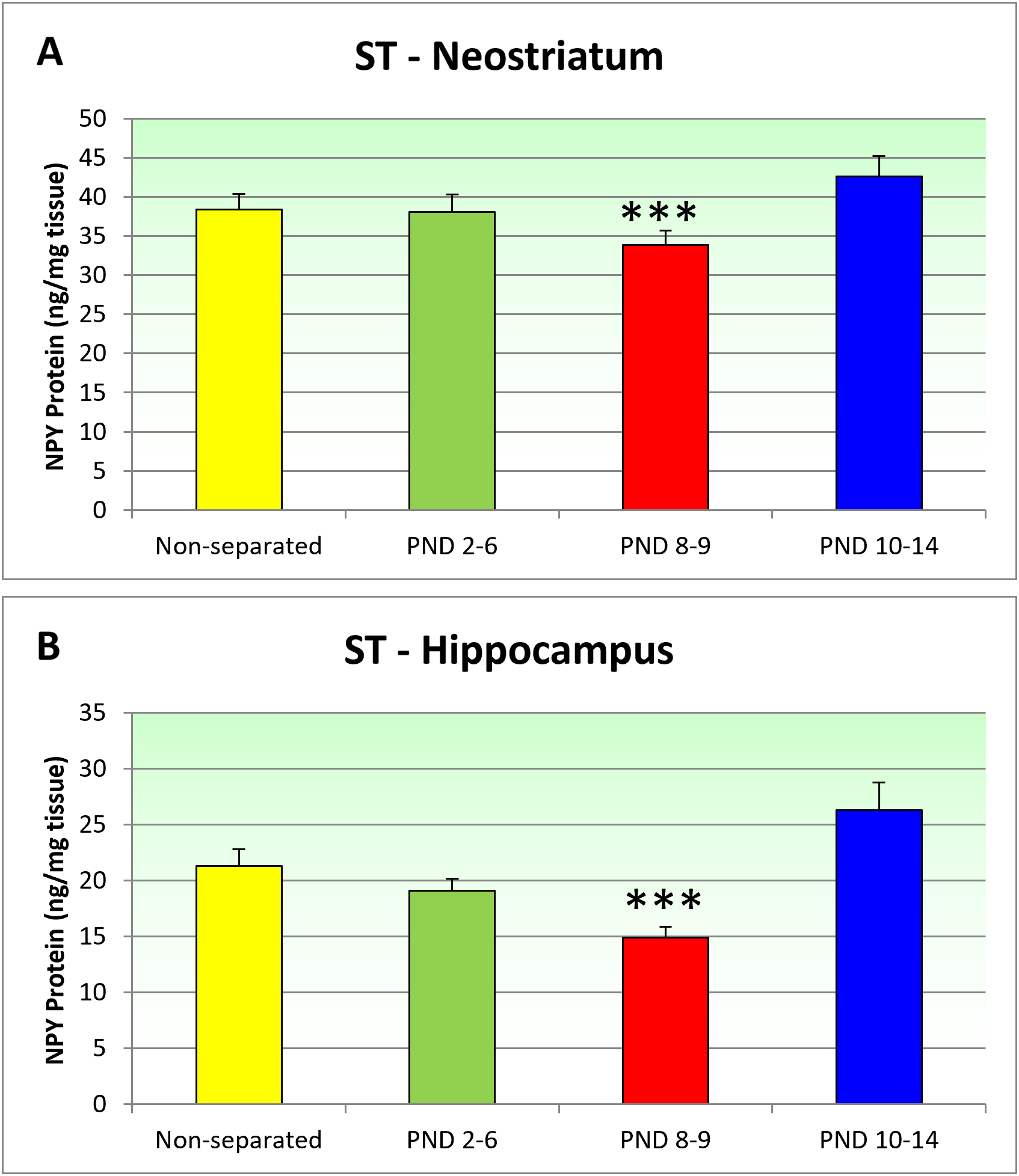
The effects of ST separation on levels of NPY are shown in Figure 3. Statistical analysis showed that ST separation induced significant changes in the neostriatum (F (3,62) = 3.3, p < 0.005) and the hippocampus (F (3,62) = 4.2, p < 0.01). Post-hoc analyses showed a significant reduction in NPY in the neostriatum (Figure 3A) and hippocampus (Figure 3B) after ST separation on PND8-9.
The effects of ST separation on levels of neuropeptide Y in the Neostratium (A) and Hippocampus (B). Represented are mean + SEM.

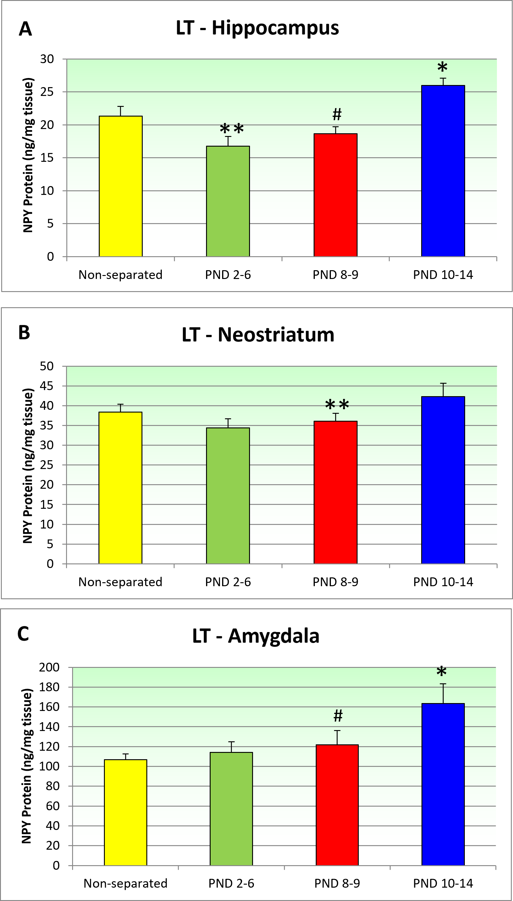
LT separation led to significant changes in the neostriatum (F (3,71) = 2.7, p < 0.05), the hippocampus (F (3,71) = 6.3, p < 0.001) and the amygdala (F (3,62) = 2.9, p < 0.05). Post-hoc analyses showed that in the hippocampus, LT separation led to a significant NPY decrease was seen after separation on days 8/9 while an increase was seen after LT separation on days 10/14 (Figure 4A). Of note, this pattern closely mimics the decrease and increase in time spent in the open arms seen after LT separation on postnatal day 8/9 respectively 10/14. Within the neo striatum (Figure 4B), a small but significant reduction in NPY was seen after LT separation on PND 8–9. Finally, within the amygdala (Figure 4C), a significant increase was seen after LT separation on PND10/14, with a trend (p = 0.09) for an increase after separation on days 8/9.
The effects of LT separation on levels of neuropeptide Y in the hippocampus (A), neostriatum (B) and amygdala (C). Represented are mean + SEM.

Discussion
The development of more effective therapies for the treatment of psychiatric disorders, including anxiety disorders, is hampered by a our lack of understanding of the underlying aetiology and psychopathology. In this study we investigated how early environmental challenges, more specifically ST (15 min) and LT (180 min) maternal separation, influence anxiety-like behaviour as well as affect NKA and NPY in the brain. Clinical studies have increasingly emphasised the critical importance of timing in the LT consequences of pre- and postnatal stressful life events (Kyle & Pichard, Reference Kyle and Pichard2006; Class et al., Reference Class, Abel, Khashan, Rickert, Dalman, Larsson, Hultman, Långström, Lichtenstein and D’Onofrio2014). Therefore, a major aspect of this study was to assess how the timing of the environmental challenge affected behaviour and neuropeptide content.
Timing crucially influences the LT outcome
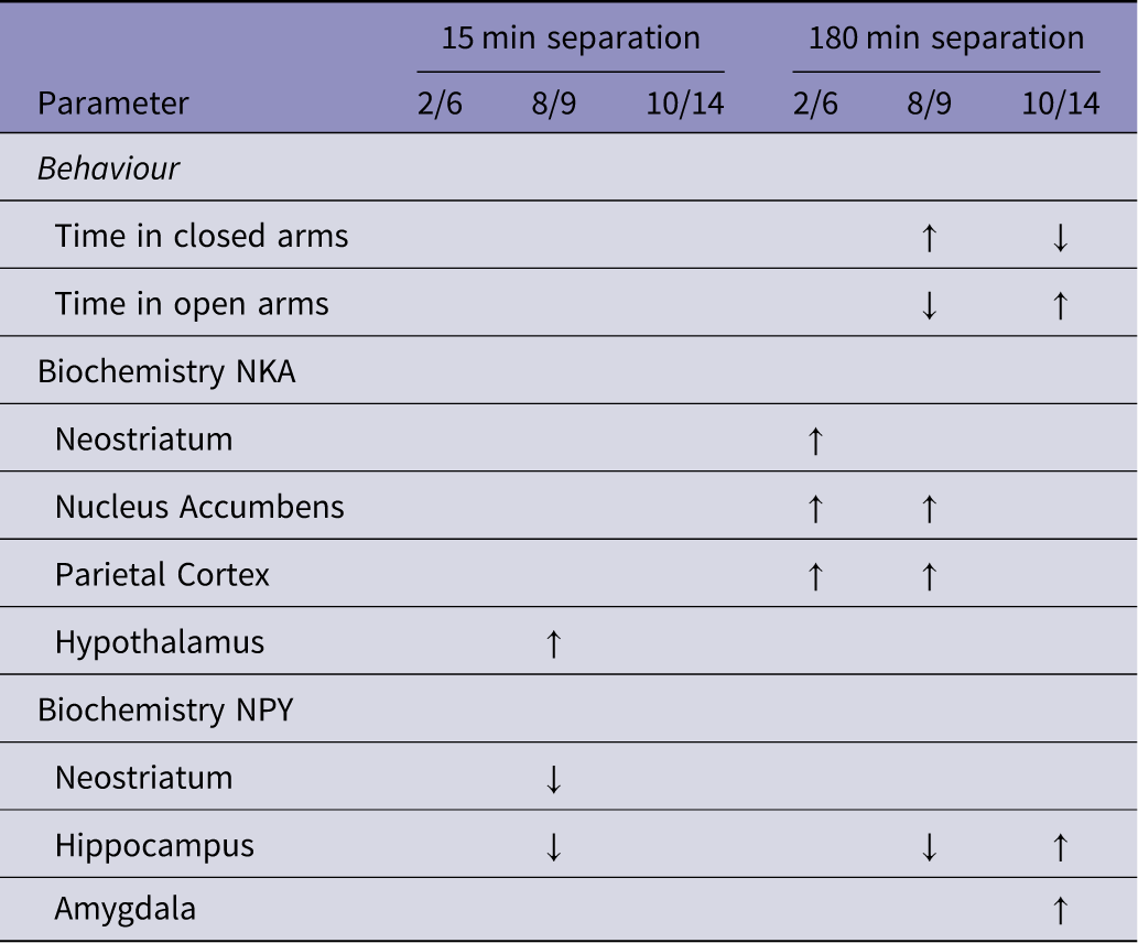
The main aim of the present study was to investigate the hypothesis that the specific time when a stressful life event occurs influences its LT outcome. The data presented support this hypothesis in two different ways. First, if we closely inspect Table 2, we can see that separation on PND8/9 has more effect on neuropeptide content than on either PND2/6 or PND10/14. In fact, for ST separation, changes in NKA and NPY were only found after exposure on PND8/9. Secondly, and more interestingly, the LT consequences of early stressful life events can even be opposite depending on the timing of the event. Thus, while LT separation on PND8/9 led to an increase in anxiety, the same separation led to a decrease in anxiety when applied between PND10 and 14 (see Figure 1). This opposite effect is paralleled by NPY changes in the hippocampus (decrease after PND8/9, increase after PND10/14).
Summary of the results. ↑ indicates a significant increase, while ↓ indicates a significant decrease compared to the non-separated controls. Empty cells mean there was no significant change

Table 2. Long description
A table with four rows and three columns comparing the effects of separation at different postnatal days (2/6, 8/9, 10/14) with 15 min and 180 min separation on behavior and biochemistry. The table has two main categories: Behavior and Biochemistry, with subcategories under Biochemistry: NKA and NPY. The Behavior category includes Time in closed arms and Time in open arms. The Biochemistry NKA category includes Neostriatum, Nucleus Accumbens, Parietal Cortex, and Hypothalamus. The Biochemistry NPY category includes Neostriatum, Hippocampus, and Amygdala. Arrows indicate significant increases or decreases compared to non-separated controls: upward arrow for increase, downward arrow for decrease, and empty cells for no significant change. For 15 min separation on 8/9, there is an increase in Time in closed arms and Neostriatum NKA, and a decrease in Time in open arms and Neostriatum NPY. For 180 min separation on 2/6, there is an increase in Time in closed arms and Hypothalamus NKA, and a decrease in Time in open arms. For 180 min separation on 8/9, there is an increase in Time in closed arms, Neostriatum NKA, Nucleus Accumbens NKA, Parietal Cortex NKA, and Amygdala NPY, and a decrease in Time in open arms, Neostriatum NPY, and Hippocampus NPY. For 180 min separation on 10/14, there is a decrease in Time in closed arms, an increase in Time in open arms, Nucleus Accumbens NKA, Parietal Cortex NKA, Hippocampus NPY, and Amygdala NPY.
These data are in line with clinical and preclinical studies indicating the importance of timing as mentioned in the introduction. Our data do not allow for a detailed discussion about the underlying neurobiological underpinnings, particularly as very little research has systematically investigated the effects of timing of stressful events on behaviour or neurochemistry. Most studies that have investigated 15 or 180 min maternal separation have used an extended (typically between 12 and 20-day) period, rather than for 2 to 5 days as was done in the present study. For example a recent study showed that maternal separation for 4 h/day from PND2 to 20 led to a significant decrease in time spent on the open arms in the EPM accompanied by a trend in reduced numbers of parvalbumin positive cells in the amygdala (Abraham et al., Reference Abraham, Schmerder, Hedtstück, Bösing, Mundorf and Freund2023).
Paradigms using a single very long (24 h) period of separation, on the other hand have provided some evidence that timing can be an important contributor to the LT effects. For example, when this manipulation was performed on PND6 or 9, rats showed a reduction in prepulse inhibition, an effect not seen after similar separations on PND3 or 13 (Ellenbroek & Riva, Reference Ellenbroek and Riva2003). Likewise, 24 h maternal separation had a much stronger effect on the HPA axis when applied on PND11 compared to PND3 (van Oers et al., Reference van Oers, de Kloet and Levine1998). Finally, 24 h separation on PND3 was much more effective in increasing immobility in the forced swim test than the same manipulation on PND 11 (Levine et al., Reference Levine, Huchton, Wiener and Rosenfeld1991). Altogether, these data clearly support the critical importance of timing.
Neurodevelopment is a prolonged process which starts early in gestation but continues well after birth in both humans and rats. Although the majority of neurogenesis occurs prenatally (Bayer et al., Reference Bayer, Altman, Russo and Zhang1993; Zeiss, Reference Zeiss2021), only 15% of granule cells in the dentate gyrus are present at birth in rats (Diamond, Reference Diamond1990). Total brain weight growth is highest around PND7 in rats (Dobbing & Sands, Reference Dobbing and Sands1979; Zeiss, Reference Zeiss2021). Altogether, although many details of brain development are still lacking, there are clear time windows during which specific developmental processes occur (or at least peak) and exposure to stressful events during those developmental vulnerability windows is likely to induce changes that are different from exposure outside of these windows.
Changes in anxiety are associated with opposite changes in NPY brain levels
Our results indicate that changes in anxiety are associated with opposite changes in levels of NPY especially in the hippocampus. Comparing Figures 1 and 4 shows that LT separation on PND8/9 leads to increases in anxiety and decreased hippocampal NPY levels, while LT separation on PND10/14 leads to the opposite: a decrease in anxiety and an increase in hippocampal NPY. This decreased anxiety also leads to increased levels of NPY in the amygdala.
There is substantial preclinical and clinical evidence showing the an inverse relationship between NPY and anxiety (Wu et al., Reference Wu, Feder, Wegener, Bailey, Saxena, Charney and Mathé2011; Thorsell & Mathé, Reference Thorsell and Mathé2017). Thus, plasma and CSF levels of NPY are lower in patients with posttraumatic stress disorder (Rasmusson et al., Reference Rasmusson, Hauger, Morgan, Bremner, Charney and Southwick2000; Sah et al., Reference Sah, Ekhator, Strawn, Sallee, Baker, Horn and Geracioti2009). In line with this, intracerebroventricular injections of NPY produce anxiolytic effects in several paradigms in rats, including Vogel’s conflict test, the EPM, the fear potentiated startle and the light-dark box (Heilig et al., Reference Heilig, Söderpalm, Engel and Widerlöv1989; Pich et al., Reference Pich, Agnati, Zini, Marrama and Carani1993; Broqua et al., Reference Broqua, Wettstein, Rocher, Gauthier-Martin and Junien1995). In addition, there is more specific evidence that hippocampal NPY is involved in anxiety-like behaviour, as both testosterone and nandrolone-induced anxiety is associated with a decrease in hippocampal NPY expression, very similar to what was observed in the present study (Joksimovic et al., Reference Joksimovic, Selakovic, Jovicic, Mitrovic, Mihailovic, Katanic, Milovanovic and Rosic2019). Likewise, 24 h maternal deprivation on PND3 led to increases in anxiety and decreases in NPY immunoreactivity in both the hippocampus and amygdala (Miragaia et al., Reference Miragaia, de Oliveira Wertheimer, Consoli, Cabbia, Longo, Girardi and Suchecki2018), partially replicating our finding, particularly after LT separation on PND10/14.
Thus, our results support the notion that an increase in hippocampal NPY is associated with a decrease in anxiety-like behaviour, while reduced hippocampal NPY levels are associated with increased anxiety-like behaviour.
Changes in anxiety are paralleled by changes in NKA brain levels
We found increases in NKA levels in the nucleus accumbens and the parietal cortex after LT separation on PND8/9, in parallel with increased anxiety. NKA is a tachykinin peptide, related to substance P (SP). While SP predominantly stimulates the neurokinin NK1 receptor, NKA has the highest affinity for the NK2 receptor. Several lines of research have suggested that NKA is involved in anxiety. Thus central administration of NKA in mice induces anxiety, by decreasing the entries into and time spent on the open arms of the EPM (Teixeira et al., Reference Teixeira, Santos, Ribeiro, Calixto, Rae and De Lima1996). Likewise, NKA reverses the anxiolytic effects of diazepam in the EPM in mice (Stratton et al., Reference Stratton, Beresford, Harvey, Turpin, Hagan and Tyers1993). Studies using NK2 antagonists have further confirmed the role of NKA in anxiety (Stratton et al., Reference Stratton, Beresford, Harvey, Turpin, Hagan and Tyers1993; Louis et al., Reference Louis, Stemmelin, Boulay, Bergis, Cohen and Griebel2008). Thus, our data can be considered as supporting the hypothesis that increased NKA is associated with increased anxiety-like behaviour. It should also be recognised that increases in NKA in both the nucleus accumbens and the parietal cortex were found after LT separation on PND2-6, which was not associated with significantly increased anxiety in the EPM. However, close inspection of Figure 1D shows that at PND2-6, there was a tendency for a reduction in time spent on the open arms, even if this did not reach statistical significance. Further studies will be needed to investigate whether increased NKA actually precedes overt changes in anxiety-related behaviour.
The duration of maternal separation is critical for its LT effects
In this study we used both ST (15 min) and LT (180 min) maternal separation. ST separation, often referred to as early handling, has been reported to reduce anxiety in several studies (Ferré et al., Reference Ferré, Núñez, García, Tobeña, Escorihuela and Fernández-Teruel1995; Kiosterakis et al., Reference Kiosterakis, Stamatakis, Diamantopoulou, Fameli and Stylianopoulou2009), although this has not been uniformly found (Durand et al., Reference Durand, Sarrieau, Aguerre, Mormède and Chaouloff1998) and in some cases, even an increased anxiety response has been reported (Jin et al., Reference Jin, Zhao, Jiang, Wang, Li, Zhang, Lian, Du, Sun and Sun2018). Virtually all these studies use daily 15-min handling separation from postnatal day 2 to 14 or even up to postnatal day 21. To the best of our knowledge this study is the first in which handling was restricted to several different (shorter) periods. Overall, we did not find any significant effect on the behaviour of rats in the EPM. Close inspection of Figure 1A and B however suggests that, if anything, ST maternal separation led to an increase in anxiety (i.e. decreased time spent on the open arms) particularly during the early separation times (PND2-6). It is possible that a significant effect could not be seen due to a floor effect, as the controls spent on average only about 10 s on the open arms, so a further (significant) reduction may be hard to obtain. On the other hand, the lack of effect of ST handling on behaviour is paralleled by a paucity of effect on neuropeptide levels. Indeed, early handling only affected NPY and NKA during one specific time point (postnatal day 8 and 9, see Table 2).
LT separation, on the other hand was much more effective in altering behaviour and neuropeptide levels, as evidenced by Table 2. Compared to the effects of ST separation, the literature on LT separation more consistently reports increases in anxiety, particularly in males (Bonapersona et al., Reference Bonapersona, Kentrop, Van Lissa, van der Veen, Joëls and Sarabdjitsingh2019). Again, as with ST separation, LT separation is typically done for an extended period of time, usually from postnatal day 2 to 14. Our results suggest that the increase in anxiety may be specifically related to separation on postnatal day 8 and 9. Several studies investigating anxiety after even longer maternal separation (24 h) on or around postnatal day 9 also reported increased anxiety in several different paradigms, including the EPM (Girardi et al., Reference Girardi, Zanta and Suchecki2014; Benmhammed et al., Reference Benmhammed, El Hayek, Nassiri, Bousalham, Mesfioui, Ouichou and El Hessni2019). On the other hand, a study that compared 24 hour separation on postnatal day 3 and 9 found increased anxiety only after exposure at the earlier time point (53), although the animals were tested at a slightly younger age than in our present study. To the best of our knowledge, no one has systematically investigated the timing of shorter (i.e. 3 hours) maternal separation.
The translational validity of early stressful life events in rats
An important question arising from our research is how our findings can be translated to the human situation. The early development of rats and humans is very different, making a direct comparison between the trajectories of both species difficult. It is generally accepted that, compared to humans, rats are born prematurely (Workman et al., Reference Workman, Charvet, Clancy, Darlington and Finlay2013; Zeiss, Reference Zeiss2021). This is reflected in the so-called precocial score (an index of how active and mobile an animal is at birth) which is 0.65 for humans but only 0.45 for rats. In an attempt to match the developmental timelines between humans and other animals, Workman and colleagues (2013) analysed 271 developmental events for 18 different species (for more details see www.translatingtime.org). While there are differences in the timelines between the individual developmental events, overall the analysis suggests that PND2/6 in rats corresponds roughly to months 5–7 of pregnancy in humans, PND8/9 to birth and PND10/14 to the first 3 to 5 months after birth. Our findings that the effects of stressors were most severe after exposure on PND8/9 is in line with findings from multiple studies that perinatal stressors and obstetric complications (such as protracted birth, preeclampsia, breech position etc.) are significant risk factors for multiple psychiatric disorders including schizophrenia (Clarke et al., Reference Clarke, Harley and Cannon2006), ASDs (Guinchat et al., Reference Guinchat, Thorsen, Laurent, Cans, Bodeau and Cohen2012) and major depression (Hompoth et al., Reference Hompoth, Pető, Fűrészné Balogh and Töreki2020).
Summary
In summary, we found that early stressful life events (ST or LT maternal separation) substantially affect anxiety-like behaviour as well as NPY and NKA levels in the brain. The effects were dependent on the intensity of the stressor, with LT stressors having significantly more effect on brain and behaviour than ST stressors. Most importantly, the LT consequences were critically determined by the timing of the stressors, with exposure at PND8/9 having a more substantial effect in adulthood than earlier or later exposure. Finally, we found that exposing rats on PND8/9 to LT maternal separation induced opposite effects to the same exposure on PND10/14. This was seen both in terms of behaviour as well as in NPY levels in the hippocampus. Taken together, the results add to the growing body of research showing the importance of timing in determining the LT consequences of early life stressors and emphasise the importance of further research into the neurobiological networks underlying these differential effects.
Acknowledgements
The authors would like to thank Teun de Vries and Luuk Lubbers for their invaluable contribution to the study.
Author contributions
BE was responsible for designing the behavioural experiments, collecting the behavioural data, data analysis, data interpretation and he took the lead in writing the manuscript.
AM was responsible for designing the in vitro experiments, collecting the neuropeptide data, data interpretation and co-writing and reviewing the manuscript.
Funding statement
This study was performed without any external funding.
Competing interests
Both authors declare that they have no conflict of interest.

 Open access

