Introduction
Congenital nasolacrimal duct obstruction is a common cause of epiphora in the paediatric population, with a reported prevalence of 1.25–12.5 per cent.Reference Paul and Shepherd1 Epiphora can lead to secondary eczematisation and impetiginisation of the eyelid. Congenital nasolacrimal duct obstruction is caused by a failure of nasolacrimal duct canalisation. The obstruction is usually present at the level of Hasner's valve, at the distal end of the duct.Reference Schnall2 In some cases, the obstruction is caused by a plug of epithelial cell debris.Reference Schnall2 This obstruction can result in conjunctivitis and dacryocystitis in some patients.
Fortunately, spontaneous resolution occurs in most cases by one year of age.Reference Ghuman, Gonzales and Mazow3 The reported rate of congenital nasolacrimal duct obstruction resolution without surgery ranges from 32 per cent to 95 per cent by 13 months of age.Reference Petersen and Robb4 For children younger than six months, non-surgical treatment is usually preferred, including antibiotic eye drops and massage of the lacrimal sac. Massage of the lacrimal sac and administration of topical antibiotics is considered the standard first-line treatment, with a cure rate greater than 90 per cent.Reference Nelson, Calhoun and Menduke5 When epiphora continues in infants aged around one year, early nasolacrimal duct probing yields good results.Reference Nelson, Calhoun and Menduke5
Two opinions exist regarding the management of congenital nasolacrimal duct obstruction in patients aged around one year or older. Some ophthalmologists report that the success rate of nasolacrimal duct probing is significantly reduced after one year of age.Reference Mannor, Rose, Frimpong-Ansah and Ezra6 Therefore, some ophthalmologists prefer to perform silicone intubation as the initial surgical procedure for older children.Reference Kushner7 Others claim that increasing age has no significant effect on the success rate of probing in older children.Reference Cifici, Akman, Sonmez, Unal, Güngör and Yaylali8 In the literature, success rates of 78–97 per cent have been reported for lacrimal probingReference Repka, Chandler, Beck, Crouch and Donahue9 and 79–96 per cent for silicone intubation.Reference Engel, Hichie-Schmidt, Khammar, Ostfeld, Vyas and Ticho10,Reference Tao, Meyer, Simon and Zobal-Ratner11
However, lacrimal probing and silicone intubation have complications, the most common of which are false passage formation and traumatic obstruction.Reference Wobig12 In addition, when these techniques are performed without an endoscope, it sometimes causes trauma to the nasal structures. Intubation adds other complications, including tubing breakage, corneal irritation, tube extrusion, punctum erosion or slitting, and pyogenic granuloma formation.
An increasing number of studies have been published in more recent years that highlight the value of using a nasal endoscope during the probing procedure.Reference Sener and Onerci13,Reference Cakmak, Yildirim, Sakalar, Keklikci and Alakus14 Despite these precautions, it is still an invasive procedure. Other techniques such as balloon catheter dilation and dacryocystorhinostomy procedures are more complicated, invasive and controversial in first intention.Reference Gioacchini, Alicandri-Ciufelli, Kaleci and Re15 A good surgical approach is a technique with a low rate of complications and a high rate of success.
This paper presents a new technique to treat congenital nasolacrimal duct obstruction: incision of Hasner's valve under endoscopic intranasal surgery. We evaluate the effectiveness and safety of this procedure for the management of congenital nasolacrimal duct obstruction.
Materials and methods
We retrospectively reviewed the medical records of patients with congenital nasolacrimal duct obstruction who underwent incision of Hasner's valve under endoscopic intranasal surgery, between April 2000 and October 2016, at the Centre Hospitalier National d'Ophtalmologie des Quinze-Vingts, Paris, France.
At the initial visit, clinical signs of congenital nasolacrimal duct obstruction (epiphora, mucous discharge and increased tear lake) were assessed by the surgeon, and reported as either present or absent. The diagnosis of congenital nasolacrimal duct obstruction was based on symptoms of epiphora or recurrent mucopurulent discharge from birth, reflux from the lacrimal sac with pressure, increased tear lake size, tear pooling, and eyelid crusting, and confirmed by a delayed dye disappearance test. All patients were examined with their parents present. Parents were asked questions related to a medical history of failed probing, conjunctivitis, dacryocystitis, and primary and secondary treatments.
Children with congenital nasolacrimal duct obstruction and a history of failed probing (secondary treatment) were included in this study. Patients with persistent epiphora who visited our hospital for a second opinion and who had undergone previous probing of the nasolacrimal duct in another centre were also included. The inclusion criteria were: children aged 11 months to 10 years, surgical indication for Hasner's valve incision, surgery performed under general anaesthesia, and informed consent from parents. The exclusion criteria were: children with Down syndrome, punctual or canalicular anomalies, previous dacryocystorhinostomy, craniofacial abnormality, loss to follow up after surgery or less than six months of follow up after Hasner's valve incision, and history of trauma to the nasolacrimal system secondary to non-iatrogenic facial injury. Although a history of failed probing can be considered trauma to the nasolacrimal system, patients with such history were included only if a surgical report of the probing procedure was available.
Different surgical options were explained to parents, and informed consent was obtained. All surgical procedures were carried out by a single oculoplastic team consisting of two surgeons at Quinze-Vingts National Center in Paris, France.
All patients had experienced failure of previous conservative therapies, which included antibiotic eye drops, lacrimal sac massage and office-based nasolacrimal duct probing under topical anaesthesia. The collected data included patient demographics, diagnosis necessitating Hasner's valve incision, Hasner's valve anatomy, surgical success, and any complications encountered and their treatment.
Surgical technique
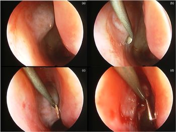
Surgery was performed under general anaesthesia. Visualisation of the nasal cavity was performed using a 2.7 mm, 30 degree, rigid Hopkins endoscope linked to a camera. Prior to visual inspection with the endoscope, cotton strips soaked with 5 per cent xylocaine naphazoline were placed medial and lateral to the inferior turbinate, where they were kept for 5–10 minutes, to ensure adequate vasoconstriction before being removed. The inferior turbinate (Figure 1) was medially displaced with a freer elevator so that the inferior meatus could be visualised. The inferior and superior canaliculi were irrigated separately with diluted fluorescein dye (permeability test) to carefully examine the Hasner's valve region. Patients were examined for anatomical abnormalities, such as synechia, cysts, follicles, imperforation and mucoceles. Probing of the nasolacrimal duct was performed under endoscopic examination. For all patients, the probing without perforating of Hasner's valve was performed prior to the incision of Hasner's valve. Finally, Hasner's valve mucosa was incised using an otorhinolaryngological lancet knife (Figure 1b–d). No tube was left in the nasolacrimal duct. Patients were prescribed post-operative antibiotic eye drops for one week and nasal washing with physiological saline for three weeks.
(a) Endoscopic visualisation of an imperforated Hasner's valve. The inferior turbinate had been displaced medially to enable visualisation of the inferior meatus and Hasner's valve region. (b–d) After probing of the nasolacrimal duct under endoscopic examination, Hasner's valve mucosa was incised with an otorhinolaryngological lancet knife.

We graded the outcome as ‘successful’ if there were no residual symptoms of nasolacrimal duct obstruction and if patients showed normal dye disappearance test results six months after surgery. We graded the result as ‘partially successful’ if the patient had symptoms of intermittent epiphora and abnormal examination results. We graded the results as ‘unchanged’ if the symptoms and examination findings were unchanged from presentation. Finally, the results were graded as ‘worse’ if the symptoms and examination revealed a complication, or if epiphora progressed to recurrent mucopurulent discharge.
The primary endpoint was the functional success rate of this procedure in all patients, as well as in those patients with no medical history of probing prior to surgery. The secondary endpoints were: Hasner's valve and inferior turbinate anatomy findings, demographic data, complication rates, and complication types.
In addition, we compared demographic data, surgical duration and Hasner's valve anatomy between the ‘successful’, ‘partially successful’, ‘unchanged’ and ‘worse’ outcome groups.
The study followed the tenants of the Declaration of Helsinki and was approved by the affiliated hospital's institutional review board. The ethics committee of the Quinze-Vingts National Center oversaw and approved this study.
Data were obtained from our computerised database. The study variables were organised using Microsoft Excel 2010® spreadsheet software. Data were statistically analysed using XLSTAT® software.
Results
A total of 484 patients were included. Forty-six patients (9.5 per cent) were excluded because of loss to follow up (Figure 2), as they did not attend the post-operative consultation. However, we managed to telephone the parents of these patients two months after surgery. All patients had neither persistent epiphora nor any complications (successful outcome). Thus, they did not return to the hospital. Among the patients who were included in the study, 45 per cent had no history of probing. Analysis of the patients’ characteristics (Table 1) showed that the mean age (± standard deviation (SD)) was 2.56 ± 1.48 years. In addition, comparison of the included patient group and the excluded patient group showed no significant differences (Table 1). The mean surgical duration was similar between the two groups.
Flow chart of study participants.

Characteristics of study population

SD = standard deviation
Concerning the primary endpoint of the study, the treatment in patients with no medical history of probing was: successful in 91 per cent, partially successful in 5 per cent, unchanged in 3.9 per cent, and worse in 0.1 per cent (Table 2). For all patients, this procedure included probing together with an endoscopic Hasner's valve incision and medicalisation of the inferior turbinate.
Primary outcome

Data represent rounded percentages (they do not always add up to 100)
The result was more successful in patients without a medical history of probing. In fact, the functional success rate of this procedure was reduced to 88.6 per cent when patients with a medical history of probing were included (Table 2). In these patients, the treatment was partially successful in 5.37 per cent, unchanged in 5.37 per cent and worse in 0.62 per cent (Table 2). Among patients with a history of probing, we identified two causes of failure at the time of the endoscopy. Firstly, the lower turbinate was found to be pressed against the lateral wall of the nasal mucosa in 78 per cent of cases, closing the lower meatus and compressing Hasner's valve. The second cause was a lack of opening of the nasal mucosa to the terminal part of the nasolacrimal system, which was found in 22 per cent of cases. Indeed, despite probing, the orifice of Hasner's valve was too small to allow the passage of dye during the permeability test. The simple incision of Hasner's valve was sufficient to restore the permeability.
The outcome was more successful in younger patients (Table 3). The mean age (± SD) of patients in the ‘successful’ group was 2.4 ± 0.4 years, versus 3.9 ± 1.8 years in the ‘unchanged’ group and 5.3 ± 1.5 years in the ‘worse’ group.
Secondary outcome: mean age comparison in each outcome group

SD = standard deviation
We performed repeat surgery on five patients in whom the Hasner's valve incision procedure was ineffective. Repeat surgery was performed using a Ritleng® bicanalicular intubation system. The results were successful for two patients, partially successful for one patient and unchanged for two patients. Figure 3 shows the procedures undertaken for each of the children in the study.
Chart showing the procedures undertaken in the children. IT = inferior turbinate; NLD = nasolacrimal duct; HV = Hasner's valve

Nasolacrimal irrigation at the beginning of surgery showed an impaired flow in 63 per cent of cases and free flow in 37 per cent of cases. Endoscopic peri-operative investigation revealed three types of anomalies causing congenital nasolacrimal duct obstruction. The first was imperforation of Hasner's valve, observed in 45 per cent of cases, in which a lack of openness of the nasal mucosa at the terminal part of the lacrimal system was found. Nasolacrimal irrigation was unsuccessful in these cases. All patients who had never previously undergone probing had Hasner's valve imperforation. The second cause was a pseudocyst of Hasner's valve, found in 35 per cent of cases with unsuccessful irrigation. This was evidenced by dilation of the nasal mucosa, and most cases presented a wall that was thick and resistant to simple incision. The final cause was anatomical abnormality of the inferior turbinate, found in 20 per cent of cases. In these cases, the inferior turbinate was easily displaced medially with a freer elevator and the inferior meatus was visualised. For these patients, nasolacrimal irrigation was successful but with a low flow.
Three types of anatomical anomalies were found on the inferior turbinate. The first was the finding that the inferior turbinate was clad against the lateral wall of the nasal mucosa, closing the inferior meatus and thus compressing Hasner's valve. This anomaly was found in 70 per cent of cases. In these cases, nasolacrimal irrigation remained permeable. The second abnormality was hypertrophic inferior turbinate, found in 15 per cent of cases. Finally, 15 per cent of cases had a Hasner's valve with follicular and inflammatory mucosa in the lower part, inducing an allergy.
From these findings, two entities of watery eyes were highlighted: watery eyes with a permeable nasolacrimal duct, usually due to an abnormal size or position of the inferior turbinate and an abnormal anatomical position of Hasner's valve, or due to follicular and inflammatory mucosa; and watery eyes with congenital nasolacrimal duct obstruction, caused by an imperforated or pseudocystic Hasner's valve, which was easily treated by a simple incision.
Tolerance and complications
The Hasner's valve incision procedure was well tolerated. Only one patient had a post-operative infection. All patients underwent general anaesthesia, and no complications related to general anaesthesia were observed. The mean (± SD) surgical duration was 13.1 ± 5.7 minutes.
Discussion
Here, we showed that incision of Hasner's valve under nasal endoscopic surgery had favourable results as a secondary treatment for persistent congenital nasolacrimal duct obstruction. To our knowledge, this study is the first to report a 91 per cent success rate with such a large cohort (484 eyes). This success rate is similar to the only previous study reporting the effectiveness of Hasner's valve incision for congenital nasolacrimal duct obstruction management; however, that study was conducted on a small cohort (55 eyes).Reference Korkmaz, Korkmaz, Karakahya and Serhatli16
Efficacy and safety
The results of this procedure are within the upper range of success achieved by other operations such as first nasolacrimal duct probing,Reference Le Garrec, Abadie-Koebele, Parienti, Molgat, Degoumois and Mouriaux17 silicone intubationReference Andalib, Gharabaghi, Nabai and Abbaszadeh18 and balloon catheter dilation.Reference Chen and Hsiao19 When we compare our success rates to those of other procedures, we can conclude that Hasner's valve incision under endoscopic intervention is a highly effective procedure.
Taking into account the safety and complication rates of all of these procedures, incision of Hasner's valve under endoscopic intranasal surgery appears to be an appropriate alternative. This study showed the safety of this procedure. Of the 484 patients who underwent Hasner's valve incision, only 1 (0.2 per cent) suffered an infection with dacryocystitis. None of the children complained of post-operative issues such as nasolacrimal system trauma, punctum laceration, corneal irritation or abrasion, and pyogenic granuloma. In addition, this procedure has no complications regarding the cornea, whereas canalicular intubation can cause corneal abrasions or ulcers in around 1.5 per cent of patients.Reference Fayet, Hurbli, Renard, Ruban, Racy and Bernard20 Incision of Hasner's valve achieves high success rates, while maintaining high safety and low complication rates. It avoids unnecessary manipulation of the canaliculi and nasolacrimal duct. Other procedures such as intubation and balloon catheter dilation appear to be more traumatic, with risks of nasolacrimal duct sequelae.
One of the major advantages of Hasner's valve incision is that no medical device is left in the nasolacrimal duct. Hence, Hasner's valve incision does not necessitate a post-operative consultation for implant removal, unlike procedures involving silicone tubes, which require a second procedure for tube removal that is cumbersome for both the child and their parents. We believe that combining nasolacrimal intubation and Hasner's valve incision is ineffective, as the result is similar with or without a silicone tube. Based on our personal experience, we recommend that intubation and balloon catheterisation are only performed in children aged over four years for the following indications: upper nasolacrimal duct fibrosis obstruction, Down syndrome and craniofacial malformations.
A key advantage of the Hasner's valve incision technique is the lack of need for clinical examination in an office setting or for follow up after surgery. In our experience, telephoning the patient's parents to ensure there are no abnormalities is sufficient during the first month of post-operative follow up. Most of our patients who were lost to follow up after surgery did not return to the office because the treatment yielded a successful result.
In addition, the surgical duration for this procedure is very short, with a mean (± SD) operation time of 13.1 ± 5.7 minutes. Thus, time under general anaesthesia was also shortened, which decreases the associated risks and complications. No studies have investigated the duration of surgery for probing or canalicular intubation. However, these procedures are not reproducible, thus the duration can never be the same.
Cost-effectiveness analysis
This procedure is cost effective. Both Hasner's valve incision and intubation or balloon catheter dilation are performed in a surgical facility under general anaesthesia. The tubes currently cost the facility between $60 and $100, and the balloon catheter costs the facility $306 for unilateral and $555 for bilateral dilations. In contrast, Hasner's valve incision only requires an otorhinolaryngological lancet knife, which costs the facility between $2 and $10, assuming the facility already has an endoscope (cost of around $5000).
Anatomical findings
Epiphora with a permeable nasolacrimal duct is less well known compared to epiphora with an imperforated Hasner's valve. We believe that this kind of epiphora is a multifactorial phenomenon caused by the presence of inflammatory and follicular mucosa around Hasner's valve, and the abnormal size and position of the inferior turbinate, which is large and attached against the lateral wall of the nasal mucosa.
In our study, follicular mucosa was seen in 15 per cent of cases. In patients with follicular mucosa around Hasner's valve, the inferior turbinate was found to be clad against the lateral wall. In this specific situation, we suspect that the inferior turbinate induces inflammation of Hasner's valve mucosa, which becomes follicular. Allergic rhinitis could be another cause of follicular Hasner's valve mucosa, as 40 per cent of patients with follicular Hasner's valve mucosa had a medical history of allergic rhinitis. Only a few epidemiological studies have reported an association between epiphora and allergic rhinitis.Reference Ohtomo, Ueta, Toyama and Nagahara21,Reference Nemet and Vinker22 Among the children in our study with no history of probing, eight had an unchanged result. All of them had follicular Hasner's valve mucosa (Figure 4) with a medical history of allergic rhinitis. Of these eight patients, five underwent repeat surgery with a Ritleng bicanalicular intubation system (Figure 3). We believe that only this kind of abnormality requires intubation.
Hasner's valve with follicular and inflammatory mucosa.

In a small number of patients, Hasner's valve was abnormally located at the angle formed by the inferior turbinate emergence and the lateral wall. In these cases, incision of Hasner's valve was not effective. This kind of anatomical localisation has not been described.
We believe that displacement of the inferior turbinate is mandatory but not sufficient for the treatment of congenital nasolacrimal duct obstruction. In fact, in cases with an imperforated Hasner's valve, canaliculi irrigation with diluted fluorescein dye showed no permeability of the Hasner's valve region, despite inferior turbinate displacement. Furthermore, many cases with a perforated Hasner's valve and inferior turbinate displacement had impaired flow when the nasolacrimal duct was irrigated. After Hasner's valve incision, we observed free flow after nasolacrimal duct irrigation for all these patients.
In the literature, the efficiency of inferior turbinate fracture for congenital nasolacrimal duct obstruction is controversial. Attarzadeh et al.,Reference Attarzadeh, Sajjadi, Owji, Reza Talebnejad, Farvardin and Attarzadeh23 Katowitz and Welsh,Reference Katowitz and Welsh24 and Rajabi et al.Reference Rajabi, Inanloo, Salabati, Rafizadeh, Tabatabaie and Bayat25 found similar success rates for probing in congenital nasolacrimal duct obstruction cases with or without inferior turbinate displacement. In contrast, Havins and Wilkins,Reference Havins and Wilkins26 Jones and Wobrig,Reference Jones and Wobrig27 and WesleyReference Wesley28 found better success rates when probing was combined with inferior turbinate fracture. Based on our experience, we recommend that both procedures always be used to treat congenital nasolacrimal duct obstruction, with Hasner's valve incision performed after inferior turbinate displacement.
During the surgery, the nasolacrimal duct was probed under endoscopic visualisation without perforating Hasner's valve before the incision. Thus, bias is reduced, and it is possible to attribute the success of the procedure to the Hasner's valve incision and not to probing.
Success rate in older children
Our study indicates that Hasner's valve incision is an effective procedure for children aged younger than three years, as children older than three years had a low rate of success. It is possible that the success of Hasner's valve incision is dictated not only by the age at surgical procedure, but also by the distribution of more severe obstructions in older age groups. These findings are similar to those of studies on nasolacrimal duct probing in older children. In those studies, the success rate decreased as age increased, especially in patients aged over two years.Reference Kushner7,Reference Kashkouli, Kassaee and Tabatabaee29 Older children are more likely to have complicated, non-membranous obstructions, which might explain the reduced success rates observed in those studies.Reference Kushner7
• Hasner's valve incision under endoscopy is an effective primary surgical treatment for congenital nasolacrimal duct obstruction
• Hasner's valve incision is more effective in children younger than three years
• Hasner's valve incision is high in safety and has low complication rates
There are two schools of thought regarding nasolacrimal duct probing management in older children. Katowitz and Welsh,Reference Katowitz and Welsh24 and BakerReference Baker30 suggest that probing failure might be the result of chronic infection and fibrosis that increases with age. Alternatively, Paul and ShepherdReference Paul and Shepherd1 propose that the apparent decrease in success for older children is the result of an accumulation of more severe obstructions after birth. We believe that there is a greater distribution of more severe obstructions in older age groups.
Conclusion
This study indicates that incising Hasner's valve after medially displacing the inferior turbinate under nasal endoscopy is an effective and adequate technique in the primary surgical treatment of congenital nasolacrimal duct obstruction. We conducted a non-comparative, non-randomised, non-interventional retrospective study, which represents a limitation. An interventional study is needed to confirm our findings.
Acknowledgement
We would like to acknowledge H. Keilani.
Competing interests
None declared