INTRODUCTION
Syllidae Grube, 1850 is one of the most complex families of polychaetes, with a high number of species and genera but easily recognizable because of its synapomorphy, the proventricle (Glasby, Reference Glasby, Beesley, Ross and Glasby2000; Pleijel, Reference Pleijel, Rouse and Pleijel2001; Aguado et al., Reference Aguado, San Martín and Siddall2012). The family is currently divided into five subfamilies: Syllinae Grube, 1850; Exogoninae Langerhans, 1879; Eusyllinae Malaquin, 1893; Autolytinae Langerhans, 1879, and Anoplosyllinae Aguado and San Martín, 2009, plus a number of Incertae Sedis genera (Aguado et al., Reference Aguado, San Martín and Siddall2012; San Martín & Aguado, Reference San Martín, Aguado and Schmidt- Rhaesa2014). The autolytines constitute a well-delineated group both in morphological, reproductive and molecular terms (Nygren, Reference Nygren2004; Nygren & Pleijel, Reference Nygren and Pleijel2010). They are characterized by their small size (from a few mm to several cm in length), lack of ventral cirri (probably blending to parapodial lobes), a slender and sinuous pharynx, provided with a trepan more or less developed without a central pharyngeal tooth (Nygren & Pleijel, Reference Nygren and Pleijel2010; Nygren et al., Reference Nygren, Sundkkvist, Mikac and Pleijel2010), the presence of a simple dorsal bayonet type chaeta (San Martín, Reference San Martín and Ramos2003; Nygren, Reference Nygren2004; Nygren & Pleijel, Reference Nygren and Pleijel2010) and its reproduction by schizogamy (with some exceptions). The subfamily is distributed worldwide inhabiting shallow waters, restricted to the shelf areas around the continents (Nygren & Pleijel, Reference Nygren and Pleijel2010), with some exceptions. They are mainly associated to calcareous algae, hydroids, bryozoans, gorgonians, balanids and other fouling organisms, which they are supposed to feed on (Martín & Alós, Reference Martín and Alós1987; San Martín, Reference Ronquist and Huelsenbeck2003; Nygren, Reference Nygren2004; Nygren & Pleijel, Reference Nygren and Pleijel2007, Reference Nygren and Pleijel2010; Nygren et al., Reference Nygren, Sundkkvist, Mikac and Pleijel2010).
The subfamily was extensively reviewed by Nygren (Reference Nygren2004) who redefined the genera Myrianida Milne Edwards, 1845, and Proceraea Ehlers, 1864, erecting several new genera, and described a number of new species. Since this revision, several new species and records were described worldwide (e.g. Nygren & Pleijel, Reference Nygren and Pleijel2007; Nygren et al., Reference Nygren, Sundkkvist, Mikac and Pleijel2010; Álvarez-Campos et al., Reference Álvarez-Campos, San Martín and Piotrowski2014).
The genus Myrianida is characterized by completely fused palps, cylindrical or flattened appendages, one or two ciliary bands on each segment, the presence of nuchal epaulettes, compound chaetae with bidentate blades, proximal tooth typically larger than the distal one, thin bayonet-shaped dorsal simple chaetae, lack of ventral simple chaetae, slender pharynx with single or multiple sinuations, trepan with several equal or unequal teeth and reproduction by gemmiparous schizogamy, although scissiparity in some species. Currently, the genus is considered monophyletic (Nygren, Reference Nygren2004).
The genus Proceraea is characterized by its colour pattern, essential for the correct identification, short dorsal cirri, the presence of nuchal epaulettes, a single thick bayonet chaetae, sometimes distally denticulated, or a single thin bayonet chaetae, a pharynx with single or multiple sinuations, a trepan with teeth arranged in 2 rings and a reproduction by scissiparous schizogamy (see Nygren, Reference Nygren2004 for further diagnostic characters). According to previous studies (e.g. Nygren & Sundberg, Reference Nygren and Sundberg2003; Nygren et al., Reference Nygren, Sundkkvist, Mikac and Pleijel2010; Álvarez-Campos et al., Reference Álvarez-Campos, San Martín and Piotrowski2014) and our own results (see Figure 6), the genus Proceraea seems to be paraphyletic, since the genera Procerastea, Imajimaea, Erseia and Virchowia appeared nested within it, but further studies are needed including more species of each genera.
During different surveys in the Peruvian coasts, several specimens of the genera Myrianida and Proceraea were collected. A new species (Myrianida paredesi sp. nov.) and two new records for Peru (Myrianida pentadentata and Proceraea micropedata) were found. A phylogenetic analysis using molecular data of the new species is also provided, in order to establish its relationships within the genus Myrianida.
MATERIALS AND METHODS
The examined material of Myrianida paredesi, sp. nov. and Proceraea micropedata (Hartmann-Schröder, 1962) was collected as part of the Biodiversity Monitoring and Assessment Program implemented by the Center for Conservation, Education and Sustainability (CCES) of the Smithsonian Conservation Biology Institute and the company PERU LNG S.R.L. Samples were obtained from pillars at the jetty of the Pampa Melchorita Plant of PERU LNG S.R.L. (Southern Lima), during 2011, at 5–10 m depth. The specimens were collected using a PVC quadrat (0.06 m2), a knife and a spatula. The specimens were removed from barnacles and other organisms, and fixed in formalin 10%. Additionally, some more specimens of M. paredesi sp. nov. were collected by the Invertebrates Laboratory of Instituto del Mar del Perú (IMARPE). Myrianida pentadentata specimens were sampled as part of the project ‘Diversity, distribution and conservation of the molluscs from tropical sea from Peru’ developed by the Biology and Systematics lab of Marine Invertebrates (LaBSIM), Universidad Nacional Mayor de San Marcos (UNMSM), Peru. Specimens were examined in the Laboratory of Marine Biology of the University Autónoma of Madrid (Spain), during a research stay of the first author, under a Nikon Optiphot microscope with a differential interference contrast system (Nomarsky), an ocular micrometer, and camera lucida drawing tube. Structures with taxonomic relevance were dissected and drawn. Length and width (at the level of the proventricle, excluding parapodia) of specimens were measured using the software (OCView Ver.2009.6.1.3.0) of a digital camera (OptixCam Summit Series OCS-9.0) attached to the optical microscope.
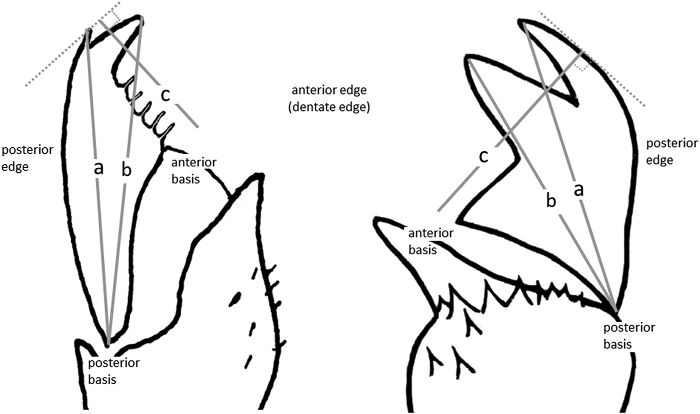
For the length of the chaetae blades, three measures were considered according to Figure 1, it was considered more of a measurement of the three blade lengths, then averaged to obtain a single value for each length (a, b and c). This was repeated in each region of the body (anterior, middle and posterior).
Measuring lengths scheme blade of compound chaetae. (a) posterior basis – distal tooth; (b) posterior basis – proximal tooth; (c) anterior basis – blade ridge (parallel to the dentate edge).

For scanning electron microscopy (SEM) examination, the specimens were critical point dried with an Emitech K850 Critical Point Dryer, gold-coated with a Q150 T-S Turbo–Pumper Sputter Coater, and examined with a Hitachi S-3000N electron microscope at SIDI (Servicio Interdepartamental de Investigación), Universidad Autónoma de Madrid (UAM).
The holotype of the new species is deposited at the scientific collection of the Biology and Systematics lab of Marine Invertebrates (LaBSIM), UNMSM; some paratypes and some specimens of the other two species, including SEM ones, were deposited at the Museo Nacional de Ciencias Naturales de Madrid (MNCN), Spain. The voucher of the specimen used for the molecular analysis was deposited in the Museum of Comparative Zoology at Harvard (MCZ).
Molecular analysis
Genomic DNA was extracted using DNAeasy Tissue Kit (Qiagen) following protocols supplied by the manufacturer. Gene fragments of 18SrDNA and Cytochrome Oxidase I (COI) markers were amplified from a paratype of Myrianida paredesi sp. nov (MCZ 25286). Inosine primers jgLCO 1490 (TITCIACIAAYCAYAARGAYATTGG) and jgHCO (TAIACYTCIGGRTGICCRAARAAYCA) (Geller et al., Reference Geller, Meyer, Parker and Hawk2013) were employed for COI. For 18S marker, three pairs of primers were used: 18S1F (TACCTGGTTGATCCTGCCAGTAG) and 18S4R (GAATTACCGCGGCTGCTGG), for the first fragment, 18S3F (GTTCGATTCCGGAGAGGGA) and 18Sbi (GAGTCTCGTTCGTTATCGGA), for the second fragment, 18Sa2 (ATGGTTGCAAAGCTGAAAC) and 18S9R (GATCCTTCCGCAGGTTCACCTAC) for the third fragment. PCR reactions were performed using 1 µL of template DNA in 25 µL reaction volumes and the following recipe for each sample: 18 µL H2O; 5 µL 5× USB Buffer, 0.25 µL each of 10 µM primers, 0.5 µL of 10mMdNTP, 0.13 µL of 1.25 U µL and 0.13 µL of GOTaq DNA Polymerase (Promega). The temperature profile for COI was as follows: 95°C/900 s; (94°C/30 s; 45°C/70 s; 72°C/90 s) × 40 cycles; 72°C/10 min; the temperature profile for 18S was: 95°C/120 s; (95°C/30 s; 47°C/30 s; 72°C/180 s) × 35 cycles); 72°C/300 s. Twenty microlitre cycle sequencing reactions were performed using 3 µL of cleaned template DNA for each primer direction using the following protocols: (9.4 µL ultrapure H2O, 3.0 µL 5× Big Dye Buffer, 0.6 µL 10 µM primer, 4.0 µL of Big Dye 3.1). The sequencing reaction temperature profile was as follows: 96°C/60 s; (96°C/10 s; 50°C/5 s; 60°C/75 s) × 15 cycles; (96°C/10 s; 50°C/5 s; 60°C/90 s) × 5 cycles; (96°C/10 s; 50°C/5 s; 60°C/120 s) × 5 cycles. Sequencing products for each primer direction were precipitated using 125 mM EDTA and ethanol. Precipitated product was denatured, followed by analysis on an ABI 3130 Genetic Analyzer (Applied Biosystems). Overlapping sequence fragments of each marker were merged into consensus sequences with Geneious 6.1.6 (Kearse, Reference Kearse, Moir, Wilson, Stones-Havas, Cheung, Sturrock, Buxton, Cooper, Markowitz, Duran, Thieber, Ashton, Meintjes and Drummond2012). Alignments were performed using the program Mafft version 7 and default parameters (Katoh & Standley, Reference Katoh and Standley2013).
Phylogenetic analysis
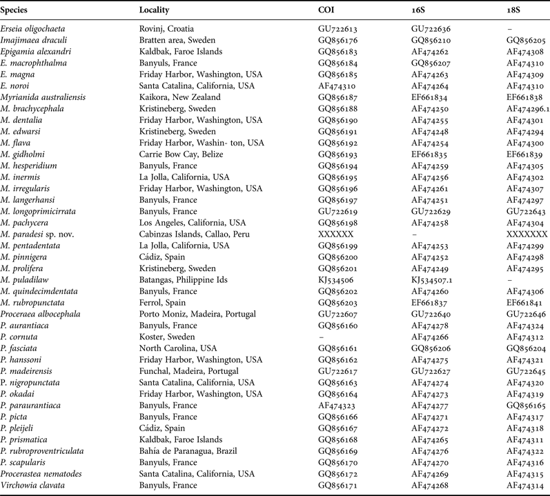
In order to establish the phylogenetic position of M. paredesi sp. nov. within the genus, the newly generated sequences were combined with nuclear (18S rDNA) and mitochondrial (16S rDNA and cytochrome c oxidase I, COI) sequences of 18 Myrianida, 14 Proceraea spp., 1 Procerastea, 1 Imajimaea, 1 Erseia and 1 Vichowia species available in GenBank. Four species of Epigamia were included as outgroups. GenBank accession numbers are provided in Table 1.
Species included in phylogenetic analyses, collection localities, and GenBank accession numbers.

Nuclear and mitochondrial datasets were analysed both individually and concatenated. The model of sequence evolution for each partition was selected implementing the Akaike information criterion (AIC) in jModeltest 2 (Darriba et al., Reference Darriba, Taboada, Doallo and Posada2012). The best model for the dataset of both mitochondrial markers was a General Time Reversible (GTR) model of sequence evolution with gamma-distributed rates across sites and a proportion of invariable sites (GTR + G + I).
Maximum likelihood (ML) partitioned analyses of the concatenated datasets were run in RAxML v. 1.3 (Stamatakis, Reference Stamatakis2006) using the GTR + G + I model. Bootstrap support values were estimated using 1000 replicates and 10 starting trees (Stamatakis et al., Reference Stamatakis, Hoover and Rougemont2008). Bayesian inference (BI) partitioned analyses were also run with the concatenated datasets and the same model, implemented in MrBayes 3.2.1 (Ronquist & Huelsenbeck, Reference Ronquist and Huelsenbeck2003). Four Markov chains were started from a random tree and run simultaneously for 20 million generations, with trees sampled every 2000 generations; the initial 25% of trees were discarded as burn-in. Convergence was tested using Tracer v1.6 (Rambaut et al., Reference Rambaut, Suchard, Xie and Drummond2014).
RESULTS
SYSTEMATICS
Family SYLLIDAE Grube, 1850
Subfamily AUTOLYTINAE Langerhans, 1879
Genus Myrianida Milne Edwards, 1845
Type species Myrianida fasciata Milne Edwards, 1845
Myrianida paredesi sp. nov.
Figures 2–5
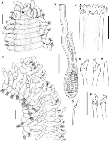
Myrianida paredesi sp. nov. (A) anterior end, dorsal view. (B) midbody segments, dorsal view. (C) pharynx and proventricle. (D) trepan. (E) compound chaetae, anterior parapodia. (F) compound chaetae, posterior parapodia. (G) dorsal simple chaeta. (H) acicula, anterior parapodia. (A), (B), (E–G) Holotype (LaBSIM-16.01/SY0001). (C), (D), Paratype (LaBSIM-16.01/SY0002). Scale: (A), (B) 0.1 mm, (C) 0.18 mm, (D–H) 20 µm.

Myrianida paredesi sp. nov. (Paratype) SEM. (A) lateral view. (B) anterior end, dorsal view. (C), (D) y (E) midbody segments with middle dorsal cirri, dorsal view. MNCN16.01/14729.

Myrianida paredesi sp. nov. (Paratype) SEM. (A) trepan, dorsal view. (B) trepan, front view. (C) midbody segments with trochs, dorsal view. (D) Compound and bayonet chaeta, median segment. MNCN16.01/14729.

Myrianida paredesi sp. nov. (A) male stolon, anterior end, dorsal view. (B) male stolon, compound chaeta. (C) female stolon, anterior end, dorsal view. (D) compound chaeta and (E) bayonet chaeta, mid-body segment. (LaBSIM-16.01/SY0004). Scale: (A), (C) 0.1 mm, (B), (D), (E) 20 µm.

MATERIAL EXAMINED
Holotype, Pampa Melchorita, 13°15′19″S–76°18′33″W, South of Lima, 10 m depth, in a Austromegabalanus psittacus and Crepipatella dilatata community, December 2011, preserved in formalin, LaBSIM-16.01/SY0001. Paratypes, seven specimens, same locality as holotype, preserved in formalin, MNCN 16.01/14729 (1 for SEM); 10 specimens, preserved in formalin, LaBSIM-16.01/SY0002; 13 specimens, preserved in alcohol, Cabinzas Islands, Callao, 12°6′52.93″S–77°12′35″W 5 m, amongst cirripeds, October 2013, LaBSIM-16.01/SY0003 (12) and MCZ 25286 (1). Stolons, Pampa Melchorita, South of Lima, 13°15′42.60″S–76°18′46″W, 10 m, July 2012, 16 male stolons and 2 female stolons chain, preserved in formalin, LaBSIM-16.01/SY0004.
DESCRIPTION
Holotype complete, well preserved specimen, with a chain of three female stolons, length without stolons 2.6 mm, including stolons 3.3 mm long, 0.24 mm width, 41 chaetigers (excluding stolons). Paratypes 2.4–5.8 mm long, 0.14–0.23 mm wide, 38–52 chaetigers. Body small, strongly flattened anteriorly (Figures 2A, 3A, B), not so flattened from midbody (Figures 2B, 3A, C–E), without colouration in preserved specimens, pale cream in alive. Prostomium subtriangular, with posterior part prolonged to peristomium (possibly a preservation artifact); two pairs of small eyes in trapezoidal arrangement (Figure 2A), anterior pair larger and two small anterior eyespots. Palps fused, about 1/3 of prostomial length. Nuchal epaulettes not seen, apparently reduced or absent (Figures 2A, 3B). Antennae thick, short (Figures 2A, 3A, B). Median antenna inserted medially on prostomium, reaching to end of chaetiger 4 (holotype) and to end of chaetiger 8 (paratypes); lateral antennae on anterior margin of prostomium, about 1–1/3 length of median antenna, reaching to chaetiger 2 (holotype) and to end of chaetiger 4 (paratypes). Dorsal tentacular cirri equal size to 1/5 longer than lateral antennae, ventral tentacular cirri 1/2 as long as dorsal pair (Figure 2A). First dorsal cirri similar in size to dorsal tentacular cirri, and twice longer than dorsal cirri of chaetiger 2. Cirrophores present on tentacular cirri and all dorsal cirri. Cirrophores from chaetigers 2 to 14 shorter than parapodial lobes, cirrostyles 1–2 times longer than cirrophores; cirrostyles ovate, up to three times longer than wide (Figure 2A). Cirri alternating in length, sometimes irregularly. From about chaetiger 15, dorsal cirri slender and elongated, with long cirrophores, up to 4 times longer than cirrostyles, bended dorsally. Cirrostyles of these segments rounded, slightly longer than wide (Figures 2B, 3C–E). Parapodial lobes rounded (Figures 2A, B, 3A, C, D). Chaetal fascicle with 5–12 compound chaetae in anterior and median chaetigers, reducing to 1–5 in posterior chaetigers. Compound chaetae typical of the genus, shafts with small spines, becoming more evident towards the posterior region; blades bidentate, with proximal tooth longer than distal one (Figures 2E, 4D), longer on posterior parapodia (Figure 2F), with short spines on margin (Figures 2E, F, 4D). Average length of blades: anterior region, a: 7.9 µm, b: 8.4 µm, c: 3.7 µm; middle region, a: 6.4 µm, b: 6.9 µm, c: 3.1 µm; posterior region, a: 5.0 µm, b: 5.6 µm, c: 2.6 µm. Bayonet chaetae slender (Figures 2G, 4D), usually beginning on chaetiger 6–8, sometimes from chaetiger 2, reaching the posterior region. Aciculae straight and pointed, in all chaetigers, 1 acicula in the first two chaetigers, 2–3 in median and 1 posterior chaetigers (Figure 2H). Pharynx with one or two sinuations extending beyond or anteriorly to proventricle (Figure 2C), sometimes without sinuation (when totally or partially everted). Trepan with variable location, may be in chaetiger 1 or 6, depending on the degree of relaxation (Figure 2A); with 14–15 equal teeth (Figures 2D, 4A, B), arranged in 1 ring. Proventricle short, throughout 3–4 segments, starting between chaetiger 10–15, with 9–10 rows of muscle cells (Figure 2C). A long dorsal, digitiform papilla, similar to dorsal cirri, on each segment, starting just after proventricle, from chaetiger 13–18, extending to the posterior region, absent in most posterior chaetigers. Dorsal papillae short in anterior segments where appear, becoming longer progressively to posterior segments, giving an appearance of having three dorsal cirri (Figures 2B, 3A, C–E, 4C). Anal cirri about as long as dorsal tentacular cirri. Ciliation as 1 trochs per segment, extending over middle-dorsal cirrus (Figure 4C).
REPRODUCTION
Schizogamous reproduction by gemmiparity behind chaetiger 41–43.
Epitokous stages
Polybostrichus (Figures 5A, B); 16 stolons, with 1.7–2.0 mm long for 16–20 chaetigers, 0.44–0.50 mm width including parapodial lobes, apparently without colouration. Prostomium rectangular, with two pairs of eyes, one pair dorsal and another ventral; ventral eyes about twice longer than dorsal ones. Two long palps, thick at bases and bifurcated at their 1/3 length, and present lateral folds at midlength (Figure 5A). Lateral antennae short, originated near anterior margin of prostomium. Median antenna slender, reaching up to chaetiger 10–11 (Figure 5A). A pair of small nuchal epaulettes extending to middle of first chaetiger (Figure 5A). Dorsal pair of tentacular cirri similar to median antenna, reaching up to end chaetiger 9–10; ventral pair shorter than lateral antennae (Figure 5A). All chaetigers with a slender dorsal cirri and a fascicle of bidentate compound chaetae (Figure 5B); single thin bayonet chaetae present from chaetiger 1; swimming capillary chaetae, beginning on chaetiger 3–4, emerging between bases of dorsal cirri and notopodial lobes. A dorsal longitudinal fold throughout body. Anal cirri equal in length to 5–6 chaetigers (not shown in Figure 5A).
Sacconereis (Figures 5C–E). One immature stolon attached to holotype and 3 mature stolons in a row unattached (detached during examination). Stolons unattached to parental with 0.46–0.58 mm long, 0.28–0.40 mm wide including parapodial lobes, 10–13 chaetigers. Anterior region wide, becoming progressively slender towards posterior region. Prostomium rectangular, wider than long, with anterior and posterior margin concave. A pair of small eyes, situated on the lateral margins of prostomium. Median antenna not seen (probably detached) (Figure 5C). A pair of short lateral antennae inserted near half of prostomium and tentacular cirri not seen (Figure 5C). All parapodia uniramous, lobes parapodial large in anterior region becoming small or absent toward posterior region. Dorsal cirri present on all segments, cirrophores 1/2 as long as lobes parapodial, cirrostyles ovate 1 and 2 as long as cirrophores. Last segments are more piled up, ending in pygidium with 2 cirri up to 4 times as long as dorsal cirri (Figure 5C). Bidentate compound chaetae similar to adult (Figure 5D). Bayonet chaetae similar to those of parentals (Figure 5E); swimming chaetae absent.
Remarks
Myrianida paredesi sp. nov. is unique among autolytines, and even all the family Syllidae, in having a longitudinal row of mid-dorsal long papillae (one on each segment), beginning immediately after the proventricle and extending to the posterior region, without reaching the last chaetigers. None of the species of this genus described in Nygren (Reference Nygren2004), and subsequent papers (Nygren & Pleijel, Reference Nygren and Pleijel2007, Reference Nygren and Pleijel2010; Nygren et al., Reference Nygren, Sundkkvist, Mikac and Pleijel2010; Álvarez-Campos et al., Reference Álvarez-Campos, San Martín and Piotrowski2014) has that row of long papillae. Also, the shape of prostomium and apparent lack of nuchal epaulettes are very characteristic of this species. Perhaps the most similar species is M. pentadentata (see below) having a similar anterior end and proventricle, but that species has well defined nuchal epaulettes, only 5 teeth in the trepan, and lack of the characteristic long dorsal papillae. Table 2 shows the morphological characters of M. paredesi sp. nov., and their most similar species. Regarding to the phylogenetic results, the new species appeared to be the sister species of the clade that includes M. puladilaw Álvarez-Campos, San Martín & Piotrowski, Reference Álvarez-Campos, San Martín and Piotrowski2014, M. gidholmi Nygren & Pleijel, Reference Nygren and Pleijel2007, M. pachycera (Augener, 1913), M. australiensis (Hartmann-Schröder, Reference Hartmann-Schröder1962a, Reference Hartmann-Schröderb), M. hesperidium (Claparède, 1868) and M. quindecimdentata (Langerhans, 1884) (Figure 6). The characters shared by this clade is the presence of 1 ciliary troch per segment (except M. pachycera; unknown in M. australiensis), moderate number of equal teeth on trepan (except M. pachycera), and the mode of reproduction.
Phylogenetic relationships of M. paredesi sp. nov. with all the Myrianida species inferred from the Maximum likelihood and BI analysis of the three concatenated genetic markers (18S rRNA, 16S rRNA and COI). Numbers above branches indicate bootstrap support values (only BS > 75% are indicated). Numbers below branches indicate posterior probabilities support values (only PP > 0.90 are indicated) inferred; white circles indicate a BS value of 100% and a PP value of 1.00.

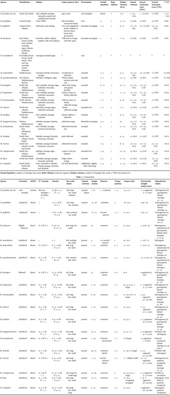
Morphological characters of Myrianida paredesi sp. nov. and all species of genus included in the phylogenetic analysis.

MDLP: Mid-Dorsal Long Papillae; N° Aciculae: A (anterior), M (medium), P (posterior); Fascicle: N°. chaetae, A (anterior), M (medium), P (posterior); Size Teeth Chaeta: sub (subdistal), dis (distal); Simple Chaetae: starting chaetiger; Pharynx: N°. Sinuations/Position Proventricle; Trepan Position: position in the chaetiger, Trepan Teeth: e (equal), u (unequal).
Table 2 shows the morphological characters of M. paredesi sp. nov., their most similar species and all species included in the phylogenetic analysis.
ETYMOLOGY
The species is named in honour of Professor Dr Carlos Paredes, for his great contribution to the knowledge of the biodiversity of marine invertebrates from Peru.
HABITAT
Among barnacles Austramegabalanus psittacus and patellogastropod Crepipatella dilatata community. Above sublittoral rocky and artificial hard bottoms.
DISTRIBUTION
Only known from the type locality (Pampa Melchorita), and Cabinzas Island, Callao. South of Lima (Peru). South East Pacific.
Myrianida pentadentata
(Imajima, Reference Imajima1966)
Figures 7 & 8
Myrianida pentadentata (Imajima, Reference Imajima1966). (A) anterior end, dorsal view. (B) trepan, dorsal view. (LaBSIM-16.01/SY0005). Scale: A:0.1 mm, (B) 20 µm.

Myrianida pentadentata (Imajima, Reference Imajima1966) SEM. (A) anterior end and midbody chaetigers, dorsal view. (B) nuchal epaulettes, dorsal view. (C) dorsal cirri, alternation in size, midbody segments. MNCN16.01/14730.

Autolytus (Autolytus) pentadentatus Imajima, Reference Imajima1966: p. 32, Figure 6A–H.
Myrianida pentadentata Nygren (Reference Nygren2004): p. 146, Figure 72A–G.
MATERIAL EXAMINED
Canoas, Tumbes, 03°54′43″S–80°53′36″W, north of Peru, 12–16 m depth, on rocky bottom covered by algae and fine sediment particles, Oct 2012, three specimens (LaBSIM-16.01/SY0005), one specimen (MNCN 16.01/14730, SEM). All specimens preserved in formalin.
DESCRIPTION
All specimens incomplete. Body small, slightly flattened, 1.2–3.3 mm long, for 18–46 chaetigers, 0.26–0.22 mm wide, (Figures 7A, 8A). Preserved specimens without colour. Prostomium rounded, slightly wider than long. Four eyes in trapezoid arrangement, anterior pair larger (Figure 7A). Palps fused, projecting 1/3–1/2 of prostomium in dorsal view. Nuchal epaulettes rounded, extending from end of peristomial segment to half of chaetiger 2, lateral margin ciliated (Figures 7A, 8B arrow). Median antenna inserted medially on prostomium, reaching chaetiger 10. Lateral antennae near anterior margin, 1/2–3/5 of median antenna length. Dorsal tentacular cirri about 1/2 length of median antenna, and ventral tentacular cirri 1/2 as long as dorsal pair. Dorsal cirri of chaetiger one 3/5 length of median antenna and twice longer than second dorsal cirri. Dorsal cirri alternating in length (Figures 7A, 8A, C). Cirrostyle of short and long cirri similar in size (Figures 7A, 8C). Cirrophores present on tentacular cirri, and all dorsal cirri. Cirrophores unequal; long cirrophores twice longer than short cirrophores, difference becoming more evident from chaetiger 9 (Figures 7A, 8A). Short cirrophores shorter or equal to parapodial lobes; long cirrophores equal or twice parapodial lobes. Short cirrophores equal in size to cirrostyle, long cirrophores up to two times longer than cirrostyles (Figures 7A, 8A, C). Parapodial lobes rounded. Single, straight acicula in all chaetigers. Chaetal fascicle with 7–11 compounds on anterior chaetigers, 5–6 in median and posterior parapodia. Compound chaetae with small distal tooth. Average length of blades: anterior region, a: 7.9 µm, b: 9.1 µm, c: 3.8 µm; middle region, a: 5.9 µm, b: 6.4 µm, c: 3.9 µm. Bayonet chaetae slender, beginning between chaetiger 1–11. Pharynx with several long sinuations, beyond and posterior to proventricle. Trepan in peristomial segment or in chaetiger 1, with 5 equal teeth, in single ring surrounded by 5 large papillae (Figure 7B). Proventricle through 4–5 segments, in chaetiger 7–12, with 19–21 rows of muscle cells.
REPRODUCTION
Posterior scissiparity (Nygren, Reference Nygren2004).
HABITAT
Subtidal rocky bottom covered by algae and fine sediment particles. Intertidal, amongst seaweed and beneath encrusting bryozans (Nygren, Reference Nygren2004).
DISTRIBUTION
North Pacific. Japan, California. First report for East Subtropical Pacific (Mancora, North coast of Peru).
REMARKS
Like Nygren (Reference Nygren2004), the annuli on long cirri, suggested by Imajima (Reference Imajima1966) in the original description, could not be confirmed; in the original description, Imajima uses the term cirri to refer to the cirrostyle, and according to Figure 6d (Imajima, Reference Imajima1966), it follows that there was a wrong delimitation of cirrophore and cirri (cirrostyle) possibly caused by an irregular surface, which was interpreted as the limit of cirrophore and cirrostyle, thereby obtaining the annuli on long cirri (cirrostyle). Therefore, the main characteristic of M. pentadentata is to have a trepan with 5 teeth.
Genus
Proceraea
Ehlers, 1864
Proceraea micropedata
(Hartmann-Schröder, 1962)
Figure 9
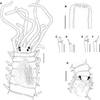
Procerarea micropedata (Hartmann-Schöder, Reference Hartmann-Schröder1962a, Reference Hartmann-Schröderb). (A) anterior end, dorsal view. (B) trepan, dorsal view. (C) Compound chaeta and (D) bayonet chaeta, anterior chaetiger. (E) Compound chaeta and (F) bayonet chaeta, midbody segment. (G) female, anterior end, dorsal view. (LaBSIM-16.01/SY0006.) Scale: (A) 0.1 mm, (B) 20 µm, (C–F) 10 µm, (G) 0.18 mm.

Odontosyllis micropedata Hartmann-Schröder, Reference Hartmann-Schröder1962a: pp. 100–103, Figures 87–92.
Proceraea micropedata Nygren (Reference Nygren2004): p. 58, Figure 17A–C.
MATERIAL EXAMINED
Pampa Melchorita, South of Lima, 13°15′19″S–76°18′33″W, 5 m, Jul 2012; five specimens, preserved in formalin, LaBSIM-16.01/SY0006. All specimens amongst Austromegabalanus psittacus and Crepipatella dilatata community.
DESCRIPTION
One complete and four incomplete specimens. Body small, 3.4 mm long, 0.27 mm wide for 39 chaetigers (Figure 9A). Preserved specimens without apparent colour pattern. Prostomium rounded, slightly wider than long (Figure 9A). Four eyes, in trapezoid arrangement (Figure 9A). Palps fused, in dorsal view projecting 1/5 of prostomial length. Nuchal epaulettes not seen. Median antenna inserted medially on prostomium, reaching to chaetiger 8. Lateral antennae similar in length to median antenna, reaching chaetiger 7. Dorsal tentacular cirri half of length of median antenna; ventral tentacular cirri about half of length of dorsal pair. First dorsal cirri 1–1.2 times as long as median antenna, about 7.5 times longer than second dorsal cirri. Sometimes, dorsal cirri of chaetiger 2 twice longer than dorsal cirri of chaetiger 3, remaining dorsal cirri similar in size, about 1/4–1/3 of body width (Figure 9A). Cirrophores of tentacular cirri and dorsal cirri of chaetiger 1 present; otherwise absent. Cirri from chaetiger 3 cylindrical; however, egg-shaped in all chaetigers or only in some chaetigers (Figure 9A). Parapodial lobes rounded, chaetigers with 1–2 aciculae. Chaetal fascicle with 10 compounds in anterior chaetigers 5–8 median chaetigers, 3–4 in posterior chaetigers. Compound chaetae with large proximal and median distal tooth in anterior chaetigers, and small distal tooth in median and posterior chaetigers (Figure 9C, E). Average length of blades: anterior region, a: 5.3 µm, b: 6.0 µm, c: 3.3 µm; middle region, a: 4.5 µm, b: 5.3 µm, c: 2.5 µm; posterior region, a: 3.8 µm, b: 4.7 µm, c: 1.9 µm. Bayonet chaetae thick (Figure 9D, F), present from chaetiger one. Pharynx with single sinuation beside to proventricle (Figure 9A). Trepan in chaetiger 1–2, with 18 unequal teeth, 9 large and 9 smaller; 1 large alternating with 1 small, arranged in 2 rings, the small teeth are about half the length of the large teeth (Figure 9B). Proventricle through 2.5–3 segments, in chaetigers 3–5, with 28–30 rows of muscle cells (Figure 9A). Anal cirri lanceolated, 1.5 times longer than dorsal cirri.
REPRODUCTION
Schizogamy by anterior scissiparity behind chaetiger 13.
Polybostrichus . One specimen incomplete. Anterior segments lost, length in preserved specimens 2.3 mm for 23 chaetigers, width 0.39 mm including parapodial lobes, without apparent pattern of colouration. Parapodial lobes equal in length to body width. Dorsal cirri slender, ovate, 3/4 as long as parapodial lobes. Chaetal fascicle with 1–3 compound chaetae. Compound chaetae with minute blades, large proximal and small distal tooth. Bayonet chaetae thick. Swimming capillary chaetae as long as parapodial lobes, absent in 6 posterior chaetigers. Anal cirri similar in form and size to adult.
Sacconereis (Figure 9G). Stolon still attached to parental 1.7 mm long, 0.42 mm wide including parapodial lobes, for 17 chaetigers (posterior region lost). Prostomium rectangular, wider than long, with anterior margin slightly concave. Two pair of eyes, with lenses, in trapezoid arrangement, anterior pair larger. Palps and nuchal epaulettes absent. Median antenna in posterior margin of protomium, reaching chaetiger 3, lateral antennae absent. Dorsal pair of tentacular cirri 2/3 as long as median antenna; ventral tentacular cirri 1/2 as long as dorsal pair. All parapodia uniramous. Dorsal cirri present from chaetiger 1; cirrophores absent. Anterior dorsal cirri similar to those of parental (Figure 9A, G), posteriorly cylindrical and slightly larger than those of parental. Chaetal fascicle with 4–5 compound chaetae. Anterior chaetigers, with minute blades, similar to those of Polybostrichus, later similarly to adult. Bayonet chaetae thick present since chaetiger 1. Swimming chaetae present from chaetiger 6.
HABITAT
Rocky bottom, amongst Austromegabalanus psittacus and Crepipatella dilatata community. Amongst algae, barnacles, Mytilus (Hartmann-Schröder, Reference Hartmann-Schröder1962a).
DISTRIBUTION
South East Pacific, from Magellan Strait to central Peru.
REMARKS
The main feature for P. micropedata remarked by Nygren (Reference Nygren2004), is the egg-shaped dorsal cirri, somewhat unstable, because in some specimens they are more or less elongated. Therefore, this feature could be variable and a product of the ontogeny of the species, which is verified on paratype cirrus drawn in the original description by Hartmann-Schröder (Reference Hartmann-Schröder1962a). Additionally, the proportions of the antennae, peristomial cirri and dorsal cirri of first and second chaetiger are irregular, with differences that might be an ontogenetic development product or by fragmentation of such structures; finding few differences in the description by Hartmann-Schröder (Reference Hartmann-Schröder1962a, Reference Hartmann-Schröderb) and Nygren (Reference Nygren2004). Nuchal organs could not be verified, they may possibly be short. For the recognition of this species, although not a constant feature, the dorsal cirri is considered as egg shaped, as suggested by Nygren (Reference Nygren2004). Proceraea micropedata shows considerable affinity with P. cornuta (Agassiz, 1862), P. okadai (Imajima, Reference Imajima1966) and P. nigropunctata Nygren & Gidholm, 2001.
DISCUSSION
Taxonomic work on polychaetes made in Peru are scarce and knowledge of the Syllidae is even scarcer, so we can mention Grube (Reference Grube1858) with the descriptions of Syllis latifrons; Kinberg (Reference Kinberg1866) with the description of Laomedora fusifera; Gravier (Reference Gravier1910) with record of Syllis palifica Ehlers (1901); Friedrich (Reference Friedrich1956) with the description of Exogone multisetosa; Hartmann-Schröder (Reference Hartmann-Schröder1962b) with the description of Typosyllis eulitoralis, Odontosyllis liniata, Sphaerosyllis parapionosylliformis and records of Syllis prolixa (Ehlers, 1901), Syllis magdalena (Wesenberg-Lund, 1961), Syllis gracilis Grube, 1840, Neopetitia amphophthalma (Siewing, 1956); Gómez et al. (Reference Gómez, Amaya, Guerra and Montes1973), with record of Syllis elongata (Johnson, 1901) and Syllis alternata Moore, 1908 and Tarazona (Reference Tarazona1974), with record of four species previously reported by Hartmann-Schröder (Reference Hartmann-Schröder1962b) and five species undetermined. Thus, in Peru there is likely a high number of syllids not yet registered, and others that have previously been described that need checking and correcting.
ACKNOWLEDGEMENTS
This publication is contribution No. 25 of the Peru Biodiversity Program implemented by the Center for Conservation, Education and Sustainability (CCES) of the Smithsonian Conservation Biology Institute. We are grateful to Carmen Yamashiro and Juan Argüelles of Invertebrates Laboratory of IMARPE for allowing the first author to participate in their monitoring activities to collect his material. Special thanks to Dr Gonzalo Giribet (MCZ) for allowing PA-C to work on his laboratory sequencing of the specimen of the new species.
FINANCIAL SUPPORT
We acknowledge financial and logistic support from PERU LNG S.R.L during the implementation of the Marine Benthic Assessment and Monitoring Protocol in Melchorita. This paper is also a contribution to the project ‘Caracterización Taxonómica y Sistemática de la familia Syllidae (polychaeta) basada en datos moleculares y morfológicos. El problema de las especies cosmopolitas y Biodiversidad en el Pacífico’, funded by the ‘Ministerio de Ciencia e Innovación’ of the Spanish Government, Project number CGL2009–12292 BOS.