As a high energy efficiency, ecofriendly, long-lifetime, and compact alternative to traditional lighting sources, solid-state lighting (SSL) in form of white light-emitting diodes (white LEDs) has received considerable attention. Nowadays, the phosphor-converted white LEDs (pc-white-LEDs) fabricated using a blue GaN chip or near-UV LED (370–420 nm) coupled with red, green, and blue phosphors are becoming the most attractive SSL devices because of its continuing improvement in the efficiency.Reference Schubert and Kim1–Reference Hashimoto, Wu, Speck and Nakamura5 The eventual performance of pc-white-LEDs devices strongly depends on the luminescence properties of the phosphors used. Therefore, the state-of-the-art SSL devices would be notably advanced by the discovery of phosphor materials that are optimized for pc-white-LEDs. The most frequently used red-emitting phosphor for near-UV LEDs is Y2O2S:Eu3+. However, Y2O2S:Eu3+ has poor absorption in near-UV region where LED chips emission occurs. A good strategy to overcome this problem is to search for a host lattice that can absorb energy efficiently in the near-UV region and transfer the energy to the activator, resulting in emission in the red region (host-sensitized emission).Reference Neeraj, Kijima and Cheetham6, Reference Neeraj, Kijima and Cheetham7 In view of practical applications, improvement of luminescence of phosphors is of great importance, especially for pc-white-LEDs while retaining the high quantum yield and high luminescence quenching temperature. Currently, modification of phosphor composition by chemical substitution is a highly effective strategy for the designing of new materials and improving luminescent properties of already known compounds.Reference Nyman, Rodriguez, Shea-Rohwer, Martin and Provencio8–Reference Xie, Yuan, Hai, Wang, Wang and Li12
Lanthanum molybdate (La2Mo2O9) has ever been studied as catalyst, fast oxide-ion conductor, and ferroelectric material.Reference Lacorre, Goutenoire, Bohnke, Retoux and Laligant13–Reference Kim and Kang15 Marrero-López et al. reported for the first time the photoluminescence properties of Eu3+-doped La2Mo2O9 phosphor.Reference Marrero-López, Núñez, Abril, Lavín, Rodríguez-Mendoza and Rodríguez16 Recently, the development of SSL has lead to a rebirth of interest in this material because it can be used as a host for optically active lanthanide ions.Reference Li, Yang, Guan, Liu and Li17, Reference Gong, Shi and Zhou18 The effects of flux on structure, morphology, and luminescent properties of La2Mo2O9:Eu3+ were investigated by Li et al.Reference Li, Yang, Guan, Liu and Li17 Gong et al. found that the emission intensity of La2Mo2O9:Eu3+ can be strengthened by doping small amounts of rare earth (Gd, Dy, Y, and Yb) ions.Reference Gong, Shi and Zhou18 However, there is no report on the luminescent enhancement of Eu3+-activated La2Mo2O9 red-emitting phosphor through chemical substitution. The aim of the present study is to explore the varying composition and attempt to improve luminescence intensity of Eu3+-activated La2Mo2O9 red-emitting phosphor. Herein, a series of novel red-emitting phosphors with nominal compositions La2(Mo1−zSiz)2O9:0.05Eu3+ (0 ≤ z ≤ 0.10) with strong near-UV absorption were prepared by solid-state process. The enhanced luminescence of La2Mo2O9:0.05Eu3+ by substituting Mo6+ with Si4+ was investigated.
La2(Mo1−zSiz)2O9:0.05Eu3+ (0 ≤ z ≤ 0.10) sample phosphors were prepared by a typical solid-state reaction. The starting materials La2O3 (99.99%), MoO3 (99.5%), SiO2 (99.90%), and Eu2O3 (99.99%) were purchased from Shanghai Chemical Reagent Ltd. Co., China, and used without any further purification. The required stoichiometric amounts of these materials were weighed and then thoroughly wet mixed in an agate mortar with acetone as the wetting medium. Prior to calcinations, the mixing samples were dried in an air oven. The obtained mixture was heated at 550 °C for 4 h in a muffle furnace, followed by the calcination at 900 °C for 8 h. Finally, the samples are ground into powder for characterizations. For obtaining the samples with almost the same size and narrow particle distribution, we separated powders by particle sizes using the multilevel sieves after above route,Reference Nazarov19 with the aim to avoid the differences in luminescence intensities due to the size effects.
The samples were characterized by x-ray powder diffraction (XRD), UV–vis diffuse reflectivity, and photoluminescence spectrum. XRD was carried out with a Japan Rigaku D/max 2500 diffractometer, using Ni-filtered Cu Kα radiation. A scan rate of 0.02°/s was applied to record the patterns in the 2θ range 10–90°. UV–vis diffuse reflectivity spectra of phosphors were measured with Cary 500 UV–vis–NIR spectrophotometer using a polytetrafluoroethylene standard as reference. The excitation and emission spectra of powders were recorded using fluorescence spectrofluorometer (Varain Cary-Eclipse). For the comparison of excitation and emission intensity, the quantity of the phosphor samples has been normalized and measurement conditions (i.e., the packing density of samples, width of slit of the excitation and emission monochromators, the photomultiplier tube detector voltage, and sensitivity) were kept consistent from sample to sample in the measurements. All the measurements were performed at room temperature.
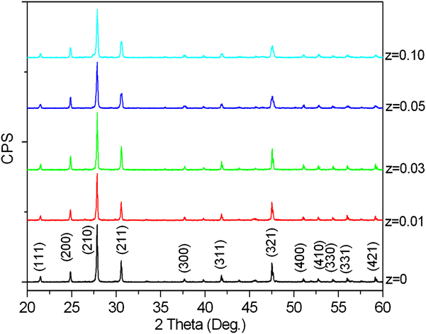
The body colors of as-obtained phosphor powders are white. La2Mo2O9 has two different structures: α- La2Mo2O9 and β- La2Mo2O9, which belong to the monoclinic phase and the cubic phase, respectively. XRD patterns of La2(Mo1−zSiz)2O9:0.05Eu3+ (0 ≤ z ≤ 0.10) series phosphors are presented in Fig. 1. As the Si4+ contents (i.e., z value) increase, XRD profiles are similar without showing discernable shifting. No impurity phases related to the starting materials and intermediates were found, clearly indicating that the compounds formed are of single phase. This also illuminates that the effective incorporation of Si4+ into host lattice have been achieved. Si4+ was supposed to occupy the Mo6+ sites in this phosphor because of the almost identical ionic radii between Mo6+ (0.041 nm) and Si4+ (0.040 nm). All diffraction peaks can be indexed to the cubic phase La2Mo2O9, which is in agreement with JCPDS card no. 28-0509. The strong and sharp diffraction peaks suggest that these samples are highly crystallized which is advantageous to luminescence.
X-ray powder diffraction patterns of La2(Mo1−zSiz)2O9:0.05Eu3+ (0 ≤ z ≤ 0.10) phosphors.

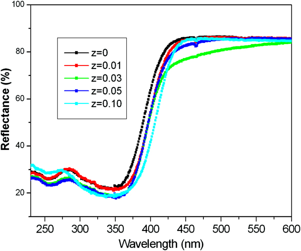
UV–vis diffuse reflectivity spectra of La2(Mo1−zSiz)2O9:0.05Eu3+ (0 ≤ z ≤ 0.10) phosphors are represented in Fig. 2. All samples show a broad absorption band in the near-UV region with the steep absorption edges in the range of 350–430 nm. With increasing of Si4+ doping content, the absorption band tends to shift toward longer wave length, i.e., the optical band gap energy (E g) decreased, which also indicates the successful incorporation of Si4+ into host lattice and is in agreement with the XRD results. It is well established that there is a direct dependence between the E g values and the presence of intermediary energy levels within the band gap.Reference Hank, Barnes, Benjamin and Patrick20 Generally, the origin of these energy states is caused by the formation of oxygen vacancies into the structure. For La2(Mo1−zSiz)2O9:0.05Eu3+ phosphors, the increasing of Si4+ content (i.e., z value) induces the increase of oxygen vacancies and consequently increasing the concentration of intermediary electronic states within band gap and decreasing the E g value. Furthermore, the shoulder band beside the absorption edge for the Si4+-containing phosphors was then observed in the long wave length side, especially in the case of z = 0.03. However, the shoulder band was not observed in the phosphor without Si4+-doping. These results indicated that this band is related to introduction of Si4+ into La2Mo2O9:Eu3+ lattice. Based on the above results, it can be concluded that substitution of Si4+ for Mo6+ into host lattice can modulate the band structure of La2Mo2O9:Eu3+ phosphors by generating oxygen vacancies.
UV–vis diffuse reflectivity spectra of La2(Mo1−zSiz)2O9:0.05Eu3+ (0 ≤ z ≤ 0.10) phosphors.

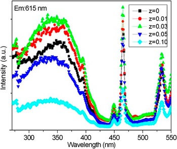
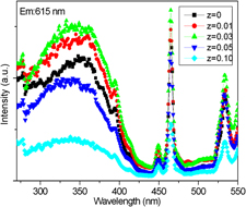
It was reported that the red emission of CaMoO4:Eu3+ phosphor can be enhanced by substituting Mo6+ with Si4+.Reference Ci, Wang, Zhang and Sun11, Reference Xie, Yuan, Hai, Wang, Wang and Li12 A similar improvement in luminescence of LiEuMo2O8 phosphor by introduction of Si4+ ion was observed by Dai et al.Reference Dai, Zhang, Li, Wang, Zhao and Liu21 To improve luminescence of La2Mo2O9:0.05Eu3+ phosphor, Si4+ was introduced into host lattice as partial substitution for Mo6+. Figure 3 depicts that the excitation spectra of La2(Mo1−zSiz)2O9:0.05Eu3+ (0 ≤ z ≤ 0.10) phosphors monitored the 5D0 → 7F2 transition of Eu3+ at 615 nm. The spectrum is comprised of a strong broadband from 250 to 430 nm and several sharp peaks at 395, 465, and 533 nm. The strong broad band with peak at 350 nm is assigned to charge transfer (CT) transitions of O2−–Eu3+ and O2–Mo6+. Sharp lines at 395, 465, and 533 nm can be ascribed to 7F0 → 5L6, 7F0 → 5D2, and 7F0 → 5D1 transitions of Eu3+ ions, respectively. The intensity of CT band was comparable to that of 4f–4f transition of Eu3+ ions. The presence of the strong broadband in the excitation spectrum indicates that there exists an energy transfer from host to Eu3+ ions. With increasing of Si4+ content (i.e., z value), both of the CT band and sharp peaks gradually increase and reach a maximum when the doping concentration of Si4+ is 0.03 mol. In addition, the CT band is considerably broadened and red-shifted, which are consistent with those observed in the above UV–vis spectra (shown in Fig. 2). The enhancement of CT band indicated that the Si4+ incorporation improved the host-sensitized emission of Eu3+. When the z value is bigger than 0.03, the excitation intensities of CT band and sharp peaks reduce, which may be due to excess defects formed by the introduction of superfluous Si4+ ions.Reference Ci, Wang, Zhang and Sun11
Excitation spectra of La2(Mo1−zSiz)2O9:0.05Eu3+ (0 ≤ z ≤ 0.10) phosphors (λem = 615 nm).

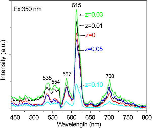
The emission spectra of La2(Mo1−zSiz)2O9:0.05Eu3+ (0 ≤ z ≤ 0.10) phosphors upon excitation with CT band are depicted in Fig. 4. The emission spectra contain some sharp peaks at 535, 554, 587, 615, and 700 nm, which are assigned to 5D1 → 7F1, 5D1 → 7F2, 5D0 → 7F1, 5D0 → 7F2, and 5D0 → 7F4 transition of Eu3+, respectively. However, no emission corresponding to host is observed under excitation of CT band. The phosphors exhibit characteristic emission of Eu3+, which clearly indicates the energy transfer from host to Eu3+ ions. With increasing of Si4+ content (i.e., z value), the emission intensity increases and reaches a maximum when doping concentration of Si4+ is 0.03 mol. However, when the Si4+ concentration is higher than 0.03 mol, the luminescent intensity decreases.
Emission spectra of La2(Mo1−zSiz)2O9:0.05Eu3+ (0 ≤ z ≤ 0.10) phosphors under excitation of CT band (λex = 350 nm).

The reasons for the enhanced luminescence under excitation of CT band by Si4+ doping may be attributed to the following aspects. As indicated above, Si4+ was supposed to occupy the Mo6+ site in host because of the almost identical ionic radii between Mo6+ and Si4+. Si4+ ions with low valence occupying Mo6+ sites in the lattices generate extra negative charges, (SiM0)··, which are supposed to generate oxygen vacancies, (VO)·· for charge neutrality. The presence of the oxygen vacancies generated by the introduction of Si4+ can promote the energy transfer from excited carriers in host lattice to Eu3+.Reference Ryu, Singh, Bartwal, Brik and Kityk22–Reference Wang, Jiang, Shen, Fang, Zheng, Wu and Shen24 However, when the Si4+ content exceeds a certain amount, excessive oxygen vacancies will be created, which leads to the luminescence quenching.Reference Ci, Wang, Zhang and Sun11
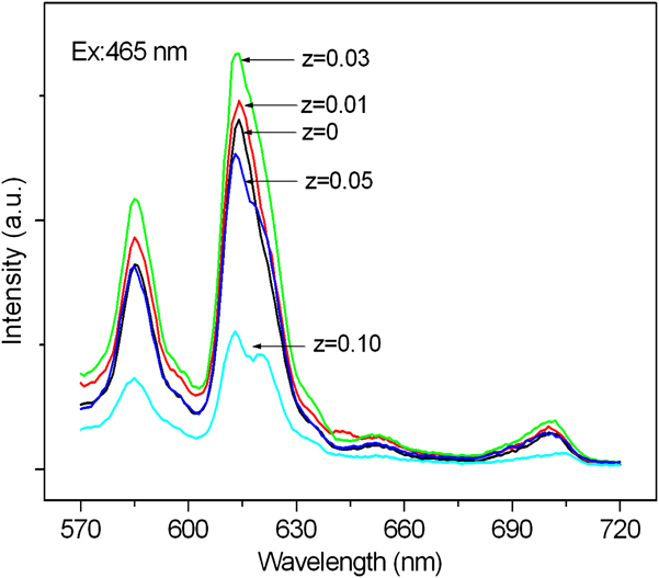
Figure 5 shows the emission spectra of Si4+-doped series samples upon excitation with direct 4f–4f transition of Eu3+ ions. The emission spectra were also dominated by 615-nm peak due to 5D0 → 7F2 transition of Eu3+, which clearly implies that Eu3+ occupies a noncentrosymmetric site. With increasing of Si4+ content, the luminescence enhancement was accompanied by the further splitting of 5D0 → 7F2 transition. While z value reaches 0.10, two peaks located at 615 and 620 nm due to 5D0 → 7F2 transition of Eu3+ can be observed. This hints that the local symmetry around Eu3+ ions was to some extent lowered by the incorporation of Si4+.25 Hence, it is deduced that more odd rank crystal field components are induced, and more opposite parity components (4f55d states) are mixed into 4f6 transitional levels of Eu3+, which leads to the enhancement of 5D0 → 7F2 transition.Reference Gong, Shi and Zhou18 Consequently, the partial substitution of Mo6+ by Si4+ increases the fluorescence of the as-obtained phosphors.
Emission spectra of La2(Mo1−zSiz)2O9:0.05Eu3+ (0 ≤ z ≤ 0.10) phosphors upon excitation with direct 4f–4f transition of Eu3+ ions (λex = 465 nm).

Eu3+-activated La2(Mo1−zSiz)2O9 (0 ≤ z ≤ 0.10) phosphors were prepared by high-temperature solid-state reaction. A significant enhancement of red photoluminescence has been demonstrated for La2Mo2O9:0.05Eu3+ phosphor by chemical substitution. XRD results show that Si4+ was effectively incorporated into host lattice and the obtained samples are of single phase. The excitation spectrum included a strong broadband ranging from 250 to 430 nm due to the CT transitions of O2−–Eu3+ and O2–Mo6+, and sharp peaks at 395, 465, and 533 nm corresponding to intra-4f transitions of Eu3+. Upon excitation with near-UV and blue light, the as-obtained phosphors exhibited a satisfactory red emission peaked at 615 nm. Partially replacing Mo6+ with Si4+ can enhance the red emission under excitation of CT band as well as direct 4f–4f excitation-mediated luminescence. The former is mainly attributed to the oxygen vacancies as sensitizer for the effective energy transfer from host to Eu3+, while the latter may be result from the relatively lower local symmetry around Eu3+ ions. Our results show that the chemical substitution is of great significance for the development of highly efficient phosphors used in solid-state lighting equipment.
ACKNOWLEDGMENTS
The authors thank Dr. Jinping Huang from Shanghai Normal University for assistance with the XRD measurements. This work was financially supported by the Natural Science Research Project of the Jiangsu Higher Education Institutions (08KJD150014), the QingLan Project of the Jiangsu Province (2008), and the Basic Research Fund of Jiangsu Teachers University of Technology (KYY09031).