Introduction
Cystic echinococcosis (CE) is a parasitic disease resulting from the infection of various organs by the encysted larvae of Echinococcus granulosus. This tapeworm is prevalent in most parts of Iran (Ahmadi & Dalimi, Reference Ahmadi and Dalimi2006). The life cycle of E. granulosus involves dogs and other carnivores such as jackals, foxes and wolves as the definitive host, and sheep and other herbivores as the intermediate host. The oncosphere hatches from ingested eggs in the intestine and enters the portal circulation in the intermediate host. The liver, being the first filter, is the most commonly involved organ, followed by the lungs. Up to 70% of hydatid cysts are located in the liver and 25% in lungs. Once within the systemic circulation, the larva may settle and develop into a hydatid cyst in other organs of the body. Although hydatid cysts can occur in any organ, it is very rare to see a cyst in the brain (Mandal & Mandal, Reference Mandal and Mandal2011). Cerebral hydatid disease is very rare in humans, representing less than 1% of all cerebral space-occupying lesions, even in countries where the disease is endemic (Gana et al., Reference Gana, Skhissi, Maaqili and Bellakhdar2008; Nemati et al., Reference Nemati, Kamgarpour, Rashid and Sohrabi Nazari2010). It has also been reported rarely in animal species (Singh et al., Reference Singh, Sharma Deorani and Srivastava1988; Ruas et al., Reference Ruas, Schild, Fernande, Ferreira and Müller1997).
It has been shown that E. granulosus has wide intra-specific variation, affecting the epidemiology, morphology, pathology, genetics, biology, physiology and control of cystic echinococcosis (Thompson & McManus, Reference Thompson and McManus2002). According to the mitochondrial DNA (mtDNA) sequences, ten distinct genotypes of E. granulosus, including G1 (common sheep), G2 (Tasmanian sheep), G3 (water buffalo), G4 (horse), G5 (cattle), G6 (camel), G7 (pig), G8 (cervid), G9 (human) and G10 (Fennoscandian cervid) have previously been recognized (McManus, Reference McManus2002; Lavikainen et al., Reference Lavikainen, Lehtinen, Meri, Hirvelä-Koski and Meri2003; Maravilla et al., Reference Maravilla, Andrew Thompson, Palacios-Ruiz, Estcourt, Ramirez-Solis, Mondragon-de-la-Peña, Moreno-Moller, Cardenas-Mejia, Mata-Miranda, Aguirre-Alcantara, Bonilla-Rodriguez and Flisser2004; Hüttner et al., Reference Hüttner, Nakao, Wassermann, Siefert, Boomker, Dinkel, Sako, Mackenstedt, Romig and Ito2008).
With regard to the recent taxonomic revisions, E. granulosus has been classified into four species, namely E. granulosus sensu stricto (G1–G3), E. equinus (G4), E. ortleppi (G5) and E. canadensis (G6–G10) (Nakao et al., Reference Nakao, McManus, Schantz, Craig and Ito2006; Romig et al., Reference Romig, Dinkel and Mackenstedt2006). Echinococcus felidis (lion strain) is a distinct species that has been shown to be phylogenetically closely related to E. granulosus sensu stricto (Hüttner et al., Reference Hüttner, Nakao, Wassermann, Siefert, Boomker, Dinkel, Sako, Mackenstedt, Romig and Ito2008). Cattle are mainly susceptible to E. ortleppi (G5 genotype), although infections with G1 and G6 genotypes have also been recorded (Thompson & McManus, Reference Thompson and McManus2002).
In Iran, previous studies have mainly described the morphological and molecular characterization of E. granulosus isolates from sheep, goats, cattle, camels and humans, collected from the liver and lungs (Hosseini & Eslami, Reference Hosseini and Eslami1998; Ahmadi & Dalimi, Reference Ahmadi and Dalimi2006; Sharifiyazdi et al., Reference Sharifiyazdi, Oryan, Ahmadnia and Valinezhad2011; Pezeshki et al., Reference Pezeshki, Akhlaghi, Sharbatkhori, Razmjou, Oormazdi, Mohebali and Meamar2012) but there is no molecular study dealing with cerebral hydatid cysts in cattle. This study describes the pathological and molecular characteristics of a cerebral hydatid cyst (G1 genotype) obtained from a cow originating from southern Iran. To the best of our knowledge, this is the first report on the basis of pathological and molecular information on cerebral hydatidosis in cattle.
Materials and methods
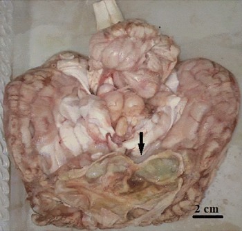
A rare cerebral hydatid cyst with sharply defined borders, occupying the frontal region of the brain, just below the cerebral cortex, was obtained from a cow slaughtered at Shiraz slaughterhouse, Fars Province, southern Iran. The exposed brain appeared greenish, and revealed the light brown wall of the cyst (fig. 1). The affected portion of the brain was fixed in 10% neutral buffered formalin. Following a routine histological tissue-processing procedure, the sample was embedded in paraffin blocks and serial sections were cut at a thickness of 4–5 μm and stained with haematoxylin and eosin (H&E). The cyst wall was recovered from the brain, rinsed five times with sterile phosphate-buffered saline (PBS) and preserved in 70% (v/v) ethanol until DNA isolation. Following removal of the ethanol, DNA was extracted from the cyst, using a commercial DNA extraction kit (DNAeasy, Qiagen, Valencia, California, USA), according to the manufacturer's protocol. The mitochondrial DNA was amplified using specific primers for cytochrome c oxidase 1 (cox1): JB3/JB4 (5′-TTT TTT GGG CAT CCT GAG GTT TAT -3′/5′-TAA AGA AAG AAC ATA ATG AAA ATG-3′) (Bowles et al., Reference Bowles, Blair and McManus1992). The 50 μl polymerase chain reaction (PCR) mixture contained 10 mm Tris-HCl, pH 8.4, 50 mm KCl, 1.5 mm MgCl2, 200 μm deoxynucleoside triphosphates (dNTPs), 20 pmol of each primer, 2 U Taq polymerase (Fermentas, Glen Burnie, Maryland, USA), 8 μl of DNA extracted as template and sterile distilled water as the negative control. PCR was performed in a thermocycler with the following program: an initial denaturation step at 95°C for 5 min; 35 cycles of denaturation at 94°C for 1 min, annealing at 55°C for 1 min and extension at 72°C for 1 min; followed by a final extension at 72°C for 5 min. The amplified products were purified with a PCR product purification kit (Bioneer, Alameda, California, USA) and sequenced directly using the capillary DNA analyser (ABI 3730; Applied Biosystems, Foster City, California, USA). Both forward and reverse nucleic acid sequence data were used to construct a concatenated sequence of cox1. Nucleotide sequence analysis was undertaken, using the National Center for Biotechnology information BLAST (Basic Local Alignment Search Tool) programs and databases. Also, the cox1 DNA sequences from different genotypes of E. granulosus, i.e. G1 (AY686562, AY686564, AY686565), G3 (M84663), G4 (AF346403), G5 (AB235846), G6 (AB208063, AF384099), G7 (AY686566, DQ062858, DQ856468), G8 (AB235848) and G10 (AB777927) existing in the GenBank database, were used for phylogenetic analysis. Multiple sequence alignments and construction of a phylogenetic tree were made with the maximum-parsimony method, using the software MEGA 4.0 (Tamura et al., Reference Tamura, Dudley, Nei and Kumar2007). The sequence profile for cox1 was submitted to GenBank with accession number KP259871.
Brain of the cow to show a large cyst occupying the frontal region.

Results and discussion
The cyst, which contained clear fluid, was dissected to reveal a cyst wall 1–2 mm thick with both germinal and laminated layers. Histopathological evaluation of tissue sections of brain stained with H&E showed granulomatous encephalitis, accompanied by eosinophilic infiltrations. Granulomatous lesions were located close to the cyst, with necrotic debris in the centre, surrounded by epithelioid macrophages and Langhans giant cells. Amorphous eosinophilic, Splendore–Hoeppli material, forming radiating clubs, was found in this case. Perivascular cuffing, gliosis, spongiosis and neural necrosis were other cerebral lesions.
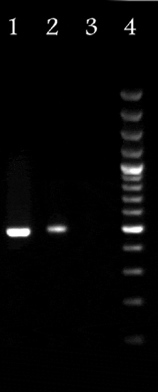
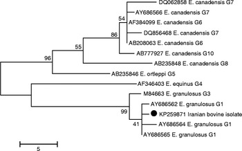
The PCR was positive for hydatid brain cyst, shown by producing a cox1 fragment of 493 bp compared with the positive control (E. granulosus) and molecular weight marker (fig. 2). The cox1 sequence similarity search using BLAST showed that the isolate belonged to a taxon of E. granulosus previously described by Bowles et al. (Reference Bowles, Blair and McManus1992) as the G1 genotype. In addition, the phylogenetic analysis of concatenated sequence data showed that this isolate was grouped with the E. granulosus sensu stricto (G1–G3 complex) cluster (fig. 3). Accordingly, the molecular results of the present report confirmed that the infection was caused by E. granulosus (G1 genotype).
Electrophoretic analysis of cytochrome c oxidase 1 (cox1) (493 bp) to show PCR amplification of the cerebral cyst sample (lane 2) compared with the molecular weight marker (lane 4, 100 bp), positive (lane 1) and negative (lane 3) controls.

Phylogenetic position of the causative agent of the cerebral hydatidosis in cattle and other Echinococcus granulosus genotypes inferred from the nucleotide sequences of partial cytochrome c oxidase 1 (cox1) using the maximum-parsimony method; numbers ranging from 41 to 99 indicate bootstrap values (%) from 1000 replicates with scale bar 5.

Molecular methods such as PCR and DNA sequencing have been used extensively to characterize strain grouping within E. granulosus (Zhang et al., Reference Zhang, Eslami, Hosseini and McManus1998; McManus & Thompson, Reference McManus and Thompson2003; Ahmadi & Dalimi, Reference Ahmadi and Dalimi2006). In the present study, we have undertaken the molecular examination of an isolate obtained from the brain tissue of cattle. The DNA sequencing of the mitochondrial cox1 gene of this isolate confirmed the occurrence of the G1 genotype of E. granulosus in the bovine host. The sheep/dog cycle (G1 genotype) is the most common cycle in Iran. Among the Iranian isolates that have been examined previously, the G1 genotype is prevalent in sheep, goats and cattle (Zhang et al., Reference Zhang, Eslami, Hosseini and McManus1998). However, sheep are the most important intermediate hosts for E. granulosus. In areas where cattle are kept together with other ruminants, cattle are likely to be exposed to the sheep strain through contact with carnivorous definitive hosts, especially dogs. Hydatid cysts are common incidental post-mortem findings in cattle in Iran, having been found most frequently in the liver and lungs. The main outcomes of this report are to emphasize the fact that hydatid cysts should be included in the differential diagnosis when a cystic brain lesion is found in animals from an endemic area for hydatid disease, and that the G1 genotype may also be found in cattle. The present case may be epidemiologically significant, so an accurate investigation, mainly at slaughterhouses, is necessary to determine the exact incidence and patterns of transmission among cattle.
In conclusion, molecular analysis concomitant to histopathological examinations is useful in the diagnosis of cerebral hydatidosis, which is reported for the first time from cattle in Iran.
Financial support
Financial support was received from Shiraz University, Shiraz, Iran.
Conflict of interest
None.
Ethical standards
This experiment was performed with the approval of the state committee on animal ethics, Shiraz University, Shiraz, Iran. Also, the recommendations of European Council Directive (86/609/EC) of 24 November 1986, regarding the protection of animals used for experimental purposes, were considered.