Introduction
One of the important diagnostic distinctions in cognitively impaired persons is between frontotemporal dementias (FTD) such as Pick disease and Alzheimer's disease (AD). Unfortunately, the ordinary office-based psychiatrist's verbal mental status examination is often not sensitive to the executive function deficits associated with frontal lobe damage, and clinicians must rely primarily on history to make the diagnosis of FTD. There are, however, nonverbal tests of executive function that can be readily performed by clinicians. These include performing simple programmed motor tasks or changing a pattern of motor activity once it has been begun (Dubois et al., Reference Dubois2000). The programmed motor tasks in current use were developed by Russian neuropsychologist Alexander Luria (1902–1977), who made extensive clinical observations that he correlated with surgical and pathological findings among brain-injured persons (Pribram, Reference Pribram, Luria and Haigh1973). One of his many observations was that persons with substantial frontal lobe lesions were unable to alter their responses to a programmed motor task when the order of actions in the task was changed. For example, a subject with a frontal lobe lesion who was asked to raise his right hand in response to one tap by the examiner and his left hand in response to two taps was able to do this without difficulty. After several repetitions, the order of tapping was changed, but the subject continued to alternately raise his right and left hands, despite being able to remember the rules for raising his hands. This indicated to Luria that the frontal lobe plays the regulatory function of facilitating and inhibiting responses, and various motor programs devised by Luria are used to help distinguish between FTD and other disorders based on this observation. Unfortunately, Luria did not investigate older adults with neurodegenerative diseases, so that the use of these motor programs in the differential diagnosis of dementing illnesses is not yet supported by data-based clinical studies.
The present study examined the utility of a more complicated programmed motor task – the three-step Luria test– to determine if difficulty performing this task helps to differentiate cognitively normal elders from persons with mild cognitive impairment (MCI; Petersen et al., 1999), FTD and AD. We also explored the relationship of the Luria test to age, education, and level of global impairment.
We hypothesized that inability to perform the Luria test correctly would differentiate normal elderly controls (NC) from persons diagnosed with FTD and AD, and that it would distinguish between early AD and FTD, but not between FTD and more advanced AD. We also hypothesized that the performance of persons with MCI would be intermediate between NC and AD subjects and that MCI subjects who showed impairment of the Luria test would be more likely to progress to dementia.
Methods
The material for this study was drawn from the database of the UT Southwestern Alzheimer's Disease Center (ADC) for the period between May 2002 and May 2009. We located the records of NC, MCI, FTD and AD subjects for whom the Luria test was recorded one or more times in addition to age, education, Clinical Dementia Rating score (CDR; Morris, Reference Morris1993), and Mini-Mental State Examination score (Folstein et al., Reference Folstein, Folstein and McHugh1975). During this time, performance of the Luria test by patients and NC had been routinely encoded in the ADC database for both clinical subjects and persons who were members of longitudinal cohorts of normal elderly control, MCI, FTD and AD.
In our clinic, the procedures of the three-step Luria test require that patients imitate three hand motions performed by the examiner. With fingers fully extended and the patient following, the examiner places his right hand with a cutting motion on his right knee or on a table, then in a fist with the knuckles down, and then palm down with fingers extended. Examiner and patient then repeat this three more times. The hand motions could be reinforced by counting from 1 to 3 along with each segment, or by saying “cut, fist, and slap.” Patients are then asked to repeat the movements unguided by the examiner. A score of 0 is recorded if the patient is unable to mimic the movement or complete three independent cycles. Luria's order of progression was fist, cut, and slap (Luria, Reference Luria and Bowden1970; Reference Luria1980); others have used the sequence of slap, fist, and cut (Cummings and Mega, Reference Cummings and Mega2003). Performance on the Luria test was scored as normal or abnormal. The test was judged to be abnormal if the hand motions differed in type or sequence from that of the examiner. A common error was having the fingers flexed instead of extended for the first movement.
All subjects underwent standardized psychiatric, neurological and neuropsychological evaluation as described elsewhere (Weiner et al., Reference Weiner1991). Control subjects were individuals without subjective memory complaint (confirmed by an informant) whose neuropsychological testing was within normal limits for age and education. Clinical diagnoses were made by a clinical algorithm based on standard criteria for MCI that included amnestic, multiple domain, and single domain non-memory MCI (Petersen et al., Reference Petersen2001). For MCI, we required cognitive task performance > 1 SD below normative population. FTD was diagnosed using the criteria of Neary et al. (Reference Neary1998) and probable AD using the NINCDS/ADRDA criteria (McKhann et al., Reference McKhann1984).
Clinical staging of subjects at the time that the test was performed was based on the CDR, which rates global impairment as 0 = unimpaired, 0.5 = questionably impaired, 1 = mild dementia, 2 = moderate dementia and 3 = severe dementia. The diagnosis of “normal” required CDR = 0; for MCI, CDR = 0.5. A diagnosis of FTD required CDR ≥ 0.5 and AD required CDR ≥ 1.0.
Statistical analyses
Two-way analysis of variance (ANOVA) compared diagnostic groups (NC, MCI, FTD and AD) and Luria test (abnormal versus normal) on the measures of education and age; Bonferroni post hoc pairwise comparisons were performed if the ANOVA was found to be significant. χ2 was used to compare groups when the data were dichotomous; when the χ2 was found to be significant, a Tukey-type multiple comparison test among proportions was performed (Zar, Reference Zar1996). Assumptions for all statistical tests were checked for violations. SPSS version 18 was used for all analyses and the significance level for all analyses was p<0.05.
Results
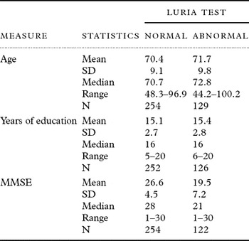
Data were available for 383 persons. Table 1 shows the age and level of education for each diagnostic group. There was essentially no difference between educational levels for any of the four groups (F(3,370) = 0.24, p = 0.866) and no relationship of education to failure in performing the Luria test (F(1,370) = 1.05, p = 0.307) as depicted in Table 2. The FTD group was significantly younger than the other groups (group effect: F(3,375) = 11.85, p < 0.001; Bonferroni post hoc maximum p = 0.014). In addition, the NC were significantly younger than the AD group (Bonferroni post-hoc p < 0.001). There was a nonsignificant effect for poorer performance on the test with advancing age (F(1,375) = 1.72, p = 0.191).
Age, education and MMSE by diagnostic group

NC = normal control; MCI = mild cognitive impairment; FTD = frontotemporal dementia; AD = Alzheimer's disease; MMSE = Mini-Mental State Examination.
Performance on Luria test by age, education, and MMSE

MMSE = Mini-Mental State Examination; SD = standard deviation.
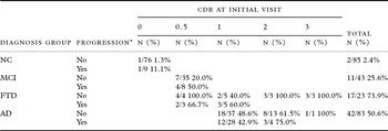
As shown in Table 3, an abnormal Luria test occurred in 2.3% of normal elderly controls, in 21.4% of those with MCI, 69.8% of those with FTD, and 54.9% of those with AD (χ2(3) = 117.47, p < 0.001). The frequency of abnormal Luria tests in all diagnostic groups increased with functional impairment as assessed by the CDR. When CDR = 3, 100% of the FTD and 72.2% of the AD subjects had abnormal Luria performance. Using a Tukey-type multiple comparisons test among proportions, all groups were pairwise significantly different from other groups (p < 0.05) except AD and FTD subjects.
Impaired Luria test by diagnosis group and Clinical Dementia Rating score for measures recorded at initial Luria test visit

NC = Normal Control, MCI = Mild cognitive impairment, FTD = Frontotemporal dementia, AD = Alzheimer disease; CDR = Clinical Dementia Rating
The number (%) of patients with abnormal Luria test results by disease progression, diagnosis group and initial CDR global scores

* Progression is based upon an increase in the CDR Global Score at a follow-up visit 9 or more months after the initial visit.
NC = Normal Control, MCI = Mild cognitive impairment, FTD = Frontotemporal dementia, AD = Alzheimer disease; CDR = Clinical Dementia Rating.
Of the 85 normal subjects with more than one visit and a minimum of 9 months between visits, nine (10.6%) progressed to CDR = 0.5 over the course of an average of 2.6 years (range 0.82 to 6.30 years); of these nine subjects, one (11.1%) had an abnormal Luria test at the first visit (Table 3). Of the 43 subjects diagnosed as MCI at their initial evaluation and for whom follow-up data were available, eight (18.6%) progressed to AD after periods of follow-up ranging from 0.75 to 7.01 years (mean 2.8 years). Of these eight subjects, four (50%) had an abnormal Luria test at the first visit. There was no difference in the percentage of abnormal Luria findings at the initial visit when the MCI group was divided into amnestic (5/21 = 23.8%), multiple domain (7/31 = 22.6%), and single non-memory domain (0/4 = 0.0%; Fishers Exact p < 0.7799).
A small number of subjects were diagnosed with purely psychiatric disorders (11 major depression, 1 schizophrenia). Of these, only 1/12 (8%) was unable to perform the Luria test correctly.
Discussion
It is not surprising that inability to perform the series of three hand motions does not readily distinguish between FTD and AD. By the time AD becomes clinically detectable, its pathology likely involves the parietal and frontal lobes (Braak and Braak, Reference Braak and Braak1991). This may explain the findings from a previous study where greater impairment on the Luria test was observed for AD relative to frontal lobar degeneration patients (Lipton et al., Reference Lipton2005). In that study it is was proposed that performance by the AD group may have been impacted by the lack of verbal instructions given during demonstration of the task. However, the present study included verbal instructions during the teaching of the Luria test and no difference between FTD and AD was found. The increasing frequency of an abnormal Luria test with increasing functional impairment, although not surprising, also makes it less useful as a differential diagnostic aid in late-stage dementia. Despite the well-known prevalence of executive dysfunction in schizophrenia (Kravariti et al., Reference Kravariti2009) and major depression (Withall et al., Reference Withall, Harris and Cumming2009), 92% of the persons with purely psychiatric diagnoses were able to perform the Luria test. The rarity of an abnormal Luria test in the elderly normal cohort suggests the utility of this measure in distinguishing persons with normal cognition from persons with early AD or FTD. We are unable to explain the appearance of an abnormal Luria test in persons with normal cognition, but just as there is a range of cognitive function among normal people, there is likely to be a range of praxis.
We obtained autopsy reports for 62 of our subjects diagnosed as probable AD or FTD. We previously reported that 86.2% of persons diagnosed in our clinic as probable AD met neuropathologic criteria for AD (Ranginwala et al., Reference Ranginwala2008). In this study, we found 90% (44/49) concordance between the clinical diagnosis and autopsy diagnosis of probable AD and 85% (11/13) concordance between the clinical and autopsy diagnosis of FTD. A potential source of bias is that although the Luria test was to have been performed on all patients, it may have been performed more often in persons whose presentation suggested frontal lobe impairment. Of the 581 persons seen from 2002 to 2010, 383 were included in this analysis. The ratio of total persons seen to total persons studied in each diagnostic group was 131/143 NC, 56/64 MCI, 43/64 FTD, and 153/183 AD subjects. The lower percentage of FTDs studied may be that following the initial diagnostic workup, their behavioral problems precluded them being followed up in clinic.
Another source of bias is that the test was not performed in exactly the same way for all subjects, but this variance was not associated with any diagnostic group.
In summary, impaired performance in the Luria test is rare in persons with normal cognition and occurs in < 10% of persons with MCI. Thus, it can be helpful in distinguishing normal and MCI subjects from AD and FTD, but does not differentiate between FTD and AD. The Luria test may be useful cross-culturally because it is non-verbal and its performance is unaffected by education and only minimally by age. We also have preliminary evidence that the Luria test may help to distinguish psychiatric from dementing illnesses, but that finding awaits further study.
Conflict of interest
None.
Description of authors' roles
M. F. Weiner designed the study, formulated the research question, supervised data collection, and participated in the preparation of the paper. L. S. Hynan assisted in the study design, performed the data analysis, and participated in preparation of the paper. H. Rossetti helped analyze the data and write the paper. J. Falkowski participated in preparation of the paper.
Acknowledgments
This study was funded by the Wallace, Barbara & Kelly Foundation and NIA AG12300.