Introduction
Tracheostomy is indicated in selected patients undergoing treatment of head and neck cancers, either at the time of surgery or during irradiation. Patients who have received prior multimodality therapy are at an increased risk of persistent tracheocutaneous fistula, and simple closure of the tracheocutaneous fistula can be ineffective in a subset of this patient population.Reference White and Smitheringale1
This report focuses on a 54-year-old male with a history of tumour–node T4N1 stage squamous cell carcinoma of the mandible, treated with primary surgery and free flap reconstruction (including tracheostomy). Following surgery, he was treated with adjuvant radiation of a standard dose and duration, and was decannulated with appropriate closure of the tracheostomy site by secondary intention. His recovery was complicated by mandibular plate extrusion and prolapse of the lower lip. He also strongly desired implant-based dental rehabilitation, for which his original fibula provided insufficient bone stock. As such, he underwent a second surgical procedure to reconstruct the mandible with a free fibular osseocutaneous tissue transfer, which also included a tracheostomy. His in-patient post-operative course was uneventful and he was decannulated prior to discharge.
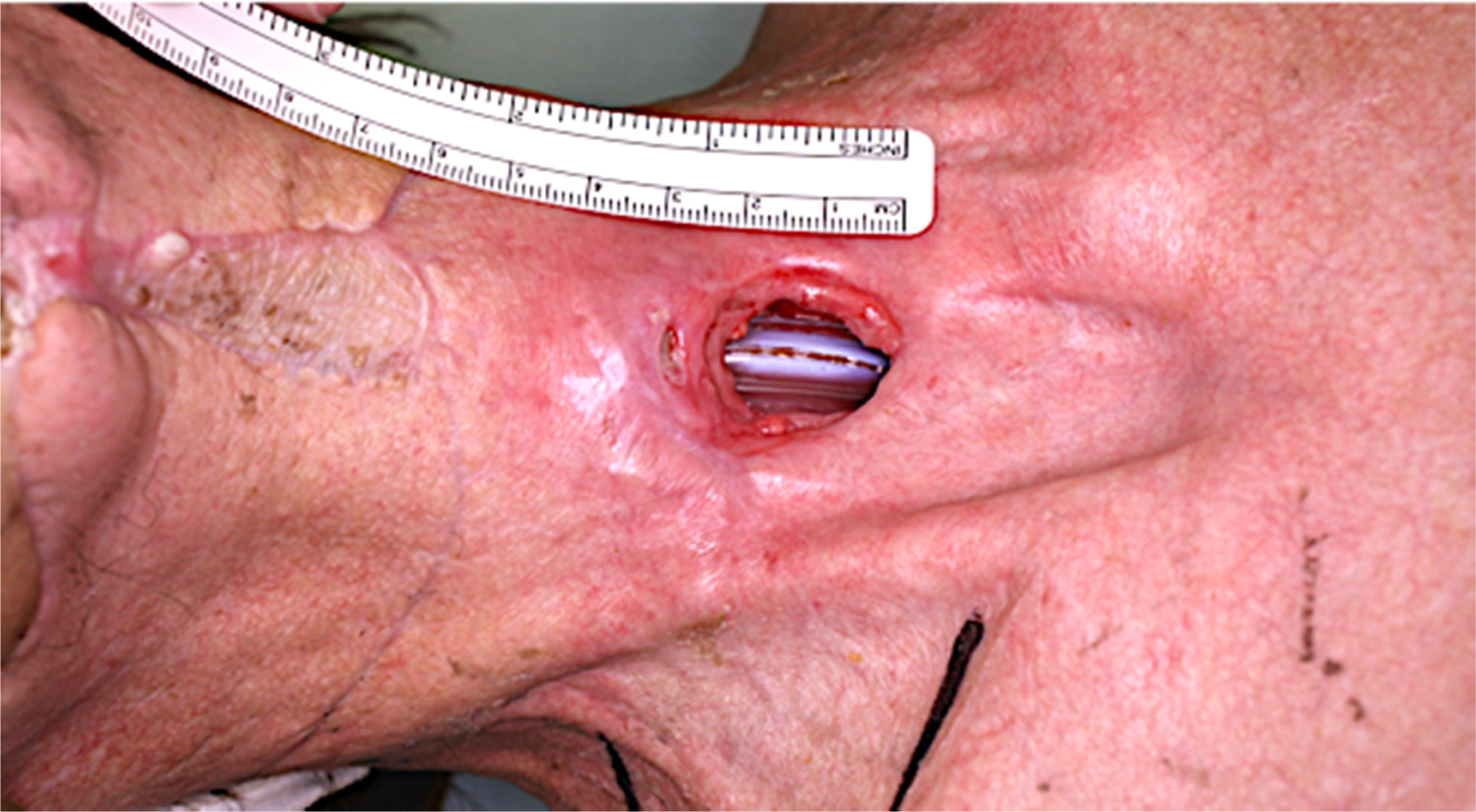
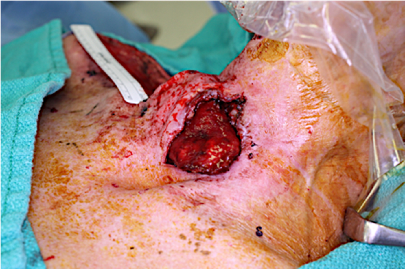
At follow up, the patient was observed to have a persistent tracheocutaneous fistula. He was taken back to the operating theatre for repair of the tracheocutaneous fistula. The tracheal defect itself was closed. Bilateral, pedicled sternohyoid rotational flaps were used to bolster the repair. Unfortunately, the patient suffered a surgical site infection. This led to a breakdown of the repair and the trachea itself, leaving a 3 cm tracheocutaneous fistula (Figure 1). A return to the operating theatre was then planned, for staged repair using conchal bowl cartilage and a supraclavicular flap.
Persistent tracheocutaneous fistula.

Materials and methods
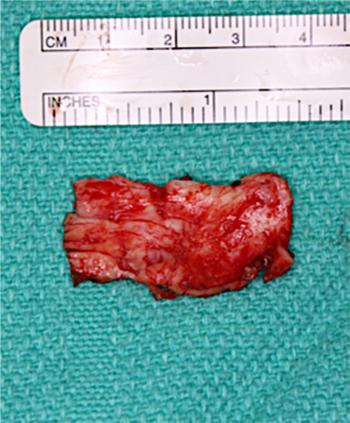
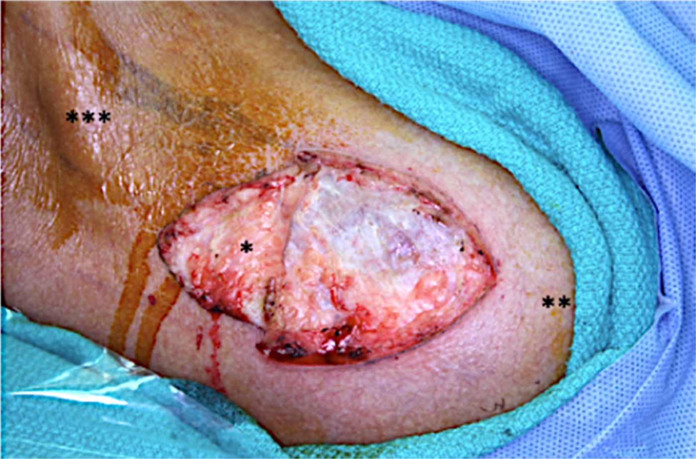
During the first stage of repair, a 2 cm × 3.5 cm conchal bowl cartilage graft was harvested from the patient's left ear (Figure 2). The right transverse cervical artery was then identified with Doppler ultrasound, and traced over the clavicle and acromioclavicular joint. A 5 cm × 5 cm wedge-shaped area at the distal aspect of the planned supraclavicular flap was then elevated (Figure 3). The cartilage graft was then placed within this tissue pocket with the convex side down, and was fixed to the underlying fascia using monofilament suture. Closure of the skin was then performed.
Conchal bowl cartilage graft.

Tissue pocket to house conchal bowl cartilage graft. *Partially elevated supraclavicular flap; **acromioclavicular joint; ***trapezius muscle

The second stage of repair took place three weeks later. This entailed the harvest of a 2 cm × 3 cm section of septal mucosa. Next, the course of the right transverse cervical artery was traced using Doppler ultrasound. The supraclavicular flap was partially raised and the cartilage was located by palpation. The mucosal graft was then centred over the cartilage graft and secured into place with monofilament sutures. A bolster dressing was subsequently placed over the surgical site.
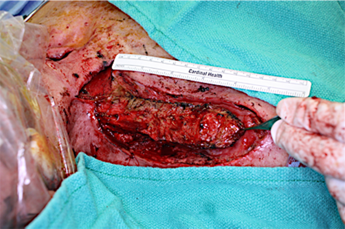
The third and final stage of repair occurred after a further three weeks had passed. The patient was taken to the operating theatre and intubated transorally. Once again, the course of the transverse cervical artery was traced out and marked. The supraclavicular flap containing the conchal cartilage and septal mucosa graft was then elevated from distal to proximal, with care taken to preserve its blood supply (Figure 4).
De-epithelialised supraclavicular flap with conchal bowl cartilage and septal mucosa graft visualised at distal edge.

A subcutaneous tunnel was then created between the proximal portion of the flap and the site of the tracheocutaneous fistula. Once the edges of the tracheocutaneous fistula were freshened, the cartilage graft was sutured into place, with the septal mucosa and concave surface facing inwards, achieving watertight closure (Figure 5).
Inset of supraclavicular flap with cartilage and septal graft (on deep surface) into tracheocutaneous fistula site.

A split thickness skin graft from the thigh was used to cover the de-epithelialised portion of the flap. The supraclavicular donor site was closed primarily. The patient was transferred to the intensive care unit, where he was kept anaesthetised and intubated for 7 days. Extubation was carried out successfully at the end of this period.
Results
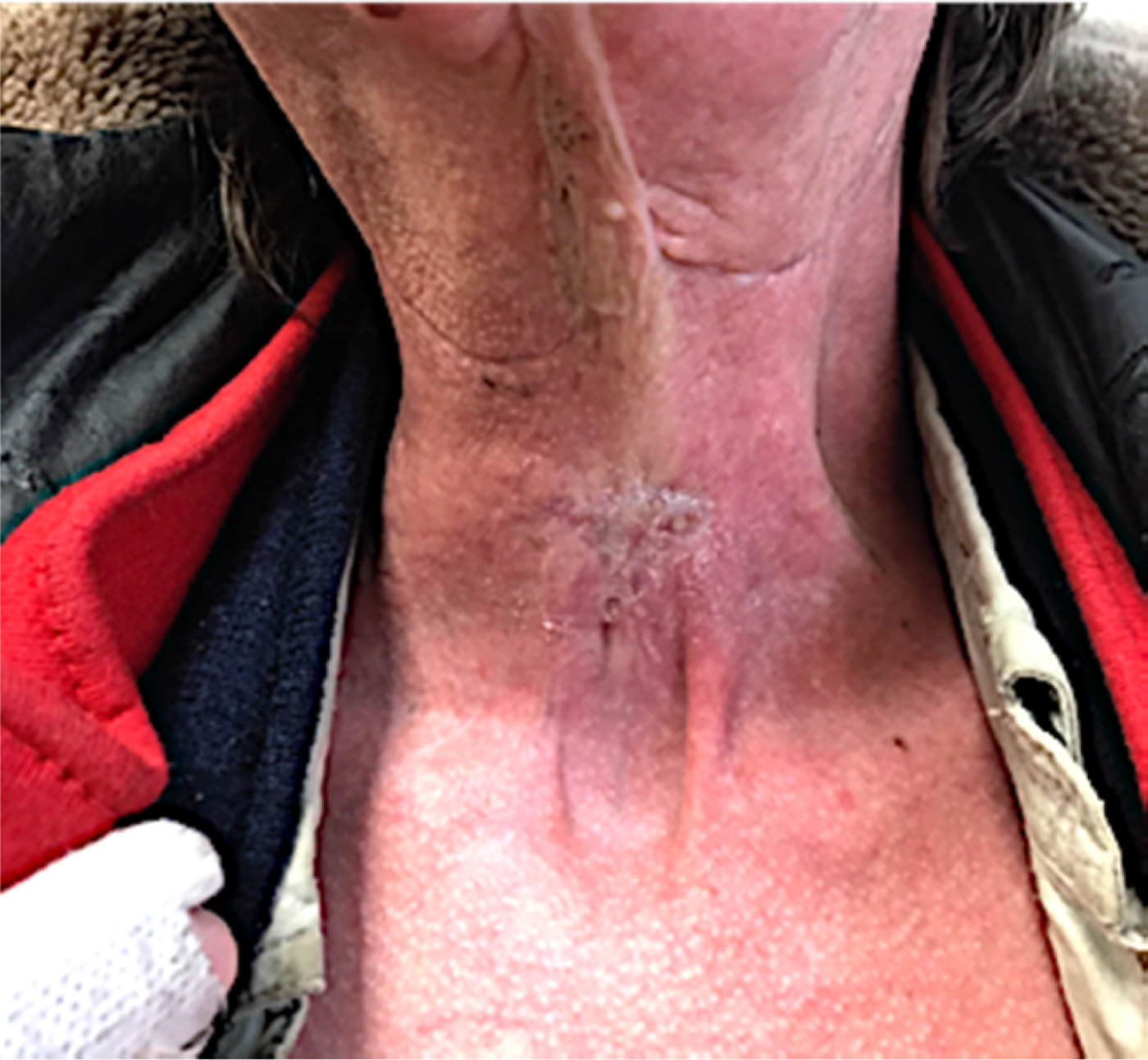
The patient's post-operative course was uneventful. The follow-up visits over the two years since surgery have revealed no dehiscence of the tracheocutaneous fistula repair and no signs of persistent fistulisation (Figure 6). The supraclavicular donor site has healed well, with no residual effect on shoulder function.
Photograph of patient at post-operative follow up.

Discussion
Tracheocutaneous fistula rates in the paediatric population range from 11 to 43 per cent after decannulation.Reference Joseph, Jani, Preece, Bailey and Evans2–Reference Tasca and Clarke4 However, this is not the case in adults, as most tracheostomy sites do heal without a persistent tracheocutaneous fistula. Small tracheocutaneous fistulas can be managed conservatively with excision of the fistulous tract, followed by simple or multi-layered closure with local rotational flaps including sternocleidomastoid, sternohyoid, platysma and skin flaps. When a large portion of the anterior tracheal wall is missing, a cartilage graft may be used for closure and support. In a patient such as ours, with multiple previous tracheostomies and irradiation to the head and neck, local tissue healing is often ineffective, leading to frequent wound breakdowns.
One of the options for repair of a tracheocutaneous fistula is to excise the tract and complete an end-to-end anastomosis of the tracheal ends.Reference Rob and Bateman5,Reference Grillo6 In patients who have undergone previous irradiation and have demonstrated poor wound healing, this is a high-risk surgical option. For the same reasons, local flaps often fail in these patients.
Jackson and Babcock, in 1934, described a lined, bipedicled flap reinforced with conchal cartilage for tracheocutaneous fistula.Reference Jackson and Babcock7 Since then, there have been various reports of using a combination of local or free flaps with or without cartilage for repair of tracheocutaneous fistula.Reference Drezner and Cantrell8–Reference Shinohara, Yuzuriha, Matsuo, Kushima and Kondoh14
Fasciocutaneous flaps with prefabricated cartilage to provide anterior wall support have been described. Shinohara et al. first described the use of a deltopectoral flap with costal cartilage and a palatal mucosal graft for the repair of tracheal stenosis. In the two cases they presented, the patients did not undergo irradiation, and the exact defect size was unclear but was at least 35 mm. Riedel et al. described the use of a deltopectoral flap with auricular cartilage.Reference Shinohara, Yuzuriha, Matsuo, Kushima and Kondoh14 Their series included two patients who had previously undergone irradiation, in whom the tracheocutaneous fistula was successfully closed with the deltopectoral flap. Royer et al. described the use of a radial forearm free flap with conchal cartilage and buccal mucosa for tracheocutaneous fistula closure.Reference Royer, Royer, Ting, Weisberger and Moore11 The patient had undergone previous irradiation to the neck, but the vascular anatomy was unaltered, thus allowing for a free flap.
Costal cartilage has also been used previously to support the anterior tracheal wall in the closure of large tracheocutaneous fistulas. As outlined by Royer et al., costal cartilage for a short segment defect is not ideal as it does not have the natural curve of the native tracheal cartilage. Additionally, harvesting costal cartilage has associated potential complications, including pneumothorax, hemothorax and post-operative pain, resulting in atelectasis and pneumonia.
Here, we describe a novel technique using a supraclavicular flap with prefabricated conchal cartilage for the closure of a tracheocutaneous fistula in a previously irradiated patient. Additionally, the patient had undergone two previous free tissue transfers; as such, he had depleted neck vasculature, making an additional free tissue transfer challenging. The fistulous tract was excised, followed by a watertight tracheal repair. The curve of the conchal cartilage was oriented to support the anterior tracheal wall, with the concave aspect facing the interior aspect of the trachea. Use of conchal cartilage also avoided the use of costal cartilage, thus avoiding its associated complications. Given the widespread use and reliability of the supraclavicular flap, we believe this technique can be readily adopted for this rare problem.
Competing interests
None declared