Case presentation
The clinical case describes a 13-year-old boy (62 Kg, 165 cm) with a history of transposition of the great arteries, ventricular septal defect, and pulmonary stenosis. He underwent a neonatal Rashkind procedure, followed for 1 year of age by surgical correction using the Réparation à l’Etage Ventriculaire (REV) procedure combined with the Lecompte maneuver. The following year, at the age of 2, he developed severe subaortic obstruction, requiring reoperation for subaortic tunnel reconstruction, myectomy, and reimplantation. During this procedure, due to severe combined valvular dysfunction involving the residual right ventricular outflow tract, a 19 mm No-React Injectable BioPulmonic prosthesis (Biointegral Surgical Inc., Ontario, Canada) was implanted.
During cardiology follow-ups in 2023, at the age of 12, the patient began to report a reduction in exercise tolerance. Echocardiograms revealed severe stenosis and insufficiency of the pulmonary bioprosthesis, along with evidence of right ventricular dilation, marked hypertrophy, and mild dysfunction. Consequently, in January 2024, the patient was admitted for diagnostic cardiac catheterisation, which confirmed a severe increase in right ventricular systolic pressures, a significant peak-to-peak gradient across the pulmonary bioprosthesis, and normal pressures in the pulmonary artery.
The case was presented at a multidisciplinary discussion, which recommended attempting a transcatheter pulmonary valve implantation using a valve-in-valve technique.
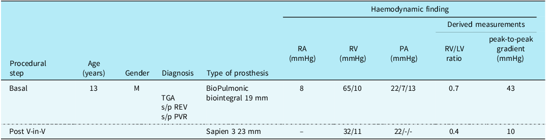
The procedure was performed in May 2024 under general anaesthesia. A baseline diagnostic catheterisation was conducted, revealing a severe peak-to-peak gradient of 43 mmHg. Balloon dilation of the biological prosthesis was then performed using Atlas balloons 20 × 20 mm (Bard Medical Inc., Tempe, AZ). A simultaneous aortography did not show any signs of coronary compression. Following this, further dilation was carried out with an Atlas 22 × 20 mm balloon, resulting in good expansion of the valve. A subsequent right ventriculography ruled out any angiographically detectable complications. Given the satisfactory expansion, the decision was made to implant a 23 mm Sapien 3 valve (Edwards Lifesciences Corp., Irvine, California). The transcatheter heart valve was implanted using a 24 Fr 65 cm long DrySeal long introducer (W. L. Gore & Associates, Inc., Flagstaff, Arizona). Post-implant pulmonary arteriography excluded any intra-perivalvular regurgitation. Post-procedure haemodynamic assessment showed a residual peak-to-peak gradient of 10 mmHg, with systolic right ventricular pressures below half of systemic pressure. There were no operative or postoperative complications. Table 1 provides a summary of pre- and post-procedural parameters. Figure 1 illustrates some key steps of the procedure.
Summary of demographic characteristics and pre- and post-procedural haemodynamic findings

TGA= Transposition of the Great Arteries (TGA). s/p REV= Status post Réparation à l’Étage Ventriculaire (REV) procedure. s/p PVR= Status post Pulmonary Valve Replacement (PVR). V-in-V: Valve-in-valve (V-in-V) procedure.
(a) Shows a fluoroscopic image in lateral projection. The image depicts the 19 mm no-react injectable BioPulmonic prosthesis (Biointegral Surgical Inc.) in an anterior position. The patient, affected by transposition of the great arteries, ventricular septal defect, and pulmonary stenosis, underwent correction with the REV procedure combined with the Lecompte manoeuvre. The image illustrates an occlusion test of the bioprosthesis using an Amplatzer Sizing Balloon II (Abbott) with simultaneous aortography. The concurrent aortography allowed for the assessment of the coronary circulation’s relationship with the bioprosthesis before proceeding with high-pressure balloon pre-dilation. The coronary position was found to be remote from the bioprosthesis. (b) Shows a fluoroscopic image post-transcatheter valve-in-valve implantation of the Sapien 3 valve. The image depicts a post-implantation pulmonary angiography, demonstrating the continent 23 mm Sapien 3 valve with a satisfactorily expanded frame. Adequate frame expansion was achieved through pre-dilation of the 19 mm no-react injectable BioPulmonic prosthesis (Biointegral Surgical Inc.) using high-pressure balloons. The peak-to-peak transvalvular gradient recorded after the procedure was 10 mmHg.

The patient was discharged on the third postoperative day, with antiplatelet therapy consisting of low-dose aspirin and clopidogrel. The pre-discharge echocardiogram documented a competent pulmonary bioprosthesis with a maximum peak gradient of 20 mmHg. At the 6-month follow-up after the procedure, the post-procedural peak echocardiographic gradient across the pulmonary prosthesis was measured at 28 mmHg, with no evidence of regurgitation.
Discussion
Clinical benefits of percutaneous valve implantation in degenerated valve or conduits are well established.Reference Lurz, Nordmeyer and Giardini1–Reference Hascoët, Bentham and Giugno4 Generally, the valve-in-valve procedure involves the implantation of a transcatheter heart valve inside the frame of a previously implanted biological prosthetic valve. Implanting a transcatheter heart valve inside a bioprosthesis with a small internal diameter relative to the patient’s Body Surface Area may result in a prosthesis with an inadequate effective orifice area, leading to patient-prosthesis mismatch. Indeed, the internal diameter of the rigid frame of a bioprosthesis can limit the size of the transcatheter-implanted prosthetic valve. Placing a percutaneous valve within the previously degenerated valve further narrows the ultimate effective orifice size.
However, intentional bioprosthetic pulmonary valve fracture or modification to enlarge the true inner diameter using high-pressure balloons has been reported as a means of facilitating further expansion of the valve also in pulmonary position, showing a significantly wider inner diameter of the valve and a decreased right ventricular outflow tract gradient in the fracture group, with no fracture-related adverse events.Reference Shahanavaz, Asnes and Grohmann5,Reference Tanase, Grohmann, Schubert, Uhlemann, Eicken and Ewert6 Unlike valves with rigid rings, such as the Magna Ease (Edwards Lifesciences Corporation, Irvine, California), the Injectable BioPulmonic prosthesis features a self-expandable nitinol frame without a rigid ring. This structural difference may affect both the response to high-pressure balloon dilation and the degree of achievable expansion. For this reason, we adopted a stepwise approach using progressively larger balloon diameters to gradually modify the bioprosthetic frame, rather than attempting intentional valve frame fracture with a high-pressure balloon.
No-React Injectable BioPulmonic prosthesis consists of a porcine pulmonary valve covered with a No-React-treated bovine pericardium sleeve and mounted on a self-expandable Nitinol stent. To our knowledge, the literature does not report the effectiveness of intentional fracture or frame modification for these valves in anticipation of a valve-in-valve procedure.
Considering the lack of data in the literature, we performed a pre-treatment in vitro test to study the response of the No-React Injectable BioPulmonic prosthesis to pre-dilation with high-pressure balloons (Figure 2).
(a) photograph of the 23 mm no-react injectable BioPulmonic prosthesis (Biointegral Surgical Inc.). The three leaflets can be seen in the closed position in the image. (b) The same 23 mm no-react injectable BioPulmonic prosthesis (Biointegral Surgical Inc.) from image 2.A after dilation with a 26 mm high-pressure Atlas Gold balloon (Bard Medical). One of the three leaflets of the bioprosthesis appears retracted, showing a clear central coaptation defect. (c) Fluoroscopic image (in cranio-caudal projection) of the same 23 mm no-react injectable BioPulmonic prosthesis (Biointegral Surgical Inc.) shown in image 2.A. The radiopaque nitinol frame can be recognised in the fluoroscopic view. (d) Fluoroscopic image (in cranio-caudal projection) of the same 23 mm no-react injectable BioPulmonic prosthesis (Biointegral Surgical Inc.) shown in image 2.A after multiple dilations with a 26 mm high-pressure Atlas Gold balloon (Bard Medical). One can see irregularities and distortion of the bioprosthesis’s nitinol frame, although there are no fractures apparent upon inspection.

The only prosthesis available for our in vitro test was the No-React Injectable BioPulmonic 23 mm. The bioprosthesis had an internal stent diameter of 23 mm and a height of 23 mm. The bioprosthesis was subjected to balloon dilation using a non-compliant balloon with a diameter of 26 mm, under fluoroscopic and direct visual control. Despite the non-compliant balloon Atlas Gold 26 × 20 mm achieving full expansion to its nominal diameter during dilation at 10 ATM, we observed partial recoil of the bioprosthesis, with post-dilation inner diameters of 25 mm. Furthermore, it was evident that after high-pressure balloon dilation, one leaflet appeared retracted with a noticeable coaptation deficit. No stent fracture was demonstrated. The outcome of the in vitro test demonstrated the potential modification and dilation of the frame of the No-React Injectable BioPulmonic prosthesis (Biointegral Surgical Inc.) using high-pressure balloons, supporting us in planning the transcatheter pulmonary valve implantation procedure. In vitro tests often differ from in vivo outcomes due to tissue elasticity, dynamic haemodynamics, and anatomical constraints. In our case, unlike the in vitro assessment, the stepwise increase in balloon diameter was a deliberate strategy to gradually evaluate the valve’s response and avoid sudden structural damage. Additionally, another difference between the in vitro and in vivo settings—although difficult to quantify—is that, despite some degree of recoil observed in vitro, the authors perceive the extent of recoil in vivo to be significantly greater.
Conclusion
Our in vitro test demonstrated the feasibility of modifying the frame of the No-React Injectable BioPulmonic prosthesis with high-pressure balloon dilation to increase its true inner diameter. This strategy, applied in a complex valve-in-valve procedure, resulted in excellent short-term outcomes in a 13-year-old patient. While long-term data are still needed, these findings encourage the use of intentional frame dilation in selected cases where a small internal diameter might otherwise compromise the success of transcatheter valve-in-valve implantation.