

Introduction
The growing issue of microbial resistance and the lack of new antimicrobials entering the market has led to concern regarding the over use and application of antibiotics in the feed industry and subsequent effects on human health. Antibiotics have been given at sub-therapeutic levels as in-feed supplements to a broad spectrum of livestock (Li, Reference Li2017), most notably in pigs and poultry. Antibiotics and other antimicrobials are mainly used prophylactically in feed against gastrointestinal diseases such as diarrhoea which reduces mortality (Thacker, Reference Thacker2013). As a result, the food and drug administration (FDA) in the USA made recommendations to limit medically important antimicrobials to therapeutic use in animals and deemed the use of antimicrobials for in feed purposes to improve animal weight gain and feed efficiency as an inappropriate application (FDA-CMV, 2012). Subsequently the Preservation of Antibiotics for Medical Treatment Act, 2017 phased out the use of antibiotic use in food-producing animals in the USA (Slaughter, Reference Slaughter2017). Under the act, medically important antibiotics cannot be administered in feed at sub-therapeutic levels to food-producing animals for disease control. However, if there is a significant risk of disease or infection transmission from the farm premises to the animal, it is permissible to use antibiotics. This legislation change followed the prohibition of antibiotic use as a growth promotor in animal feed by the European union (European-Commission, 2005) due to the risk of resistance being passed from livestock through the food chain to human consumers (Phillips et al., Reference Phillips, Casewell, Cox, De Groot, Friis, Jones, Nightingale, Preston and Waddell2003).
The World Health Organisation covering South East Asia region (SEAR), is a region that uses intensive farming methods but lack regulations and restrictions on antibiotic use in food producing animals that is seen in the EU and USA. Several challenges and requirements for implementing policy changes need to be addressed to combat antimicrobial resistance in food producing animals in each of the SEAR countries (Goutard et al., Reference Goutard, Bordier, Calba, Erlacher-Vindel, Góchez, de Balogh, Benigno, Kalpravidh, Roger and Vong2017). The increased restrictions and regulations on antibiotic usage has exacerbated farmers concerns on issues of controlling livestock infections and poor feed conversions (Rosen, Reference Rosen2004) which have a direct impact on farmers profitability and animal health (Coffman et al., Reference Coffman, Augsburg, Beran, Colten, Greig, Halloran, Hayes, Kaneene, MacDonald, McNutt, Meeker, Nickerson and Seay1999; Cervantes, Reference Cervantes2015).
The demand for an alternative to counter infection in livestock and in humans has led to increased research and application of alternative methods to control pathogenic bacteria. This has come in the form of antimicrobials (Guani-Guerra et al., Reference Guani-Guerra, Santos-Mendoza, Lugo-Reyes and Teran2010; Leonard et al., Reference Leonard, Affolter and Bevins2012), feed enzymes that influence microflora (Halas and Nochta, Reference Halas and Nochta2012), probiotics (Anadón et al., Reference Anadón, Martínez-Larrañaga and Aranzazu Martínez2006) and prebiotics including mannan oligosaccharides (MOS; Spring et al., Reference Spring, Wenk, Connolly and Kiers2015) and fructooligosaccharides (FOS; Ricke, Reference Ricke2015). Prebiotics, such as MOS products derived from Saccharomyces cerevisiae, take advantage of the means by which pathogenic bacteria invade the host via the gut through binding to mannan molecules that coat the intestines (Halas and Nochta, Reference Halas and Nochta2012).
Certain strains of ETEC are responsible for causing food borne diseases in humans and enteric diseases such as diarrhoea and dysentery in animals. These pathogenic bacteria bind to intestinal epithelial cells through external projections from their cell wall known as pili or fimbriae. E. coli expressing type-1 fimbriae have a specific α-D-mannan binding tip which facilitates their adhesion and invasion of epithelial cells, causing disease (Ofek et al., Reference Ofek, Mirelman and Sharon1977; Hartmann et al., Reference Hartmann, Papavlassopoulos, Chandrasekaran, Grabosch, Beiroth, Lindhorst and Röhl2012). The mannoproteins isolated from the yeast competitively bind to the type-1 fimbriae thus preventing the ETEC from binding to mannose residues on cells. The E. coli strain used in this study possesses H fimbriated mannose binding regions (Fein, Reference Fein1981; Kennan and Monckton, Reference Kennan and Monckton1990), that facilitates binding to mannose residues present on human and pig intestinal cells. Previous findings demonstrated the ability of the E. coli strain used here to invade intestinal porcine epithelial cells (IPEC; Horgan et al., Reference Horgan, Madden and Jacques2018).
Humans and pigs are infected in the same manner by similar enteric microbes which makes this study examining the control of infection applicable to both species (Yuan et al., Reference Yuan, Ward, Rosen, To and Saif1996; Steele et al., Reference Steele, Feng, Parry and Tzipori2010; Brady et al., Reference Brady, Radhakrishnan, Liu, Magoun, Murphy, Mukherjee, Donohue-Rolfe, Tzipori and Leong2011) as symptoms for infected pigs are very similar to humans (Meurens et al., Reference Meurens, Summerfield, Nauwynck, Saif and Gerdts2011). Human and pig intestines are structurally conserved, which enables strong correlations to be derived between pig and human studies in vivo (Vitovec and Koudela, Reference Vitovec and Koudela1992; Nedrud, Reference Nedrud1999; Zhang et al., Reference Zhang, Widmer and Tzipori2013) and in vitro (Geens and Niewold, Reference Geens and Niewold2011; Brosnahan and Brown, Reference Brosnahan and Brown2012). Pigs and humans share similar microflora colonisation of the large and small intestines (Miller and Ullrey, Reference Miller and Ullrey1987; Pang et al., Reference Pang, Hua, Yang, Ding, Che, Cui, Jia, Bucheli and Zhao2017).
Piglets have a greater susceptibility to severe infections during their weaning phase, most notably in the form of PWD caused by E. coli infection of the intestines which features prominently in the first two weeks of weaning (Rhouma et al., Reference Rhouma, Fairbrother, Beaudry and Letellier2017). Infection can result in poor growth due to dehydration and a lack of food absorption that ultimately can lead to death, which is costly to pig producers (Amezcua et al., Reference Amezcua, Friendship, Dewey, Gyles and Fairbrother2002; Fairbrother et al., Reference Fairbrother, Nadeau and Gyles2005). Weaned piglets have long been administered antibiotic-supplemented feed to control harmful intestinal infections during such succeptible periods (Verstegen and Williams, Reference Verstegen and Williams2002). In-feed antibiotics have an added effect of promoting animal growth (Cromwell, Reference Cromwell2002). MOS has demonstrated substantial health benefits and has significantly improved piglet outcomes during their susceptible weaning period in the absence of conventional antibiotics (Smith et al., Reference Smith, Harris, Johnson, Silbergeld and Morris2002; White et al., Reference White, Newman, Cromwell and Lindemann2002; Burkey et al., Reference Burkey, Dritz, Nietfeld, Johnson and Minton2004; Castillo et al., Reference Castillo, Martin-Orue, Taylor-Pickard, Perez and Gasa2008; Liu et al., Reference Liu, Piao, Kim, Wang, Shen, Lee and Li2008; Zhao et al., Reference Zhao, Jung and Kim2012), without the risk of antimicrobial resistance (AMR) developing.
Feeding MRF derived from MOS has resulted in improved weight gain, feed conversion and intestinal health, while decreasing pathogen load and mortality rates (Hooge, Reference Hooge2004; Spring et al., Reference Spring, Wenk, Connolly and Kiers2015) in piglets. MRF has similarly shown the ability to bind to and reduce intestinal colonisation by E. coli and Salmonella spp. in poultry (Biggs et al., Reference Biggs, Parsons and Fahey2007; Luquetti et al., Reference Luquetti, Furlan, Alarcon and Macari2012). A similar trend in AMR among humans, in the form of gastrointestinal infections, is a growing issue which may benefit from alternative measures such as prebiotic applications of MRF (Al Naiemi et al., Reference Al Naiemi, Heddema, Bart, de Jonge, Vandenbroucke-Grauls, Savelkoul and Duim2006; Monira et al., Reference Monira, Shabnam, Ali, Sadique, Johura, Rahman, Alam, Watanabe and Alam2017). MRF may offer similar effectiveness in humans against gastrointestinal infections, due to the similar mode of infection seen in pigs and humans.
The work presented here aimed to determine the impact of MRF at an intestinal cell level through limiting ETEC infection in vitro using human colorectal adenocarcinoma cells (HT-29). Assessment of gene pathways associated with bacterial infection and resultant protein abundance changes was carried out following treatment of the cells with MRF and E. coli infection. The physical barrier impact MRF had on blocking infection in the intestinal cells was determined.
Materials and Methods
MRF reagent preparation
MRF was obtained from Alltech Inc. (Nicholasville, KY, USA) and an enzymatic digestion was performed on the MRF. Digestion was adapted from a three-step in vitro technique by Boisen and Fernandez (Reference Boisen and Fernández1997), including pepsin digestion carried out at pH 2 for 2 h at 39°C followed by a pancreatin digestion at pH 6.8 for 4 h at 39°C, using experimental procedures outlined by Meunier et al. (Reference Meunier, Manzanilla, Anguita, Denis, Pérez, Gasa, Cardot, Garcia, Moll and Alric2008). Digests were filtered in 10kDa spin filters (Vivaspin® 20) to remove pepsin and pancreatin following adjustment to a final protein concentration of 16 mg/ml of MRF before application. The same concentration of MRF was applied to all experiments, which was a preoptimised concentration (Horgan et al., Reference Horgan, Madden and Jacques2018).
Mammalian cell culture
Undifferentiated HT-29 cells were grown in sterile Dulbecco's McCoys 5A media that was supplemented with 10% (v/v) foetal bovine serum (FBS) and 1% (v/v) L-glutamine, Gibco® (Grand Island, NY, USA). A stock of HT-29 cells was taken up from frozen in liquid nitrogen at passage 168. Cells were split using Dulbecco's trypsin-EDTA and were maintained at 37°C with a 5% CO2 atmosphere. HT-29 cells were seeded onto 12 well cell culture plates at a density of 2 x 10⁵ cells per well in McCoys 5A supplemented media. Cells were left to attach to the surface overnight at 37°C in a 5% CO2 atmosphere prior to use.
Escherichia coli strain: culture conditions
Type 1 fimbriated E. coli (O141: K85:H4) NCTC number 10674 (ACDP grade 2), a mannose sensitive strain known to infect swine, was isolated from swine oedema and used for the duration of these experiments. Cells were grown in liquid nutrient broth (Avantor, Difco™ BD, 90002-660) at 37°C and 150 rpm overnight. Liquid cultures were plated on nutrient broth agar at 37°C overnight and stored at 4°C prior to sub-culturing, plates were stored for a maximum of 14 days. Fresh overnight E. coli cultures were sub-cultured and incubated for 2 h at 37°C to enable isolation of E. coli in the exponential growth phase. E. coli was washed in 10 mM phosphate buffered saline solution (PBS) by centrifugation at 8500 rpm. Optical densities of E. coli were measured using a UV spectrophotometer set at 650 nm and adjusted to an optical density (OD) of 0.2 (1 x 10⁸ CFU/ml).
Bacterial adhesion assay
A 12 well cell culture plate was seeded at 2 x 10⁵ HT-29 cells per well and left overnight for cells to adhere to the plate. The HT-29 cells were removed from the incubator the following day and media was replaced with 10% (v/v) FBS and 1% (v/v) L-glutamine supplemented CO2 independent media containing 1×10⁸ CFU/ml E. coli cells with and without MRF or, for controls, MRF on its own in media (treatment control) or media alone (negative control). Following the addition of treatments to the 12 well cell culture plates they were incubated at 37°C for 2 h.
The HT-29 monolayer was washed with CO2 independent media prior to lysing intestinal cells with the addition of 1 ml of sterile PBS containing 1% (v/v) TritonX-100 for 5 min at room temperature (RT) followed by pipette mixing. The cell suspension (100 μl) was plated on MacConkey agar plates and incubated overnight at 37°C. Colonies were enumerated to determine the number of E. coli cells that had adhered to the HT-29 cells. Three biological replicates of adhesion assays were carried out on separate days.
RNA isolation
Cells were washed after exposure to E. coli in the presence and absence of MRF, wells were suspended in 350 μl of RLT lysis buffer (Qiagen, Germany) and scraped with a cell scraper. The cell suspension was ruptured for 30 s with a tissue rupture probe on ice to generate a homogenous mixture. The RNA was isolated following the RNeasy Micro Kit (Qiagen, Germany) procedure, as detailed in the user manual, and RNA quality was determined via an RNA Nano 6000 LabChip kit using an Agilent 2100 Bioanalyzer (Agilent Technologies, CA, USA). RNA integrity number (RIN) values of 7.8 and above and RNA concentrations of 0.88 μg and above were obtained.
Quantitative PCR gene array analysis
Reverse transcription was performed on RNA (0.88 μg) acting as a template to synthesise cDNA using SuperScript® III First-Strand Synthesis System (Invitrogen, USA) prior to PCR array analysis being performed. PCR array analysis on gene expression changes associated with bacterial infection was analysed using the antibacterial response PCR Array (PAHS-148Z) and RT2 Profiler PCR array system (SABiosciences, Frederick, MD, USA) according to the manufacturer's protocol. Real-time PCR was performed in triplicate using a two-step cycling programme, consisting of a heat activation step (95°C for 10 min) and a cycling step (40 cycles, 95°C for 15 s and 60°C for 1 min; ABI 7500 Fast; Applied Biosystems, USA). Results were analysed using the SABiosciences PCR array data analysis (https://www.qiagen.com/kr/shop/genes- and-pathways/data-analysis-center-overview-page/) with cells treated with or without MRF in the presence or absence of E. coli compared to untreated HT-29 cells. Criteria for significant changes in gene expression was set at P ≤ 0.05. RNA used later was supplied by Ambion and SYBR Green supplied by SABioscience, Frederick, MD, USA.
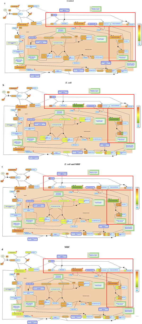
Analysis of the qPCR array results was assessed using pathway analysis software Reactome (https://reactome.org/) to determine gene expression changes within pathways. Fold changes from PCR array results were uploaded into Reactome and changes in expression were marked by colour changes, with dark brown /red representing increased fold expression and green representing reduced gene expression seen in the scale bar on the right side of images.
Western blotting
Protein targets LY96 and TICAM-1 ascertained from the gene expression changes of PCR arrays were assessed by Western blotting. Protein was isolated from HT-29 cells following washing the monolayer with cells resuspended in lysis buffer (7M Urea, 2M Thiourea, 4% CHAPS, 30mM Tris) containing a protease inhibitor cocktail (Sigma Aldrich, P2714) and scraped with a cell scraper. Protein was quantified by Bradford (BIORAD, 500-0006) using a bovine serum albumin standard. Protein (48 μg) was loaded in each well of a 12.5% polyacrylamide gel, run at 70–90 V for 15 min and 110 V until tracking dye reached the end of the gel. Polyacrylamide gels were transferred to a PVDF membrane at 25 V for 45 min on a semi-wet transfer sandwich (Thermo Scientific Pierce G2 Fast Blotter).
Resulting blots were Ponceau stained to check for correct transfer of equal protein prior to blocking in 1% BSA/5% Milk protein resuspended in PBS Tween 20 (0.005%) for 30 min. Blots were probed with target antibody separately for using Rabbit Anti-human LY96 (Sigma, SAB 1410439) or Mouse Anti-human TICAM-1 (Santa Cruz, SC514384) and, for housekeeping, protein actin using Mouse Anti-human Actin (Abcam, AB123034). Each primary antibody was individually incubated for 1 h at RT. Secondary Horseradish Peroxidase (HRP) linked antibody for Anti-mouse-HRP (Sigma Aldrich, A9044) or Anti-rabbit-HRP (Abcam, AB191866) were probed for 1 h at RT to bind to the primary antibody and washed for 10 minutes in triplicate. Resulting Western blotting images were developed by chemiluminescence and analysed using the LICOR scanner and analysis software.
TNFα analysis
TNFα ELISA compatible with cell lysates and cell supernatants was supplied by Sigma Aldrich (RAB1089). All reagents and samples were brought to RT (18–25°C) before use. All standards and secreted supernatant samples were run in duplicate and 100 μl of each standard and sample was added into appropriate wells and incubated for 2.5 h at RT with gentle shaking (150 rpm). Solutions were discarded, and wells washed four times with 1x wash solution between all applications of new reagents. A total of 100 μl of 1x detection antibody was added to each well and incubated for 1 h at RT with gentle shaking. Solutions were removed, and wells washed prior to addition of 100 μl of Streptavidin solution to each well. Wells were covered and incubated for 45 minutes at RT with gentle shaking. Following washing, 100 μl of tetramethylbenzidine (TMB) was added to each well and incubated for 30 minutes at RT in the dark with gentle shaking. Stop solution was added (50 μl) prior to reading at 450 nm absorbance.
Statistics
Statistical analysis of Western blots, ELISA and adhesion experiments were analysed using Students t-test and Tukeys post-hoc test with results determined significant at P ≤ 0.05, MiniTab 18. Bar charts are represented as the mean average and standard error mean (SEM).
Results
qPCR gene array analysis of MRF on bacterial infection
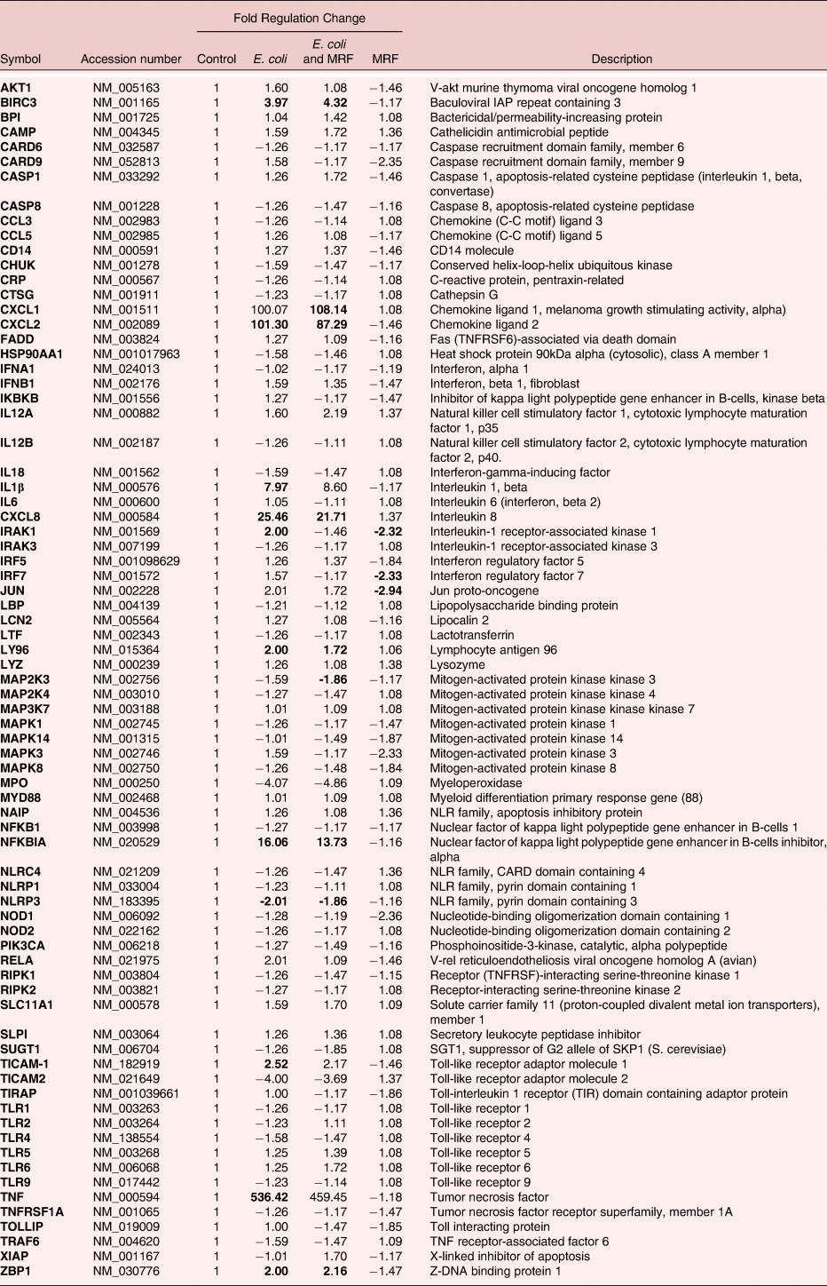
Gene expression was assessed following exposure of intestinal cells to E. coli in the presence and absence of MRF to determine any variance in the immune response. Expression changes greater or less than the control was deemed significantly different at P < 0.05 (Table 1). E. coli treated HT-29 cells had 10 genes expressed at levels significantly higher than the control and one lower than the control. The E. coli exposed to HT-29 cells treated with MRF had six genes expressed at levels significantly higher than the control and two significantly lower than the control. MRF treated HT-29 cells alone had three genes with significantly lower expression than the control.
Table of gene fold changes of HT-29 cells with or without E. coli exposure in the presence or absence of MRF. Qiagen array analysis calculates p values based on a Student's t-test of the replicate 2^(- Delta CT) values for each gene in the control group and treatment groups, and p values less than 0.05 are indicated in bold (n=3).

A number of inflammatory genes were significantly upregulated in HT-29 cells in response to E. coli exposure (IL-1β, TNFα, and ZBP1) when compared to the control (Table 1). Similarly, immune cell attraction to the sight of infection in the form of chemo-attractants were significantly elevated for HT-29 cells exposed to E. coli alone for both CXCL2 and CXCL8, while CXCL1 had a 100-fold increase over the control, but was not significantly different. Immune cell signalling for a number of important intracellular signalling molecules demonstrated either a significant increase (IRAK1) or higher expression (IRF7) in HT-29 cells that were exposed to E. coli alone. BIRC3, an anti-apoptotic gene, was significantly increased in response to E. coli. Genes responsible for upstream pathogen recognition through toll-like receptor 4 (TLR4) were significantly elevated for HT-29 cells following exposure to E. coli (LY96 and TICAM-1) when compared to the control.
E. coli exposed to HT-29 cells in the presence of MRF demonstrated significant increases in the inflammatory gene ZBP1, chemo-attractants CXCL1, CXCL2 and CXCL8, pattern recognition protein LY96 and anti-apoptotic gene BIRC3 compared to control cells. The MAP kinase signalling gene MAP2K3 was significantly decreased in MRF treated HT-29 cells exposed to E. coli. MRF treatment alone on HT-29 cells demonstrated significantly decreased expression of immune signalling genes IRAK1, IRF7 and JUN when compared to the control. E. coli infected HT-29 cells treated with MRF demonstrated greater reductions in gene expression for immune signalling genes IRAK1 and IRF7 compared to the HT-29 cells exposed to E. coli only. The addition of MRF to E. coli infected HT-29 cells reduced expression of inflammatory genes IL-1β and TNFα to non-significant levels when compared to the E. coli infected intestinal cells alone. The expression of proinflammatory marker NLRP3 was significantly reduced in both HT-29 infected groups with and without MRF when compared to the control.
CXCL2 and pathogen recognition gene CD14. Proinflammatory gene Caspase-1 (CASP1) expression was reduced by MRF addition to -1.46 compared to the control. Similarly, a number of genes demonstrated decreased expression of equal to or greater than (−1.45) fold compared to the control for inflammatory associated genes IRF5, ZBP1, IFNB1, TNFRSF1A, immune signalling genes NOD1, RELA, MAPK1, MAPK14, MAPK3, MAPK8, TIRAP, TICAM-1, CARD9, chemokine gene.
Western blotting of HT-29 cell protein following E. coli infection and treatment with MRF
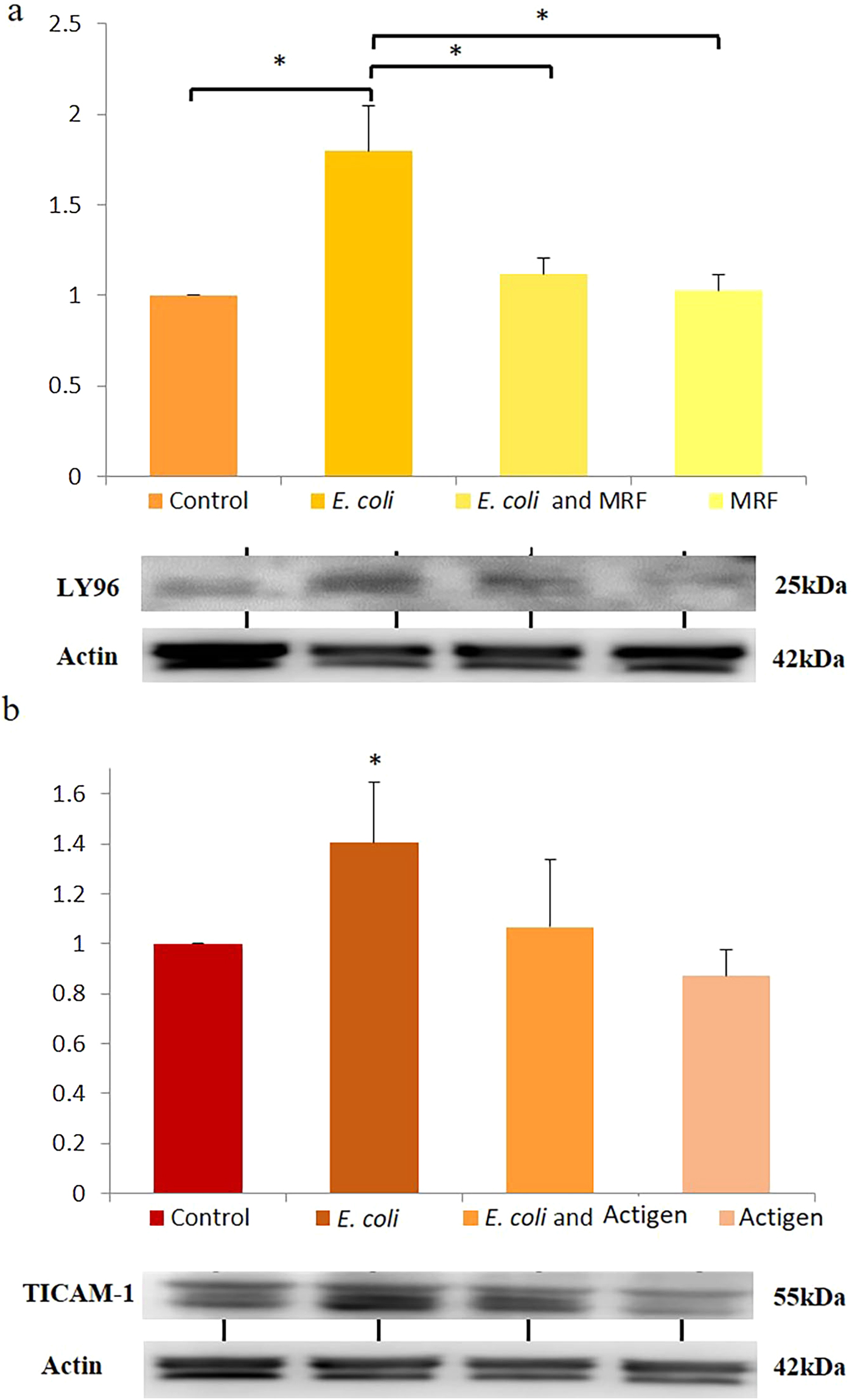
Protein targets LY96 and TICAM-1 were selected due to the significant change in HT-29 gene expression in response to E. coli infection. Both TICAM-1 and LY96 proteins have important functions in TLR4 pathogen recognition, with changes in these protein abundances serving as indicators for the level of infection in the presence or absence of MRF. Target antibodies demonstrated binding to both TICAM-1 and LY96 with developed bands corresponding to respective molecular weights 55 kDa and 25 kDa. LY96 protein abundance normalised to the actin control demonstrated a significant rise in abundance of LY96 in the E. coli treated HT-29 cells (1.79±0.25) over the control (1.0 ± 0, P ≤ 0.05) and the treatment control (1.02 ± 0.09, P ≤ 0.05; Figure 1a). E. coli exposed to HT-29 cells in the presence of MRF (16 mg/ml) demonstrated a non-significant rise over the control to 1.12 ± 0.09 and the MRF treatment control showed no change to the negative control at 1.02 ± 0.09. The addition of MRF to E. coli infected HT-29 cells (1.12 ± 0.09, P ≤ 0.05) significantly reduced LY96 abundance compared to the HT-29 cells infected with E. coli alone (1.79 ± 0.25). TICAM-1 demonstrated a similar trend to LY96 abundance, with a significant rise seen only in the E. coli (1.40 ± 0.24, P ≤ 0.05) treated HT-29 cells over the control (1.0 ± 0.0). HT-29 cells exposed to E. coli with MRF (1.064 ± 0.27) demonstrated no change in TICAM-1 abundance to the control (Figure 1b).
Western blotting of HT-29 cell protein following E. coli infection and treatment with MRF. LY96 protein abundance graph 1a, TICAM-1 protein abundance graph 1b normalised to Actin housekeeping protein abundance and western blotting representative images for TICAM-1, LY96 and Actin. Data was analysed using a One-way ANOVA with Tukey's multiple comparison test and significance marked as * (p < 0.05), (n = 3).

TNFα secretion from HT-29 cells exposed to E. coli in the presence and absence of MRF
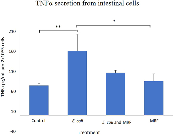
TNFα, a proinflammatory marker indicative of infection was measured for extracellular secretions using a TNFα ELISA. The HT-29 cells exposed to E. coli demonstrated significantly higher TNFα secretion of 161.7 ± 42.0 pg/ml (P ≤ 0.01), compared to control TNFα levels (74.89 ± 4.25 pg/ml). The same trend was seen when comparing E. coli exposed HT-29 cells alone, having significantly higher TNFα levels (161.7 ± 42.0 pg/ml; P ≤ 0.05) over cells treated with MRF only (85.96± 17.7 pg/ml; P ≤ 0.05) (Figure 2). HT-29 cells exposed to E. coli in the presence of MRF (107.23 ± 6.13 pg/ml) had higher than the negative control and the MRF only levels of TNFα, but these were not significantly different (Figure 2).
TNFα secretion from HT-29 cells exposed to E. coli in the presence and absence of MRF. ELISA measurement of TNFα secretion from HT-29 cells exposed to E. coli in the presence or absence of MRF and controls with media (Control) or MRF treatment only (MRF). Data was analysed using a One-way ANOVA with Tukey's multiple comparison test and significance marked as * (p < 0.05), ** (p < 0.01), (n = 3).

Bacterial adhesion of HT-29 cells treated with MRF
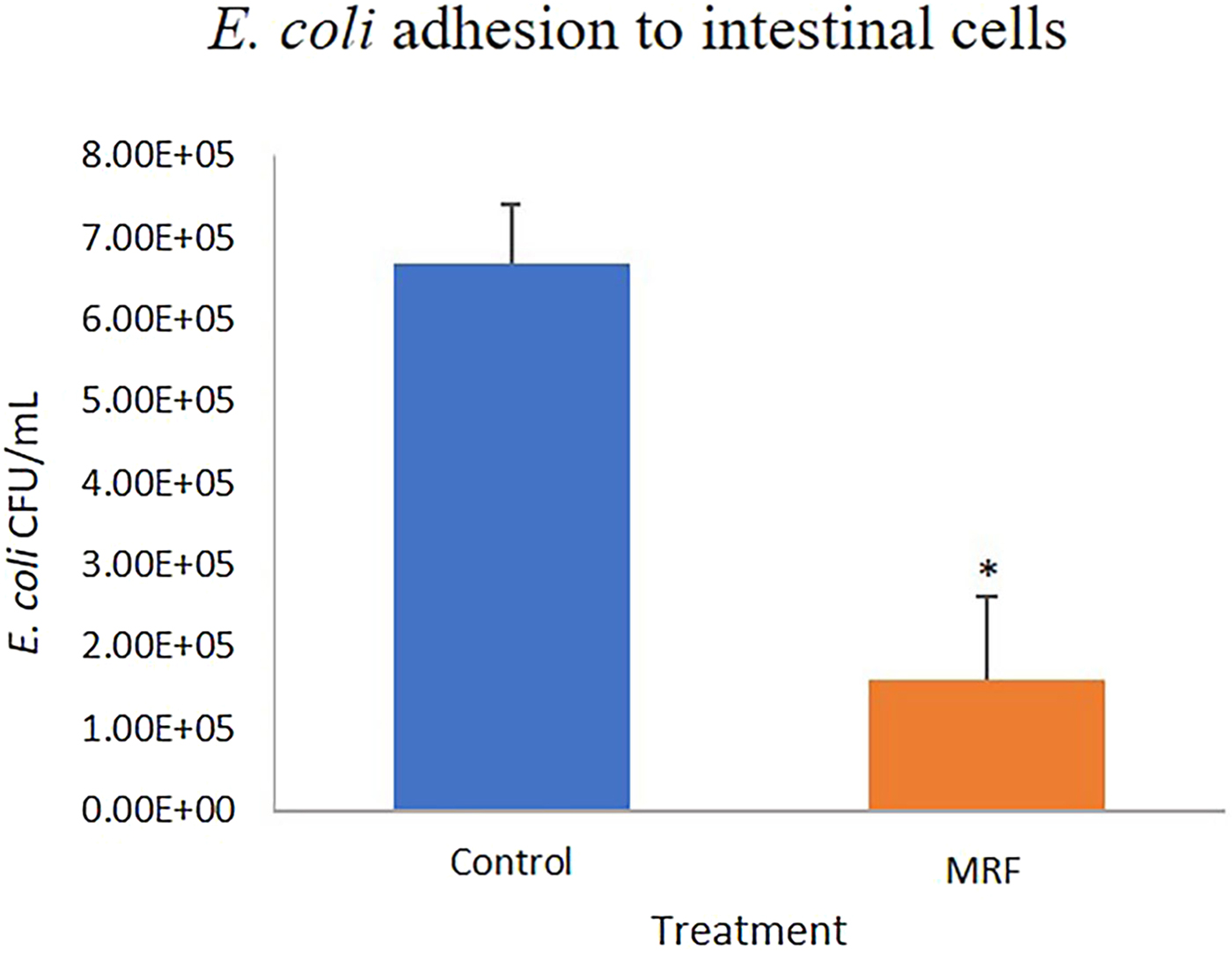
In a comparison of adherence of E. coli to HT-29 cells exposed for 2 h with and without MRF highlighted that treatment of cells with MRF reduced the ability of E. coli to bind to the HT-29 cells by 78% at 1.59×10⁵ ± 1.02 x10⁵ CFU/ml compared to the control (E. coli only) at 6.69×10⁵ ± 7.25×10⁴ CFU/ml with no MRF (Figure 3).
Bacterial adhesion to HT-29 cells treated with MRF. Attachment of E. coli to human intestinal HT-29 cells in the presence of MRF (E. coli and MRF) or absence of MRF the positive control (E. coli). Statistical analysis carried out using a one-tailed students t-test with significance marked as * (p < 0.05), (n = 4).

Discussion
Increases in both inflammation and apoptosis are key host responses to E. coli intestinal infection (Wong Fok Lung et al., Reference Wong Fok Lung, Pearson, Schuelein and Hartland2014). The expression of genes for the chemo-attractants CXCL2 and CXCL8 used by infected host cells to attract immune cells to the sight of infection were significantly higher in HT-29 cells exposed to E. coli alone compared to cells exposed to E. coli and MRF. Exposure of HT-29 cells to E. coli significantly increased LY96 and TICAM-1 genes associated with pathogen recognition and intracellular signalling of the TLR4 pathway. Both of these genes are essential components to the TLR4-led recognition of Gram-negative bacteria such as E. coli (Rajaiah et al., Reference Rajaiah, Perkins, Ireland and Vogel2015; Kuzmich et al., Reference Kuzmich, Sivak, Chubarev, Porozov, Savateeva Lyubimova and Peri2017). It was evident that MRF in combination with E. coli reduced the expression of TICAM-1 to non-significant levels and LY96 to lower levels than were observed in the HT-29 cells exposed to E. coli alone. Reduced detection of bacterial infection via the TLR4 pathway by HT-29 cells exposed to E. coli with MRF may indicate the ability of MRF to impair adhesion of the E. coli to the surface of intestinal cells, thus lowering infection rates.
TICAM-1 gene expression appeared to show a similar trend to protein abundance, as HT-29 cells treated with E. coli has a significant increase in TICAM-1 in both gene and protein abundance (Figure 1b) over the negative control. Yet TICAM-1 protein abundance was not significantly changed for HT-29 cells exposed to E. coli with MRF treatment. Similarly, LY96 protein abundance was significantly elevated in HT-29 cells, responding to the E. coli treatment alone (Figure 1a), but not for intestinal cells exposed to E. coli with MRF. Interestingly the addition of MRF to E. coli infected HT-29 cells reduced LY96 abundance to non-significant levels when compared to HT-29 cells infected with E. coli alone. A comparable study of broilers fed yeast-derived, mannan rich carbohydrates demonstrated lower TLR4 gene expression (Munyaka et al., Reference Munyaka, E. H, Yitbarek, Camelo Jaimes, Sharif, Guenter, House and Rodriguez-Lecompte2012). Although this result was not duplicated in TLR4 gene expression in the current study, a similar trend was observed with genes and proteins associated to the TLR4 complex when MRF was combined with E. coli and for the MRF treatment control group (Table 1, Figure 1a and 1b).
Assessment of the TLR4 associated pathways by Reactome software depicted greater significant changes in gene expression for HT-29 cells exposed to E. coli only but not for treatments including E. coli combined with MRF or MRF alone when compared to the negative control (Figure 4).
representing fold changes in genes for TLR4 Pattern recognition and signalling for (a) Control, (b) E. coli, (c) MRF with E. coli and (d) MRF treated HT-29 cells respectively. Scale bar of fold expression changes ranges from +2.52-4 fold with red box highlighting change of interest.

MRF treatment alone demonstrated a much lower activation of genes associated with the TLR4 pathway over the control, which has been previously demonstrated in poultry (Xiao et al., Reference Xiao, Power, Mallonee, Crowdus, Ao, Pierce and Dawson2010; Brennan et al., Reference Brennan, Graugnard, Xiao, Spry, Pierce, Lumpkins and Mathis2013), piglets (Che et al., Reference Che, Song, Liu, Johnson, Kelley, Van Alstine, Dawson and Pettigrew2012; Samuel and Brennan, Reference Samuel and Brennan2012), and fish (Zhao et al., Reference Zhao, Li, Beck, Zhang, Thongda, Davis and Peatman2015). Zhao et al., (Reference Zhao, Li, Beck, Zhang, Thongda, Davis and Peatman2015) suggested that dietary mannose priming of fish switched expression of inflammatory cytokines to mechanisms for resolution and repair, which may explain the impact of MRF on HT-29 cells on its own and when combined with E. coli in the current experiment. Although no evidence of priming was evident in this study when MRF was added on its own.
MRF in combination with E. coli affected HT-29 gene expression specifically in reducing the expression of genes involved in inflammation, bacterial recognition and immune signalling when compared to HT-29 cells exposed to E. coli only. Overall gene markers for the signs and severity of E. coli infection in the form of inflammation (IL-1β and TNFα) and apoptosis (BIR3) were decreased with addition of MRF in comparison to the E. coli exposed to HT-29 cells alone (Table 1).
TNFα gene expression was significantly raised in HT-29 cells exposed to E. coli alone over the control but not in the HT-29 cells exposed to E. coli combined with MRF (Table 1, Figure 5).
Representing fold changes in genes for TNFR1 signalling pathway for (a) Control, (b) E. coli, (c) MRF with E. coli and (d) MRF treated HT-29 cells respectively. Scale bar of fold expression changes ranges from +536-1.59 fold. TNF receptor activation through TNFa binding and downstream signalling.

This suggested that MRF conferred a protective effect on cells from E. coli infection. TNFα plays a central role in regulation of both the immune response and inflammation (Wang et al., Reference Wang, Czura, Tracey and Lotze2003). Further assessment of protein levels of TNFα secretion demonstrated a strong correlation with the gene expression, as E. coli exposure to HT-29 cells triggered a significant release of TNFα compared to the control. Interestingly MRF combined with E. coli and exposed to HT-29 cells demonstrated a non-significant rise in TNFα over the control in both protein secretion and gene expression. This again highlighted MRFs potential capacity to protect cells from the effects of E. coli infection that results in lower TNFα production and release. Previous studies assessing MRF similarly highlighted its ability to lower inflammation while blocking harmful intestinal bacteria (Xiao et al., Reference Xiao, Power, Mallonee, Crowdus, Ao, Pierce and Dawson2010; Che et al., Reference Che, Song, Liu, Johnson, Kelley, Van Alstine, Dawson and Pettigrew2012; Munyaka et al., Reference Munyaka, E. H, Yitbarek, Camelo Jaimes, Sharif, Guenter, House and Rodriguez-Lecompte2012; Samuel and Brennan, Reference Samuel and Brennan2012).
The adhesion of E. coli to its cellular target has been reported to trigger a signalling pathway response (e.g., MAPK and PI3K) leading to CXCL8 synthesis, which induces the migration of neutrophils to the epithelial site of infection (Cane et al., Reference Cane, Ginouves, Marchetti, Busca, Pouyssegur, Berra, Hofman and Vouret-Craviari2010). This response has been reported to follow the stimulation of epithelial cells to synthesise TNFα and IL-1β (Betis et al., Reference Betis, Brest, Hofman, Guignot, Kansau, Rossi, Servin and Hofman2003) which was observed here in both HT-29 cell treatment groups exposed to E. coli. The expression of CXCL8, TNFα and IL-1β, were significantly increased in response to E. coli exposure, but not for TNFα and IL-1β expression in response to E. coli treated with MRF. Reduced inflammatory markers from HT-29 cells exposed to E. coli and MRF when compared to HT-29 cells exposed to E. coli alone demonstrated the potential reduction of infection through MRF addition.
Research has previously shown that yeast mannans have a positive influence on the immune status of sows and their newly weaned piglets (Duan et al., Reference Duan, Chen, Zheng, Tian, Wang, Mao, Yu, He, Li, Huang, Ao and Yu2016) when sow diets were supplemented. The effect of yeast mannans on the immune response of cells when challenged with ETEC has been examined by Hermes et al. (Reference Hermes, Manzanilla, Martin Orue, Perez and Klasing2011). It was subsequently demonstrated that the activation of TLR-4, TLR-5 and the levels of inflammatory markers, such as IL-1β and TNFα, were reduced similarly to what was observed in the current experiment, although only for the TLR4 pathway.
The anti-inflammatory effects of MRF alone were highlighted by Munyaka et al. (Reference Munyaka, E. H, Yitbarek, Camelo Jaimes, Sharif, Guenter, House and Rodriguez-Lecompte2012) in broilers, demonstrating the downregulation of gene expression of the proinflammatory marker interferon. Similarly, it was shown in the current experiment that MRF treatment alone significantly reduced HT-29 cell expression for IRF7 and IRAK1 activators of proinflammatory markers TNFα and IL-1β respectively, when compared to the control levels (Table 1). Further evidence of anti-inflammatory effects of MRF was seen by the number of inflammatory genes that were reduced in expression compared to the control. For example, caspase1, an enzyme responsible for cleaving protein precursors to their active inflammatory forms such as IL-1β (Thornberry et al., Reference Thornberry, Hg, Calaycay, Chapman, Howard, Kostura, Miller, Molineaux, Weidner, Aunins and Aunins and1992), was reduced in expression compared to the control.
Adhesion of E. coli to HT-29 intestinal cells facilitates infection, which was demonstrated to be significantly impaired by the presence of MRF (Figure 3). MRF functions as a decoy molecule to E. coli blocking the normal interaction by which the bacteria bind to the intestinal cell lining through mannan surface molecules. Interestingly, previous findings demonstrated significantly reduced invasion of IPEC by the same E. coli strain used here when MRF was added compared to a control (Horgan et al., Reference Horgan, Madden and Jacques2018). The method of physically blocking bacterial invasion exploits the bacteria's reliance on the mannan-dependent means of invasion. This method of invasion that yeast mannan targets, appears to be evolutionarily difficult for the bacteria to adapt to and generating alternative means against invasion. This is unlike targeting the bacteria via antimicrobials, which act as a selective pressure allowing bacteria to evolve to overcome such pressure and attain resistance (Sandegren, Reference Sandegren2014). Thus, the method of utilising a mannan decoy via MRF to block adhesion may prove more difficult for bacteria such as E. coli and Salmonella spp. to negate or block.
Previous work on MRF demonstrated beneficial effects in limiting infection by E. coli and Salmonella spp. in poultry (Biggs et al., Reference Biggs, Parsons and Fahey2007; Luquetti et al., Reference Luquetti, Furlan, Alarcon and Macari2012), as observed here with HT-29 intestinal cells exposed to pathogenic E. coli (Figure 3). Intracellularly, the characteristic markers for infection were elevated in response to E. coli infection but were reduced with MRF addition. MRF exposure in the absence of infection demonstrated potential anti-inflammatory effects on cells, as reported in previous research (Munyaka et al., Reference Munyaka, E. H, Yitbarek, Camelo Jaimes, Sharif, Guenter, House and Rodriguez-Lecompte2012). Yet the MRF didn't demonstrate any priming of intestinal cells to either fungal or bacterial gene pathways (Figure 4d). A balance between pro and anti-inflammatory changes is required, since impairing inflammation can affect the hosts capacity to fight infection, while too strong an inflammatory response results in excessive tissue damage, which could lead to poor nutrient absorption from the gut.
Conclusions
The changes in gene and protein biological markers in response to intestinal infection may be utilised to determine reoccurrence of pig infections and the effectiveness of in feed prebiotics such as MRF. The incorporation of MRF in feed to improve PWD outcomes in pigs could similarly be assessed by changes in key biomarkers observed here for the severity of infections through measuring inflammatory, apoptotic and immune signalling changes. Feed additives such as MOS and MRF offer the potential to prevent colonisation of the animal's gut by pathogenic bacteria, leading to lower incidence of PWD and ultimately providing a means to improve gut health naturally, without the need for antimicrobial usage on farms.