Introduction
Signaling between vertebrate photoreceptors and horizontal cells is believed to be mediated by the release of glutamate from photoreceptor synaptic terminals (Copenhagen & Jahr, Reference Copenhagen and Jahr1989). Glutamate release by photoreceptors is highest in the dark and results in the depolarization of horizontal cells by activation of ionotropic glutamate receptors (Ayoub et al., Reference Ayoub, Korenbrot and Copenhagen1989; Copenhagen & Jahr, Reference Copenhagen and Jahr1989). Based largely on pharmacological criteria, ionotropic glutamate receptors have been classified broadly into three families: NMDA receptors, kainate-activated receptors, and AMPA-preferring receptors. NMDA receptors have long been known to permit the influx of calcium when open, which is believed to play a key role in the postsynaptic effects of glutamate mediated by this receptor. Kainate/AMPA receptors are characterized by particularly rapid desensitization to glutamate, and the relative permeability of kainate/AMPA receptors to calcium can differ depending on their particular subunit composition (Fletcher & Lodge, Reference Fletcher and Lodge1996; Ozawa et al., Reference Ozawa, Kamiya and Tsuzuki1998; Dingledine et al., Reference Dingledine, Borges, Bowie and Traynelis1999). A number of new pharmacological tools, including more selective agonists and antagonists of kainate and AMPA receptors, as well as agents that affect selectively the desensitization of the two receptor subclasses, now permit clearer distinctions to be made between different classes of glutamate receptor subtypes (Fletcher & Lodge, Reference Fletcher and Lodge1996; Ozawa et al., Reference Ozawa, Kamiya and Tsuzuki1998; Dingledine et al., Reference Dingledine, Borges, Bowie and Traynelis1999).
Electrophysiological examinations of the ionotropic glutamate receptors of horizontal cells of tiger salamander, catfish, carp, stingray, cat, rabbit, and human have revealed the presence of kainate/AMPA subtypes on cells of these species (Hals et al., Reference Hals, Christensen, O’Dell, Christensen and Shingai1986; O’Dell & Christensen, Reference O’Dell and Christensen1989a,Reference O’Dell and Christensenb; Qin & Pourcho, Reference Qin and Pourcho1996; Yang et al., Reference Yang, Maple, Gao, Maguire and Wu1998; Blanco & de la Villa, Reference Blanco and de la Villa1999; Okada et al., Reference Okada, Schultz, Geurtz, Hatt and Weller1999; Shen et al., Reference Shen, Finnegan and Slaughter2004). In addition, cone-driven horizontal cells of the catfish appear to possess NMDA receptors (Hals et al., Reference Hals, Christensen, O’Dell, Christensen and Shingai1986), as do the cone-driven H1 horizontal cells of the carp (Shen et al., Reference Shen, Zhang, Jin and Yang2006). The large horizontal cells of the skate (Raja erinacea and R. ocellata) have long been used as a model system to examine the roles that horizontal cells play in information processing within the retina (Dowling, Reference Dowling1987). The photoreceptor complement of the skate retina is composed entirely of rods (Szamier & Ripps, Reference Szamier and Ripps1983), providing an excellent and simple model system in which to study the processing of rod-generated signals. Glutamate has been shown previously to depolarize skate horizontal cells in a concentration-dependent manner (Lasater et al., Reference Lasater, Dowling and Ripps1984; Kreitzer et al., Reference Kreitzer, Andersen and Malchow2003). However, a detailed characterization of the type(s) of glutamate receptors present on skate horizontal cells has yet to be conducted.
The nature and regulation of excitatory postsynaptic responses mediated by glutamate receptors will have a profound effect on visual signals within the retina. Ionotropic glutamate receptors show a wide diversity in sensitivity to changes in extracellular H+ concentrations. For example, half-maximal inhibition of NMDA receptors of cerebellar granule cells occurs when extracellular pH dropped to 7.3, quite near the normal range of physiological pH, while kainate-induced responses are inhibited only when pH dropped to 5.76 (Traynelis & Cull-Candy, Reference Traynelis and Cull-Candy1991). Such differences in sensitivity to alterations in extracellular acidity have the potential to impact significantly on the processing of neural signals within the retina. For example, Devries (Reference DeVries2001) has shown that extracellular changes in proton concentration resulting from vesicular release of neurotransmitter from photoreceptors can be sufficient in magnitude to inhibit further release of neurotransmitter from photoreceptor synaptic terminals. Depending on the nature of the glutamate receptor subunits expressed in horizontal cells, such changes in extracellular pH could potentially have significant effects on the glutamate-induced currents of horizontal cells. Another ion implicated in the regulation of ionotropic glutamate receptors is zinc. Zinc is localized at the photoreceptor synaptic terminals in the retina and may be coreleased with glutamate in synaptic vesicles from these terminals (Wu et al., Reference Wu, Qiao, Noebels and Yang1993). Regulatory effects of zinc on ionotropic non-NMDA glutamate receptors could have significant effects on fast excitatory synaptic transmission in the outer retina. Non-NMDA ionotropic glutamate receptors can be either positively or negatively regulated by zinc in a concentration-dependent manner; the nature of the effect and its dose dependence depends on receptor subunit composition (Rassendren et al., Reference Rassendren, Lory, Pin and Nargeot1990; Bresink et al., Reference Bresink, Ebert, Parsons and Mutschler1996).
The purpose of the present study was to characterize using pharmacological tools the ionotropic glutamate receptor(s) responsible for fast excitatory transmission in skate horizontal cells. Our data suggest that the current induced by glutamate in enzymatically isolated skate horizontal cells results from AMPA glutamate receptors, with a significant contribution from calcium-permeable isoforms. Our studies also show that H+ and Zn2+ are potential regulators of the glutamate receptors present in these cells.
Materials and methods
Skate (R. erinacea and R. ocellata) were obtained from the Marine Biological Laboratory in Woods Hole (MA) and maintained for up to 3 weeks in a circulating artificial seawater tank kept at 14°C. Prior to enucleation, animals were anesthetized for 5 min by placing them in approximately 1 gallon of water containing 1 g/gallon MS-222 (tricaine), then cervically transected, and double pithed.
Cell dissociation
The dissociation protocol used in this study was identical to that reported in Kreitzer et al. (Reference Kreitzer, Andersen and Malchow2003). Isolated retinae were bubbled for 45 min in a skate-modified L-15 media at pH 7.6 containing 2 mg/ml papain (Calbiochem-Novabiochem, San Diego, CA) and 1 mg/ml l-cysteine. The L-15 media had been modified to meet ionic and osmotic requirements for a marine elasmobranch by adding 3.0 g NaCl, 10.5 g urea, 2.5 g glucose, 0.5 g HEPES, and 1.0 ml penicillin–streptomycin (P0781; Sigma Chemical) to 500 ml of L-15 solution. Following bubbling, the retinal pieces were rinsed thoroughly with modified L-15 lacking papain or l-cysteine and triturated in a 5-ml glass graduated sterile pipette until the tissue had broken apart. Cells were dropped into 35-mm Falcon-3001 tissue culture dishes (Becton Dickinson, Franklin Lakes, NJ) and stored for up to 3 days at 4°C.
Electrophysiology
Electrical currents were measured from isolated external horizontal cells of skate using whole-cell patch clamp as described previously (Kreitzer et al., Reference Kreitzer, Andersen and Malchow2003). Two- to four-micrometer-diameter microelectrodes with resistances of 2–4 MΩ were made using a P-97 Micropipette Puller (Sutter Instruments, Novator, CA) to pull Kovar capillary tubing (7052; Garner Glass, Claremont, CA). Microelectrodes were filled with (mm) 204 CsCl, 0.05 CaCl2, 0.5 EGTA, 2 MgCl2, and 10 HEPES, pH 7.6, and contained 0.5% phenol red. The electrode was connected to a HEKA EPC-9 patch-clamp amplifier by a silver chloride wire, while the return pathway went through a silver/silver chloride wire contacting the solution in the media dish. Series resistance was not compensated but was monitored during the course of all trials, and recordings in which the series resistance changed by more than 50% were discarded. Typical series resistances were between 15 and 30 MΩ. Data were acquired using the program Pulse (version 8.3) at a rate of 250 Hz.
External solutions were applied using a Warner Instrument three-barrel perfusion system that expelled solutions at a rate of 2 ml/min with solution being removed by a suction pipette in the dish. Spacing between the three-way square glass (3SG700-5) barrels was 0.7 mm. Complete solution exchange occurred in 19 ± 3.6 ms as determined by measuring liquid junction potential changes upon switching from normal skate Ringer’s solution to a solution in which all sodium had been replaced with N-methyl-d-glucamine. Except where mentioned, the skate Ringer’s solution consisted of (mm) 270 NaCl, 6 KCl, 1 MgCl2, 4 CaCl2, 360 urea, 10 glucose, and 5 HEPES at pH 7.6. The urea levels are common to all elasmobranch Ringer’s solutions, and these concentrations are typically detected in the plasma levels of these species, including the skate. In experiments examining the current–voltage relation of glutamate-gated currents, cells were superfused with a Ringer’s solution that contained 10 mm cesium, 10 mm 4-aminopyridine, 20 mm tetraethylammonium, 100 μ m nifedipine, and 1 μ m tetrodotoxin to block voltage-gated conductances. I–Vs were obtained by subtracting currents in the presence of glutamate from currents obtained in the absence of glutamate. All chemicals were obtained from Sigma Chemical (St. Louis, MO), unless otherwise noted. Data are presented throughout the paper as the mean ± s.e.m. Parameters for the dose–response relationships were determined using an iterative nonlinear regression statistical package incorporated in the SigmaPlot version 7 program (Jandel Scientific, San Rafael, CA). Student’s t-tests were used throughout to determine statistical significance, with a criterion of <0.01 selected as indicating significantly different distributions.
Immunolabeling
After dissociation, isolated cells were fixed for 30 min with 4% paraformaldehyde in phosphate-buffered saline (PBS), pH 7.4, and washed 3 times in PBS. Whole retinas were fixed in 4% paraformaldehyde for 2 h at 4°C, washed 3 times in PBS, and cryoprotected by stepping through cold 20 and 30% sucrose overnight. The tissue was mounted with HistoPrep (Fisher Scientific, Pittsburgh, PA), sectioned at 12 μm, and picked up on Superfrost Plus Slides (Fisher Scientific). Cells and tissue slices were immersed for 1–2 h in blocking solution (4% goat serum, 2% bovine gamma globulin, and 0.3% Triton-X in PBS) and then incubated overnight at room temperature in solutions containing primary antibody diluted 1:100 in blocking solution. Four types of polyclonal antibodies (GluR 1, GluR2, GluR2/3, and GluR4; Chemicon, Temecula, CA) were tested in this study. Control experiments were performed in which the primary antibody was omitted; no obvious staining was observed under these conditions. After three washes in PBS, cells were incubated in darkness for 2 h with a secondary antibody (Goat Anti-Rabbit IgG Cy3 conjugate; Jackson Labs, West Grove, PA) diluted 1/500 and washed 3 times over 30 min with PBS. Coverslips were mounted on slides with VectaShield (Vector Labs, Burlingame, CA). Isolated cells were examined using a Zeiss Axioskop 2 plus microscope equipped with an Atto-ARC mercury lamp. Retinal slices were analyzed using a Zeiss confocal laser scanning microscope (LSM 510) equipped with an Ar/Kr laser. Images were prepared for publication using Adobe Photoshop 7.0. In all cases, digital processing was confined to adjusting the origin and slope of the linear input–output curve, thus manipulating the brightness and contrast of the images collectively as a whole.
Results
When the membrane potential was held at −70 mV, application of externally applied glutamate and several other glutamate agonists to whole-cell voltage-clamped skate external horizontal cells induced currents in a concentration- and voltage-dependent manner. Previous studies have shown that skate horizontal cells respond to glutamate in a steep concentration-dependent manner (Kreitzer et al., Reference Kreitzer, Andersen and Malchow2003). The concentration dependence of such glutamate-induced currents was fit to a Hill equation of the form:

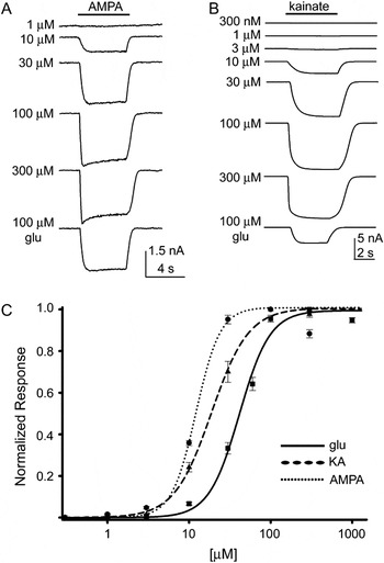
where R is the response induced by a glutamate concentration of I, R max is the maximum response, s is the concentration of glutamate producing a half-maximal response, and n is a coefficient affecting the steepness of the curve (Hill coefficient). The glutamate dose–response curve was best fit with a half-maximal concentration of 41.5 ± 1.4 μ m and a Hill coefficient of 2.32 ± 0.16 and is plotted as the solid line in Fig. 1C.
(A). Electrical currents obtained from 5 s applications of 1, 10, 30, 100, and 300 μ m AMPA followed by a subsequent application of 100 μ m glutamate to a horizontal cell whole-cell voltage clamped at −70 mV. (B). Electrical currents obtained from 5 s applications of 300 nm, 1, 3, 10, 30, 100, and 300 μ m kainate followed by a subsequent application of 100 μ m glutamate to a horizontal cell whole-cell voltage clamped at −70 mV. (C). Dose–response relationships for electrical currents elicited by glutamate (solid trace, squares, seven cells), AMPA (dotted trace, circles, seven cells), and kainate (dashed trace, triangles, seven cells).

Application of AMPA (a-amino-3-hydroxy-5-methyl-4-isoxazolepropionic acid), a non-NMDA ionotropic glutamate receptor agonist, also produced a concentration-dependent response. Fig. 1A shows traces elicited from an individual cell voltage clamped at −70 mV. At AMPA concentrations of 100 μ m and greater, a small decline in the initial current followed by a steady-state response was observed. AMPA receptor responses obtained from other neurons typically display a very rapid and large desensitization, with the time constant for desensitization being on the order of several milliseconds (Dingledine et al., Reference Dingledine, Borges, Bowie and Traynelis1999). Given the nature of our superfusion system, which permits complete solution exchange in 20 ms, the AMPA-mediated currents measured from horizontal cells likely reflect partially desensitized responses. The currents obtained from the different AMPA concentrations in seven cells were fit to the Hill equation [eqn. (1)] with responses from 100 μ m AMPA being used as the R max, and the curve was plotted as the dotted line in Fig. 1C. When fit to the Hill equation, the AMPA responses revealed a half-maximal response of 12.18 ± 0.32 μ m and Hill coefficient of 2.92 ± 0.24.
Kainate, another non-NMDA glutamate receptor agonist that activates both kainate and AMPA GluR1s (Bettler et al., Reference Bettler, Boutler, Hermans-Borgmeyer, O’Shea-Greenfield, Deneris, Moll, Borgmeyer, Hollmann and Heinemann1990; Keinanen et al., Reference Keinanen, Wisden, Somer, Werner, Herb, Verdoorn, Sakmann and Seeburg1990; Egebjerg et al., Reference Egebjerg, Bettler, Hermans-Borgmeyer and Heinemann1991; Sommer et al., Reference Sommer, Burnashev, Verdoorn, Keinanen, Sakmann and Seeburg1992; Partin et al., Reference Partin, Patneau, Winters, Mayer and Buonanno1993), also produced inward currents in horizontal cells voltage clamped at −70 mV. Kainate currents were large and sustained, much larger than the steady-state currents observed when either glutamate or AMPA was applied to isolated skate horizontal cells (Fig. 1B). Kainate elicits a nondesensitizing response on AMPA receptors (cf. for review, Fletcher & Lodge, Reference Fletcher and Lodge1996; Dingledine et al., Reference Dingledine, Borges, Bowie and Traynelis1999), which likely accounts for the large size of the observed currents. Kainate responses from seven cells were fit to the Hill equation [eqn. (1)] with responses from 100 μ m kainate designated as the R max and plotted as the dashed line in Fig. 1C. The half-maximal value for the dose–response relationship for kainate was 18.8 ± 0.94 μ m and the Hill coefficient was 1.90 ± 0.15. The dose–response relation for glutamate- and AMPA-induced currents was significantly steeper (i.e., have a higher Hill coefficient) than that for kainate.
NMDA failed to elicit a current in skate external horizontal cells. Trials performed on seven cells bathed in a Ringer’s solution containing 0 mm Mg2+ and 1 mm glycine failed to elicit a current with 100 μ m NMDA application when cells were voltage clamped at either −70 or +30 mV. In the same cells, a large inward current was readily induced by 100 μ m glutamate. One Hundred micromolar DAP-5, an NMDA receptor antagonist, was also unable to significantly reduce the glutamate-elicited currents, which were 95 ± 3.1% of control values. These data suggest that NMDA receptors do not contribute to the current elicited by glutamate and infer the absence of NMDA receptors on skate horizontal cells.
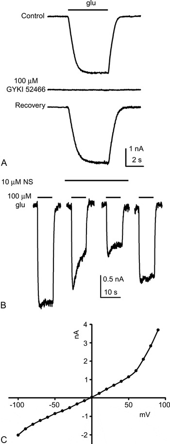
Previous findings by Kreitzer et al. (Reference Kreitzer, Andersen and Malchow2003) showed that glutamate-induced currents of skate horizontal cells could be completely abolished with coapplication of the ionotropic glutamate receptor blocker 100 μ m CNQX, suggesting the glutamate currents in these cells were due to the activation of AMPA/kainate receptors. The development of pharmacological agents that selectively target AMPA receptors now permits a clearer determination of which receptors are responsible for glutamate-induced currents. 1-(4-Aminophenyl)-4-methyl-7,8-methylenedioxy-5H-2,3-benzodiazepine, or GYKI 52466, is an antagonist of AMPA receptors (Donevan & Rogawski, Reference Donevan and Rogawski1993; Fletcher & Lodge, Reference Fletcher and Lodge1996; but see Shen et al., Reference Shen, Finnegan and Slaughter2004). Fig. 2A shows that GYKI 52466 completely abolished the response to glutamate obtained from a single cell. In seven cells, 100 μ m GYKI 52466 reduced 100 μ m glutamate-induced currents by 99.4%. The response to 20 μ m AMPA was reduced 91.0% by 100 μ m GYKI 52466; responses to 20 μ m kainate were reduced by 89.5% (N = 7) in the presence of this antagonist.
(A). Effects of GYKI 52466 on glutamate-elicited currents. Traces resulting from application of glutamate onto an individual cell voltage clamped at −70 mV. The top trace shows the current elicited by application of 100 μ m glutamate. The cell was then bathed for several seconds in GYKI 52466. The second trace shows the current obtained when glutamate was coapplied with 100 μ m GYKI 52466; the glutamate-elicited current was abolished under this condition. The bottom trace shows recovery of the glutamate-elicited current following washout. (B). Effects of naphthylacetyl spermine (NS) on glutamate-induced currents of skate horizontal cells. Traces from a cell challenged with glutamate in the presence of 10 μ m NS. The first trace is a control current elicited by a 10 s application of 100 μ m glutamate. Two current traces resulting from coapplication of NS and glutamate are shown in the second and third traces. The final trace shows the glutamate-induced current following washout of NS. (C). I–V relationship for the glutamate-gated current from one horizontal cell. The graph reflects the current induced by glutamate at a series of voltage steps subtracted from currents remaining at those voltages in the absence of glutamate. The intracellular pipette contained 300 μ m spermine, and the extracellular solution contained 10 mm cesium, 10 mm 4-aminopyridine, 20 mm tetraethylammonium, 100 μ m nifedipine, and 1 μ m tetrodotoxin to reduce voltage-gated conductances. Similar results were obtained from four additional cells.

Several subunits of AMPA receptors, typically designated GluR 1 through 4 and stemming from a single gene family, are known to exist, each possessing distinct functional and pharmacological characteristics (Dingledine et al., Reference Dingledine, Borges, Bowie and Traynelis1999). Functional characterization of GluR3 and GluR4 receptors examined in a number of expression systems indicates that these receptors permit the direct flux of calcium through the activated receptors (Ozawa et al., Reference Ozawa, Kamiya and Tsuzuki1998). Previous studies using Ca2+ fluorescent measurements in horizontal cells of skate (Kreitzer et al., Reference Kreitzer, Andersen and Malchow2003), as well as catfish, carp, and rabbit (Linn & Christensen, Reference Linn and Christensen1992; Okada et al., Reference Okada, Schultz, Geurtz, Hatt and Weller1999; Rivera et al., Reference Rivera, Blanco and de la Villa2001), demonstrate direct influx of Ca2+ along with Na+ through the non-NMDA ionotropic glutamate receptors present in these cells. An additional useful pharmacological agent for further addressing the issue of Ca2+ permeability of AMPA receptors is 1-naphthylacetyl spermine (NS), an agent that specifically blocks Ca2+-permeable AMPA receptors in a use-dependent manner (Koike et al., Reference Koike, Iino and Ozawa1997). Fig. 2B shows that 10 μ m NS induced a use-dependent block of the glutamate-induced response. Glutamate currents were reduced to 38.3 ± 1.5% in the presence of 10 μ m NS (N = 7), 15.5 ± 0.9% in the presence of 100 μ m NS (N = 6), and 10.1 ± 2.7% in the presence of 250 μ m NS (N = 5). The finding that NS significantly reduces glutamate-elicited currents suggests that glutamate responses in the horizontal cells of skate are mediated to a very large degree by Ca2+-permeable AMPA receptors.
Fig. 2C shows the current–voltage relation for the glutamate-induced current obtained from one skate horizontal cell when the intracellular pipette contained 300 μ m spermine. Polyamines such as spermine are known to induce a rectifying current–voltage relation when responses are mediated by calcium-permeable AMPA receptors (Ozawa et al., Reference Ozawa, Kamiya and Tsuzuki1998). The fact that the current–voltage relationship demonstrated rectification is additional support for the presence of calcium-permeable AMPA receptors in the skate horizontal cells. Similar results were obtained in four additional cells. The inward rectification we detect is not as extensive as observed in some other studies (cf. Itazawa et al., Reference Itazawa, Isa and Ozawa1997; Liu & Zukin, Reference Liu and Zukin2007, for review). Furthermore, the degree of rectification was similar when pipettes did not contain 300 μm spermine. In this latter regard, it is worth mentioning how large skate horizontal cells are—150 μm tip to tip is not uncommon, and it may well be that cells were not depleted of endogenous spermine during the time course of our recordings. It is also possible that the results with and without spermine reflect a species difference, with calcium-permeable AMPA receptors of skate possessing less rectification compared to those of other species, or it might also be indicative of the presence of both calcium-permeable and calcium-impermeable AMPA receptor types in the skate retinal horizontal cells.
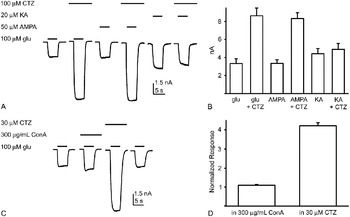
The effects of cyclothiazide (CTZ), which is known to remove the desensitization component of AMPA receptors but not of kainate receptors (Partin et al., Reference Partin, Patneau, Winters, Mayer and Buonanno1993; Shen et al., Reference Shen, Zhou and Yang1999, Reference Shen, Finnegan and Slaughter2004), provided additional evidence that glutamate currents in skate horizontal cells were mediated by AMPA receptors. In the presence of CTZ, sustained AMPA receptor responses generated by either glutamate or AMPA will be significantly larger due to the removal of receptor desensitization by CTZ. On the other hand, activation of AMPA receptors by kainate is not expected to be affected by the addition of CTZ since kainate induces a nondesensitizing response of AMPA receptors. If the glutamate response of skate horizontal cells is mediated principally by AMPA receptors, it would then be expected that responses to glutamate and AMPA would be enhanced in the presence of CTZ, while CTZ would have little effect on kainate-induced currents. Fig. 3 shows that these expectations were borne out. In these trials, the effects of 100 μ m CTZ were examined on currents elicited by application of 100 μ m glutamate, 50 μ m AMPA, and 20 μ m kainate. The first trace shows application of glutamate in normal Ringer’s solution for 5 s. Following removal of glutamate, the cell was bathed in Ringer’s solution containing CTZ for several seconds. The second trace shows the greatly enhanced glutamate-elicited current obtained in the presence of CTZ. Following washout of CTZ, the process was repeated in the third and fourth traces for the agonist AMPA and in the fifth and sixth traces for the agonist kainate. In the presence of CTZ, the response to 50 μ m AMPA was greatly enhanced, while the peak current elicited by kainate was virtually unaltered. Average responses for six cells shown in Fig. 3B demonstrate a consistent potentiation of both glutamate and AMPA responses by CTZ—potentiation of 260% (P = 0.0001) and 249% (P < 0.0001) in the presence of CTZ, respectively—while little effect was observed with kainate—110% of control currents in the presence of CTZ (P = 0.1660). The potentiation of both glutamate and AMPA responses further suggests that the currents elicited are due primarily to activation of AMPA receptors.
Effect of cyclothiazide (CTZ) on glutamate-, AMPA-, and kainate-elicited currents of skate horizontal cells. (A). Responses from a single horizontal cell voltage clamped at −70 mV. Leftmost trace shows control response to 100 μ m glutamate. The second trace shows the response to glutamate in the presence of 100 μ m CTZ; a large increase in the inward current was observed. Following washout of the CTZ, the cell was then challenged with 50 μ m AMPA (third trace). The fourth trace shows the large increase in response to the same concentration of AMPA in the presence of CTZ. The final two traces show kainate-elicited currents in the absence and presence of CTZ. (B). Average current sizes are shown for six cells challenged with glutamate, AMPA, and kainate in the absence and presence of CTZ. (C). Concanavalin A (con A) failed to significantly alter the size of glutamate-induced currents. Traces from a single cell voltage clamped at −70 mV. The first trace is a control 5 s application of 100 μ m glutamate. The second trace shows the current elicited during coapplication of 300 μg/ml of con A and glutamate for 5 s. Following washout of con A and glutamate, the cell was bathed in a Ringer’s solution containing 30 μ m CTZ, and the cell was again challenged with glutamate in the presence of CTZ (third trace); a large increase in inward current was observed. The final trace shows a control current elicited from a second application of glutamate following washout of the CTZ. (D). Average responses of six cells in the presence of con A or CTZ, normalized to the initial 100 μ m glutamate response.

Concanavalin A (con A) eliminates the desensitization of kainate receptors but has little effect on desensitization of AMPA receptors (Partin et al., Reference Partin, Patneau, Winters, Mayer and Buonanno1993). The effects of con A on glutamate-induced responses of the external horizontal cells were tested by extracellular application of 300 μg/ml of con A. Fig. 3C shows representative traces from a single cell voltage clamped at −70 mV. The first trace is a control 5 s application of 100 μ m glutamate. Following washout of glutamate, the cell was bathed in a Ringer’s solution with 300 μg/ml of con A, and the cell again challenged with glutamate in the presence of con A (second trace); little alteration in the size of the glutamate response was observed. After washout of con A and glutamate, the cell was bathed in a Ringer’s solution containing 30 μ m CTZ, and the cell was again challenged with glutamate in the presence of CTZ (third trace). A large increase in the size of the glutamate-induced current was noted. Fig. 3D shows the average responses of six cells in the presence of con A and CTZ, normalized to the initial 100 μ m glutamate response. Con A had little effect on the glutamate-induced responses (P = 0.1091), while CTZ had a large potentiating effect on the glutamate current (P = 0.0001). The potentiation of glutamate response observed with 30 μ m CTZ (435%; Fig. 3D) is much larger than those observed with 100 μ m CTZ (260%; Fig. 3B).
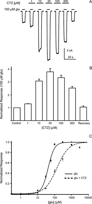
The dose dependence for potentiation of glutamate-induced currents by CTZ is shown in Fig. 4. Electrical currents from an individual cell voltage clamped at −70 mV and challenged for 5 s with 100 μ m glutamate in absence and in the presence of increasing concentrations of CTZ (1, 10, 30, 100, and 300 μ m) are shown in Fig. 4A, and average responses from seven cells are shown in Fig. 4B. The largest potentiation of the glutamate-induced current occurred at a CTZ concentration of 30 μ m, which induced a 380 ± 25% increase in the glutamate-elicited current. Potentiation declined when the concentration of CTZ was increased to 100 and 300 μ m, a phenomenon previously observed for AMPA receptors expressed in oocytes (Partin et al., Reference Partin, Patneau, Winters, Mayer and Buonanno1993) and in retinal neurons (Shen et al., Reference Shen, Zhou and Yang1999). We next examined the effects of 30 μ m CTZ on the dose–response relationship for glutamate-induced responses. Concentration-dependent glutamate responses were obtained in normal Ringer’s solution and in the presence of CTZ from horizontal cells from the same dissociation. The half-maximal response of glutamate was shifted to a higher concentration, increasing from a value of 41.5 ± 1.4 μ m in normal Ringer’s solution to 137.8 ± 0.93 μ m in the presence of 30 μ m CTZ. Moreover, the presence of 30 μ m CTZ also produced a flattening of the glutamate dose–response curve, decreasing the value of the Hill coefficient from 2.32 ± 0.16 in normal Ringer’s solution to 1.25 ± 0.01 in the presence of 30 μ m CTZ. The dose–response relationship for glutamate alone and in the presence of 30 μ m CTZ is shown in Fig. 4C.
Effects of cyclothiazide (CTZ) on glutamate-gated currents of skate horizontal cells. (A). Traces from a single cell voltage clamped at −70 mV and challenged with 5 s applications of 100 μ m glutamate in the presence of increasing concentrations of CTZ (1, 10, 30, 100, and 300 μ m). Following washout of CTZ, the glutamate current returned to control levels (seventh trace). (B). Average responses from seven cells to 100 μ m glutamate in the presence of increasing concentrations of CTZ; responses are normalized to the response to 100 μ m glutamate without CTZ present. (C). Dose–response relationship for electrical currents elicited by glutamate (solid trace, circles, seven cells) and glutamate in the presence of 30 μ m CTZ (dashed trace, triangles, seven cells). CTZ broadens the glutamate dose–response curve and shifts it to the right.

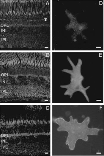
The distribution of AMPA receptors in the skate retina was also examined by immunostaining with commercial antibodies. Four polyclonal antibodies (GluR 1, 2, 2/3, and 4) were tested in this study. However, the GluR1 antibody was incapable of recognizing the skate variant of the protein, as no positive staining for GluR1 could be detected in retina or brain of skate, and no positive GluR1 band observed on Western blot of skate retinal and brain tissue samples using this antibody. Specific labeling was observed in retinal slices labeled with antibodies against GluR2, 2/3, and 4. Specific labeling for the GluR2 antibody was largely restricted to a narrow punctate band at the level of the outer limiting membrane (Fig. 5A). Faint fluorescence was also discernable in the outer plexiform layer (OPL) and occasional cells in the ganglion cell layer. The outer and inner segments of photoreceptors also showed prominent fluorescence; however, this labeling was present in control slices in which the primary antibody had been omitted, demonstrating that the photoreceptor labeling was nonspecific. This nonspecific staining of outer and inner segments of photoreceptors occurred consistently in all slices examined. The GluR2/3 antibody resulted in robust specific labeling in the OPL and the inner plexiform layer (Fig. 5B). Labeling was also apparent in the somas of cells within the inner nuclear region. Labeling with the GluR4 antibody was prominent in the OPL, with little detectable fluorescence in other layers of the retina (Fig. 5C). The diffuse pattern of staining in the OPL resembled closely that seen with the GluR2/3 antibody.
Immunohistochemical localization of AMPA receptor subtypes in slices of skate retina [(A), GluR-2, (B) GluR-2/3, and (C) GluR-4] and in isolated external horizontal cells [(D) GluR-2, (E) GluR-2/3, and (F) GluR-4]. Labeling at the level of the photoreceptor inner and outer segments was determined to be nonspecific based on control experiments applying the secondary antibody alone (not shown). Asterisk in (A) shows punctate labeling at the level of the outer limiting membrane. Scale bars are 10 μm. IPL, inner plexiform layer. INL, inner nuclear layer.

Antibody localization experiments were also conducted on external horizontal cells enzymatically isolated from the retina. Fig. 5D shows that very little fluorescence was present when cells had been incubated with the antibody to GluR2. However, the external horizontal cells displayed bright labeling with the GluR2/3 and GluR4 antibodies (Fig. 5E and 5F). No fluorescent signal was detectable using the GluR1 antibody in either retina or brain of skate, suggesting that the antibody cannot recognize the skate variant of the GluR1 receptor. Given the paucity of significant labeling of GluR2 in both the OPL and the isolated external horizontal cells, these data suggest that external horizontal cells of the skate retina preferentially express the AMPA receptor subunits GluR3 and 4.
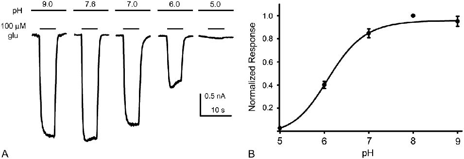
We next examined modulation of glutamate-induced responses of horizontal cells by hydrogen ions, which have been suggested to play an important role in regulating synaptic transmission within the retina generally and within the outer synaptic plexus specifically. pH is dynamic in the central nervous system and affects the functions of a variety of proteins. In catfish horizontal cells, NMDA receptors have been shown to have proton sensitivity with increasing proton concentrations reducing the size of NMDA-gated currents. These studies showed that the NMDA-gated channels have a pK of 6.5 (Wu & Christensen, Reference Wu and Christensen1996). Furthermore, currents mediated by non-NMDA ionotropic receptors of catfish horizontal cells have also been shown to be downregulated by extracellular acidification with a pK value near 6.5 for currents elicited by AMPA, kainate, and quisqualate (Christensen & Hida, Reference Christensen and Hida1990). However, studies by Tang et al. (Reference Tang, Dichter and Morad1990) showed a proton inhibition in NMDA-gated channels in embryonic rat hippocampus, while non-NMDA-gated channels were relatively unaffected by changes in extracellular pH, suggesting that different isoforms of neurotransmitter receptors may have differing levels of sensitivity to proton block. In the present study on isolated skate horizontal cells, H+ reversibly inhibited glutamate-induced currents. Changing extracellular pH to 6.0 reversibly inhibited glutamate-elicited currents to 48.1 ± 1.2% of control values (N = 7). The inhibition occurred in a concentration-dependent manner, with increasing concentrations in hydrogen ions (i.e., lowered pH) resulting in further downregulation of glutamate currents. Fig. 6A shows typical results from an individual cell. The cell was challenged with 100 μ m glutamate for 5 s while continually bathed in Ringer’s solutions at pH 9.0, 7.6, 7.0, 6.0, and 5.0, respectively. The response to glutamate was virtually abolished by solutions adjusted to pH 5.0. Fig. 6B shows average responses to seven cells fit to a Hill equation of the form shown in eqn. (1) where R max was taken as the current elicited at a pH of 7.6, s, with its negative logarithm equal to the pK, was determined to be 6.1, and the Hill coefficient n best fit with a value of 1.
pH effects on glutamate-induced currents in isolated skate horizontal cells. (A). Representative glutamate responses from a cell bathed in solutions ranging in pH from 9.0 to 5.0. Cells were challenged for 5 s with 100 μ m glutamate while being continually bathed in Ringer’s solutions at pH 9.0, 7.6, 7.0, 6.0, or 5.0, respectively. (B). Response amplitude as a function of extracellular pH is shown representing the average responses of seven cells. Responses were normalized to the glutamate-elicited current obtained at a pH of 7.6 for each cell.

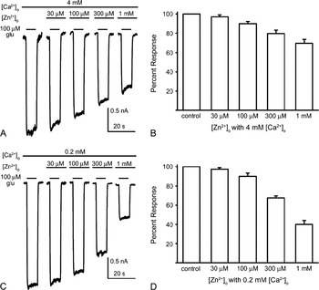
In addition to pH, divalent cations are thought to play an important regulatory role in the outer retina. It has been hypothesized that Zn2+ may be coreleased from photoreceptor synaptic terminals with glutamate (Ugarte & Osborne, Reference Ugarte and Osborne2001). Recently, Zhang et al. (Reference Zhang, Ribelayga, Mangel and McMahon2002) showed that AMPA receptor–mediated synaptic transmission from photoreceptors to horizontal cells in hybrid bass retina was inhibited by Zn2+. In the present study, Zn2+ also inhibited AMPA-mediated responses in skate horizontal cells. Fig. 7A shows five traces from a cell voltage clamped at −70 mV. The bath contained the standard 4 mm Ca2+, and cells were challenged with 100 μ m glutamate in the presence of 0, 30, 100, 300, and 1000 μ m Zn2+, respectively. Average glutamate responses in the presence of Zn2+ concentrations ranging from 30 μ m to 1 mm are shown for eight cells in Fig. 7B. Glutamate responses were reduced by Zn2+ in a concentration-dependent manner.
Zinc effects on glutamate-induced currents in isolated skate horizontal cells. (A). Five traces from a cell voltage clamped at −70 mV. The cell was bathed in a Ringer’s solution containing normal Ca2+ (4 mm), and the traces depict currents elicited when challenged with 100 μ m glutamate in the presence of 0, 30, 100, 300, and 1000 μ m Zn2+, respectively. (B). Average glutamate responses in the presence of the different Zn2+ concentrations are shown for eight cells. Responses for each cell were normalized to the glutamate response obtained in the absence of extracellular Zn2+. (C). Traces from an individual cell bathed in a Ringer’s solution containing lowered extracellular Ca2+ (0.2 mm) and subsequently challenged with 100 μ m glutamate in the presence of 0, 30, 100, 300, and 1000 μ m Zn2+, respectively. (D). Average responses from seven cells are shown; currents were normalized to the response initially obtained in a Ringer’s solution lacking extracellular Zn2+.

Sustained activation of photoreceptors in the dark has been postulated to lower extracellular calcium concentrations in the synaptic cleft (Gallemore et al., Reference Gallemore, Li, Govardovskii and Steinberg1994; Rabl, Reference Rabl and Thoreson2002; Cadetti et al., Reference Cadetti, Tranchina and Thoreson2005). Consequently, we examined Zn2+ regulation of the glutamate response in the presence of a lowered 0.2 mm [Ca2+]o Ringer’s solution. Traces from a single cell examined in this condition are plotted in Fig. 7C and suggest that the inhibitory effects of zinc at the higher concentrations are greater in the lowered calcium condition. Average responses from seven cells are shown in Fig. 7D. Statistical analysis revealed that the two highest concentrations of zinc were significantly more potent in reducing the glutamate-elicited current in the lowered calcium condition. At lower concentrations of Zn2+, glutamate-elicited currents were reduced to 97 ± 1.6% in 30 μ m Zn2+ and 89.9 ± 3.3% in 100 μ m Zn2+, which were similar to the magnitude of reduction found when trials were conducted in Ringer’s solution containing normal 4 mm [Ca2+]o (97.1 ± 2.0% and 90.0 ± 2.3%, respectively). However, at the two higher concentrations of Zn2+, glutamate-elicited currents obtained in lowered [Ca2+]o were reduced to 67.5 ± 2.0% in 300 μ m Zn2+ and 40.0 ± 4.0% in 1 mm Zn2+, significantly larger reductions than those obtained in Ringer’s solution containing normal levels of [Ca2+]o (79.5 ± 3.9% and 69.6 ± 4.0% for 300 μ m and 1 mm Zn2+, respectively, P < 0.01).
Calcium is thought to regulate a variety of processes in the OPL, such as voltage-gated Ca2+ channel function, spinule retraction, neurotransmitter release, and GABA transport (Okada et al., Reference Okada, Schultz, Geurtz, Hatt and Weller1999; Linn & Gafka, Reference Linn and Gafka2001; Kreitzer et al., Reference Kreitzer, Andersen and Malchow2003). Extracellular Ca2+ has also been shown to inhibit AMPA receptor–mediated currents in perch horizontal cells as well as in rat CA1 region hippocampal pyramidal cells (Schmidt, Reference Schmidt1999; Buldakova et al., Reference Buldakova, Bolshakov, Tikhonov and Magazanik2000). We found a similar inhibitory effect of increasing Ca2+ concentrations on the glutamate-induced currents of skate horizontal cells. Individual cells challenged with 100 μ m glutamate for 5 s in the presence of 0.4, 4, 10, and 20 mm [Ca2+]o were inhibited in a dose-dependent manner. The normal skate Ringer’s solution used in this study contains 4 mm [Ca2+]o. Reducing the [Ca2+]o from 4 to 0.4 mm increased 100 μ m glutamate responses to 118.8 ± 2.4% of control levels, while increasing [Ca2+]o to 10 mm inhibited glutamate responses to 85.7 ± 2.5% of control levels. Increasing [Ca2+]o to 20 mm further reduced glutamate responses to 74.4 ± 3.9% of control responses.
Discussion
Characterization of the glutamate receptors of skate horizontal cells
Our data are consistent with the hypothesis that fast excitatory synaptic transmission onto external horizontal cells in the skate retina is mediated largely by calcium-permeable AMPA receptors. Cells voltage clamped at −70 mV responded with large inward currents to extracellular application of glutamate, AMPA, and kainate in a concentration-dependent manner. GYKI 52466, which blocks responses mediated by activation of AMPA receptors (Tarnawa et al., Reference Tarnawa, Farkas, Berzsenyi, Patfalusi and Andrasi1990; Lukasiewicz et al., Reference Lukasiewicz, Wilson and Lawrence1997; Blanco & de la Villa, Reference Blanco and de la Villa1999), completely abolished glutamate-induced responses at a concentration of 100 μ m. The potentiation of glutamate-elicited responses by CTZ further supports the contention that AMPA receptors are the major mediators of glutamate-induced responses of skate horizontal cells since CTZ selectively potentiates responses of AMPA receptors while having little effect on kainate receptors (Partin et al., Reference Partin, Patneau, Winters, Mayer and Buonanno1993). Application of AMPA or glutamate in the presence of CTZ produced significantly larger responses than in the absence of CTZ, while little effect of CTZ was observed on kainate-induced responses. Moreover, the inability of concanavalin A, which preferentially potentiates responses of kainate receptors but not of AMPA receptors (Yang et al., Reference Yang, Maple, Gao, Maguire and Wu1998), to produce significant alterations in the currents elicited by glutamate further strengthens the argument for AMPA receptors mediating the majority of glutamatergic excitation of skate horizontal cells. There was no evidence for NMDA receptors on these cells: challenge with NMDA when Mg2+ had been removed from the bath and glycine was added failed to induce a current.
It is interesting in this latter context to compare our results with two other recent studies, one examining horizontal cells of the carp (Shen et al., Reference Shen, Zhang, Jin and Yang2006) and the other investigating the pharmacological properties of glutamate receptors in human horizontal cells (Shen et al., Reference Shen, Finnegan and Slaughter2004). Shen et al. (Reference Shen, Zhang, Jin and Yang2006) reported that the cone-driven H1 horizontal cells of carp possess NMDA receptors, while rod-driven horizontal cells (H4) of the same species lacked such receptors. Our own results clearly indicate that the rod-driven horizontal cells of the skate also lack NMDA receptors. In examining the characteristics of glutamate receptors of human horizontal cells, Shen et al. (Reference Shen, Finnegan and Slaughter2004) reported results remarkably similar to what we have found in the evolutionarily distant elasmobranch retina. In both skate and human horizontal cells, the responses to glutamate and AMPA were significantly enhanced by CTZ but only to a very small extent by concanavalin A. Moreover, the responses to direct kainate application were overall larger than for glutamate and AMPA, and kainate-induced responses were only modestly affected by coapplication with CTZ or concanavalin A. GYKI 52466 was also potent in blocking responses of glutamate agonists. Finally, similar to our findings in the skate horizontal cells, NMDA failed to induce any measurable currents in human horizontal cells. Shen et al. (Reference Shen, Finnegan and Slaughter2004) noted that their desensitization experiments with CTZ and concanavalin A, which are so similar to our own results, did not reveal a “hidden” population of kainate receptors on human horizontal cells. While we cannot completely rule out the possibility that some small fraction of the currents that we see in the skate horizontal cells might result from the activation of some kainate receptors, it is the striking similarity in the responses to glutamate, AMPA, kainate, GYKI 52466, CTZ, and concanavalin A between skate and human horizontal cells that are the most noteworthy points of comparison and argue for a particularly strong evolutionary pressure in maintaining the functional characteristics of glutamate receptor function within such close tolerances.
Molecular cloning studies have demonstrated that four different receptor subunits (GluR1–4) may combine to form hetero-oligomeric ion channels present on AMPA receptors. Differential expression patterns for these subunits have been reported in the OPL of a variety of vertebrate species, including goldfish, carp, cat, and rat (Hamassaki-Britto et al., Reference Hamassaki-Britto, Hermans-Borgmeyer, Heinemann and Hughes1993; Peng et al., Reference Peng, Blackstone, Huganir and Yau1995; Thoreson & Witkovsky, Reference Thoreson and Witkovsky1999; Hack et al., Reference Hack, Frech, Dick, Peichl and Brandstatter2001; Klooster et al., Reference Klooster, Studholme and Yazulla2001; Schultz et al., Reference Schultz, Janssen-Bienhold and Weiler2001). Antibodies to GluR1, GluR2, GluR3, and GluR4 label cat horizontal cells, while rodent horizontal cells have been found to express GluR2, GluR3, and GluR4 (Qin & Pourcho, Reference Qin and Pourcho1996; Hack et al., Reference Hack, Frech, Dick, Peichl and Brandstatter2001). AMPA receptors in horizontal cells of goldfish appear to consist of GluR2 and GluR3 subunits (Klooster et al., Reference Klooster, Studholme and Yazulla2001; Schultz et al., Reference Schultz, Janssen-Bienhold and Weiler2001). In the present study, antibodies directed against GluR2, 2/3, and 4 labeled cell processes in the OPL of the skate retina; the GluR2/3 and GluR4 antibodies also recognized cell bodies within the inner nuclear layer. The distinct labeling patterns of GluR2 and GluR2/3 indicate that the GluR3 subunit is recognized by the GluR2/3 antibody. Obvious GluR2/3 and GluR4 staining was also observed in isolated external horizontal cells of the skate. These data suggest that skate horizontal cells express the GluR3 and GluR4 forms of AMPA receptor subunits. It remains possible that the antibody for GluR2 is not particularly robust for the skate version of this AMPA receptor and that skate horizontal cells could possess receptors that are not clearly discernable with the present techniques. Moreover, it is also possible that GluR2 subunit might have been preferentially lost during the process of cellular dissociation used to produce isolated cells.
AMPA receptors containing the GluR2 subunit are reported to be impermeable to calcium, while those composed of GluR3 and GluR4 have been found to permit the direct influx of calcium into cells through the glutamate-gated channels (Fletcher & Lodge, Reference Fletcher and Lodge1996; Ozawa et al., Reference Ozawa, Kamiya and Tsuzuki1998; Dingledine et al., Reference Dingledine, Borges, Bowie and Traynelis1999). Our anatomical findings showing positive staining for GluR 2/3 and GluR4 suggest that skate horizontal cells possess calcium-permeable isoforms of AMPA receptors. In support of this anatomical evidence, previous studies using the calcium indicator dye Oregon Green have demonstrated calcium rises in skate horizontal cells that appear to be mediated by the influx of calcium directly through the glutamate-gated channels: removal of extracellular calcium abolishes the increase in fluorescence upon application of glutamate or kainate, changes in Oregon Green fluorescence (a calcium indicator dye) are prevented by prior application of the ionotropic glutamate blocker CNQX, and increases in intracellular calcium induced by glutamate occur even when voltage-gated calcium channels are blocked with nifedipine (Kreitzer et al., Reference Kreitzer, Andersen and Malchow2003). Furthermore, 1-naphthylacetyl spermine (NS), which selectively antagonizes Ca2+-permeable AMPA receptors in a use-dependent manner (Koike et al., Reference Koike, Iino and Ozawa1997), also removed a large portion of the glutamate-elicited current in a use- and concentration-dependent manner. These findings suggest that a significant proportion of the glutamate receptors present on skate horizontal cells are Ca2+-permeable forms. Huang and Liang (Reference Huang and Liang2005) suggested that calcium-permeable and calcium-impermeable AMPA receptors coexist on horizontal cells of the goldfish. The partial blockade of glutamate-elicited currents by NS leaves open the possibility that this is also the case for the horizontal cells of skate. The relatively modest rectification of the glutamate-induced currents shown in Fig. 2C and the lack of dramatic effect of intracellular spermine also suggest the possibility of contribution from other glutamate receptor types. Activation of AMPA/kainate receptors on horizontal cells of rabbit, carp, and catfish has also been reported to permit the direct influx of calcium (Linn & Christensen, Reference Linn and Christensen1992; Okada et al., Reference Okada, Schultz, Geurtz, Hatt and Weller1999; Rivera et al., Reference Rivera, Blanco and de la Villa2001). This is a striking conservation of function over such a wide range of evolutionarily distant species, ranging from the relatively primitive elasmobranchs used in the present study all the way to mammals. The presence of Ca2+-permeable AMPA receptors could allow for Ca2+-dependent regulatory roles for these receptors in synaptic processing in the outer retina. Previous studies in skate have shown that Ca2+-permeable glutamate receptors modulate GABA transport in horizontal cells (Kreitzer et al., Reference Kreitzer, Andersen and Malchow2003). In addition, Ca2+-permeable AMPA receptors have also been shown to play roles in regulation of voltage-gated Ca2+ channels in catfish horizontal cells (Linn & Gafka, Reference Linn and Gafka2001) and in the retraction of dendritic spinules present on horizontal cells in the carp retina (Okada et al., Reference Okada, Schultz, Geurtz, Hatt and Weller1999).
AMPA receptors enter a desensitized state very rapidly (within several milliseconds) following application of glutamate (Kiskin et al., Reference Kiskin, Kristal and Tsyndrenko1986; Patneau et al., Reference Patneau, Vyklicky and Mayer1993; Eliasof & Jahr, Reference Eliasof and Jahr1997). The present experiments were done using a superfusion system in which solutions were completely exchanged in about 20 ms, making it likely that the glutamate-induced responses reflect currents from largely desensitized receptors. In this regard, it is worth noting that experiments employing very rapid superfusion (1–2 ms or less) reveal dose–response values for steady-state (i.e., desensitized) responses that are shifted to the left by approximately two orders of magnitude compared to the initial rapid peak responses induced by activation of AMPA receptors (EC50s of about 50 μ m; O’Dell & Christensen, Reference O’Dell and Christensen1989b, compared to 1 mm, respectively; Lu et al., Reference Lu, Shen and Yang1998; Shen et al., Reference Shen, Zhou and Yang1999). The EC50 of 41 μ m reported here for the dose–response to glutamate suggests that the glutamate-induced responses in the present study are largely from desensitized receptors. Furthermore, addition of CTZ to nondesensitized AMPA receptors induces a shift to the left of peak AMPA receptor responses by about a factor or 2 (Patneau et al., Reference Patneau, Vyklicky and Mayer1993), whereas the dose–response curve reported here shifts significantly to the right in the presence of CTZ, again suggesting that the responses recorded here are from desensitized receptors.
One function of desensitization could be the reduction of noise, the notion being that many low-conducting channels might generate less noise than a few very large conducting channels. The observation that the dose–response relation to glutamate was significantly broader in the presence of the desensitization inhibitor CTZ than to glutamate alone suggests another potential impact of desensitization. The Hill coefficient for glutamate in the presence of CTZ was 1.25, whereas the coefficient for glutamate alone was 2.3. The dose–response relation to kainate, which elicits a response that does not desensitize, was also broader than that of glutamate. These results suggest that one function of desensitization of the glutamate receptors may be to narrow the dose–response range over which glutamate exerts an effect. This sharpening of the dose–response curve has the potential to play a role in enhancing the response of horizontal cells to changes in light intensity around a mean steady-state value when the cells are adapted to a steady-state background illumination. Photoreceptors, horizontal cells, and other postsynaptic elements of the skate retina show extensive adaptation to background light, shifting their light responses over many orders of light intensity (Green et al., Reference Green, Dowling, Siegel and Ripps1975). When adapted to a steady background light, the voltage of a horizontal cell is likely to sit near the middle of its response range, allowing the cell to respond to decrements as well as increments of light. In this adapted state, a horizontal cell possessing a steep dose–response relationship to glutamate can potentially generate a larger change in response to a dim stimulus compared to a cell having a lower Hill coefficient if the total overall response range were constant. Thus, paradoxically, desensitization of the glutamate receptors has the potential to sharpen and enhance the response of horizontal cells to small increments and decrements of light whose intensity is near that of the steady-state background. Also, as noted above, the dose–response curves for steady-state (i.e., desensitized) responses are shifted to the left by approximately two orders of magnitude compared to the initial peak responses; this leftward shift would also paradoxically suggest a greater sensitivity to glutamate when receptors are “desensitized.” On the other hand, it is very clear from many previous studies using very rapid superfusion techniques that the magnitude of the glutamate-induced current produced by desensitized receptors is significantly reduced compared to the peak nondesensitized responses (cf. Patneau et al., Reference Patneau, Vyklicky and Mayer1993; Shen et al., Reference Shen, Zhou and Yang1999). An important question is to what extent the tonic nature of neurotransmitter release from photoreceptors in the outer retina results in AMPA receptors on horizontal cells operating in a desensitized state (cf. Yang et al., Reference Yang, Maple, Gao, Maguire and Wu1998; Hirasawa et al., Reference Hirasawa, Shiells and Yamada2001) and to what degree the narrowing of the response curve and the leftward shift of the glutamate dose–response curve that accompanies the desensitized state contributes to the cells’ responses to alternations around the mean intensity of light.
Modulation of glutamate responses by H+, Ca2+, and Zn2+
Changes in the extracellular concentration of hydrogen ions have been shown to have a role in regulation of release of the neurotransmitter glutamate from photoreceptor synaptic terminals by altering Ca2+ entry into the synaptic terminal (DeVries, Reference DeVries2001). Alterations in extracellular levels of acidity have also been shown to alter a variety of postsynaptic conductances in the OPL, including glutamate responses on horizontal cells of catfish. Hydrogen ion concentration, likely through the protonation of histidine groups, has been shown to regulate both NMDA and non-NMDA receptors on catfish horizontal cells (Christensen & Hida, Reference Christensen and Hida1990; Wu & Christensen, Reference Wu and Christensen1996). In the present study on skate horizontal cells, increasing H+ concentrations exerted a depressive effect on glutamate currents. A remaining question is whether alterations in extracellular pH in the normal retina would normally approach the magnitude needed to induce significant alterations in the function of the glutamate receptors present in the skate horizontal cells. The extracellular space within the invaginating synapse where horizontal cells contact the synaptic terminals of the photoreceptors is small and rather restricted, and small changes in the amount of free hydrogen ions could result in significant alterations in the pH of this compartment. Indeed, DeVries (Reference DeVries2001) has presented evidence that the small number of protons added to the extracellular space upon the fusion of a synaptic vesicle may be sufficient to alter neurotransmitter release from photoreceptors. The effects seen by DeVries (Reference DeVries2001), however, were thought to occur at the level of voltage-sensitive calcium channels, which are much more sensitive to changes in pH than the glutamate currents we have examined. Thus, we believe that the depressive effects of decreased pH on glutamate receptors that we have observed are likely to play only a small role in the normal processing of visual information within the outer retina.
Zn2+ has been shown to be localized at the synaptic terminals of photoreceptors and may be coreleased with glutamate onto second-order retinal neurons (Wu et al., Reference Wu, Qiao, Noebels and Yang1993; Redenti & Chappell, Reference Redenti and Chappell2005; Redenti et al., Reference Redenti, Ripps and Chappell2007). Regulation of the postsynaptic effects of glutamate by Zn2+ thus has the potential to play an important physiological role in synaptic processing in the outer retina (Rosenstein & Chappell, Reference Rosenstein and Chappell2003). Zn2+ can exert either a facilitatory or an inhibitory effect on glutamate receptor–mediated currents, depending on its concentration and the particular subunit composition of the glutamate receptors. We found that extracellular application of Zn2+ consistently promoted downregulation of glutamate currents of skate horizontal cells. High levels of calcium also had a depressive effect on currents elicited by glutamate in skate horizontal cells. Thus, alterations in [Ca2+]o in the outer plexiform could affect not only the release of neurotransmitter from the presynaptic terminal but also the postsynaptic effects of glutamate as well. Previous studies have suggested that this type of downregulation of the current through glutamate receptors is likely due to direct binding of calcium to allosteric sites on the external surface of the channel and/or block of the flow of ions through the pore of the glutamate-activated channels (Buldakova et al., Reference Buldakova, Bolshakov, Tikhonov and Magazanik2000). It is also possible that some of the block in glutamate receptor current might be mediated by calcium actually permeating through the channel. In addition, the inhibition by higher concentrations of Zn2+ (300 μ m and 1 mm) was enhanced at lower Ca2+ levels in the extracellular environment of the cell. This suggests that inhibition of glutamate receptor currents by Zn2+ and Ca2+ may be occurring through effects on a common extracellular site.
In conclusion, our results suggest that the excitatory responses induced by glutamate in skate horizontal cells are mediated predominantly by AMPA receptors, of which at least part of the population of receptors on the cells are Ca2+ permeable. The receptors show a significant degree of desensitization, which may play a role in narrowing the dose–response relation of the horizontal cells to glutamate. This narrowing of the dose–response curve has the potential to enhance responses to increments and decrements of light stimuli whose intensity is near that of the steady-state background illumination. The function of these receptors can also be regulated by alterations in extracellular H+, Ca2+, and Zn2+, and such alterations have the potential to play a modulatory role in the normal processing of visual signals within the retina of the skate.
Acknowledgments
We are grateful to Indiana Wesleyan University and the Department of Biological Sciences of the University of Illinois at Chicago for their contributions in support of this manuscript. This work was also supported by a grant from the National Science Foundation (009-1281) and a Hinds Fellowship from Indiana Wesleyan University. Special thanks are due to Dr. Anthony Molina for his comments on the manuscript.