Introduction
The cestodes of Schistocephalus Creplin, 1829 represent one of the most important model systems for host-parasite interaction studies (Barber and Scharsack Reference Barber and Scharsack2010; Heins Reference Heins2017; Lester Reference Lester1971; Svensson et al. Reference Svensson, Eghbal, Eriksson and Nilsson2022). The genus Schistocephalus comprises widely distributed parasites that complete their life cycle through planktonic crustaceans, fish, and endothermic vertebrates, primarily birds (Dubinina Reference Dubinina1959). The Schistocephalus solidus (Müller, 1776) - three-spine stickleback Gasterosteus aculeatus Linnaeus, 1758 system has been most extensively studied, providing fundamental insights into host-parasite relationships (Barber and Scharsack Reference Barber and Scharsack2010; Heins Reference Heins2017; Lester Reference Lester1971; Svensson et al. Reference Svensson, Eghbal, Eriksson and Nilsson2022). To date, a total of six Schistocephalus species are recognized as valid (Caira et al. Reference Caira, Jensen and Barbeau2022). Among them, two species exhibit strict host specificity: S. solidus almost exclusively infects the three-spine stickleback, whereas S. pungitii Dubinina, Reference Dubinina1959 is a specific parasite of the ninespine stickleback (Pungitius pungitius Linnaeus, 1758) (Bråten Reference Bråten1966; Heins et al. Reference Heins, Moody, Arostegui, Harmon, Blum and Quinn2024; Nishimura et al. Reference Nishimura, Heins, Andersen, Barber and Cresko2011; Orr et al. Reference Orr, Hopkins and Charles1969; Dubinina Reference Dubinina1959). This narrow host specificity has been experimentally confirmed through both plerocercoid transplantation between hosts (Bråten Reference Bråten1966) and cross-infection studies (Orr et al. Reference Orr, Hopkins and Charles1969; Dubinina Reference Dubinina1959). However, there are molecularly confirmed records of three plerocercoids of S. solidus from P. pungitius in Alaska (Heins et al., Reference Heins, Moody, Arostegui, Harmon, Blum and Quinn2024).
Beyond sticklebacks, Schistocephalus plerocercoids have also been recorded in the body cavities of other Scorpaeniformes – cottid fishes across Holarctic regions. A distinct species, S. cotti, was described from the European bullhead (Cottus gobio Linnaeus, 1758) in Finland (Chubb et al. Reference Chubb, Seppälä, Lüscher, Milinski and Valtonen2006). Another species, S. nemachili, was reported from the cypriniform fish (noted as Nemacheilus crassus) in China (Dubinina Reference Dubinina1959), representing the only documented infection in non-scorpaeniform hosts. Additionally, S. thomasi as described from the herring gull (Larus argentatus Pontoppidan, 1763) and the common tern (Sterna hirundo Linnaeus, 1758) in the Lake Michigan area, USA (Caroian Reference Caroian1960); however, its piscine intermediate hosts remain unknown.
Prior to the description of S. cotti, plerocercoids from cottid fishes were universally attributed to S. solidus. Some authors have questioned the validity of distinguishing S. cotti, continuing to identify cottid-derived plerocercoids – with their characteristic morphological features – as S. solidus (Ieshko et al. Reference Ieshko, Shulman, Lebedeva, Barskaja and Niemela2012, Pospekhov et al. Reference Pospekhov, Atrashkevich, Orlovskaya and Mikhailova2021). A similar taxonomic uncertainty surrounds the identification of Schistocephalus plerocercoids in Lake Baikal. This unique lake, the deepest on the planet, is also believed to be the oldest (Kozhova and Izmesteva Reference Kozhova and Izmesteva1998, Sideleva Reference Sideleva2003). It represents a hotspot of biodiversity and endemism, including a unique radiation of cottid fishes. The current number of endemic sculpin-like fishes (Cottoidei) in Lake Baikal is estimated at 33–35 species. These are classified either within the single family Cottidae (e.g., FishBase) or within the three families Cottidae, Comephoridae, and Abyssocottidae (Sideleva Reference Sideleva2003; Taliev Reference Taliev1955). However, the exact species count remains debated due to ongoing taxonomic revisions and the discovery of cryptic species complexes (Kontula et al. Reference Kontula, Kirilchik and Väinölä2003; Sideleva Reference Sideleva2023). The greatest diversity occurs in the genera Asprocottus Berg, 1906 (7–8 species) and Procottus Gratzianow, 1902 (4–5 species), reflecting their adaptive radiation within the Lake Baikal’s ecosystem. Dubinina (Reference Dubinina1966) hypothesized that Baikal sculpins might host a distinct Schistocephalus species, but this remained speculative due to insufficient data. The available records identify S. solidus and S. nemachili as the only Schistocephalus species reported from Baikal sculpins (Rusinek Reference Rusinek2024; Rusinek Reference Rusinek2007). Large plerocercoids in Siberian waters have also been documented in the body cavity of Siberian sculpins, Cottus sibiricus Warpachowski, 1889, from Lake Teletskoye. It was previously identified as Ligula sp. (Titova Reference Titova1954, Reference Titova1965). Lake Teletskoye is the deepest (with 325 m maximum depth) lake in Western Siberia (Selegei Reference Selegei, Dehandschutter and Klerkx2001). Its steep depth profile, oligotrophic nature, and low water temperature align it with Lake Baikal. However, these earlier taxonomic assignments conflict with established host specificity patterns for both S. solidus (Bråten Reference Bråten1966; Heins et al. Reference Heins, Moody, Arostegui, Harmon, Blum and Quinn2024; Nishimura et al. Reference Nishimura, Heins, Andersen, Barber and Cresko2011; Orr et al. Reference Orr, Hopkins and Charles1969; Dubinina Reference Dubinina1959) and Ligula spp. (Orr Reference Orr1967; Dubinina Reference Dubinina1966). In this study, we re-evaluate the species identity of plerocercoids from sculpins in Lakes Baikal and Teletskoye using integrated morphological and molecular genetic analyses.
Materials and methods
Sample collection
Siberian sculpins were collected in the northern part of Lake Teletskoye (51°79’N; 87°30’E) in May 2024. Sculpins of the endemic genera Batrachocottus Berg, 1903, Leocottus (Taliev, Reference Taliev1955), Paracottus Taliev, in Berg, 1949, and Procottus were sampled from four sampling sites in Lake Baikal: Zama (53°26’N; 107°37’E), Bugul’deika (52°31’N; 106°02’E), Bol’shie Koty (51°53’N; 105°04’E), and Tankhoi (51°34’N; 105°11’E) villages in spring and summer 2024–2025. Collected fish were measured for the total body mass and total body length; the morphometric data of the examined specimens are provided in Supplementary 1. Plerocercoids of Schistocephalus were also examined from the stomach contents of piscivorous fishes in Lake Baikal, where they occurred alongside sculpin remains. For comparison with S. solidus (the species previously assigned to the Baikal cestodes), our analysis also included plerocercoids from G. aculeatus collected in the estuarine Nizhnee Ershovskoye Lake near the White Sea coastline (66°32’N; 33°03’E) (Figure 1).
Sampling sites and known records of Schistocephalus spp. from sculpin fishes in Siberia and the Russian Far East. Sites of original material are marked in red, while literature-based records are shown in green. 1. Lake Nizhnee Ershovskoe; 2. Lake Teletskoye; 3. Lake Keta – S. pungitii from P. knerii according to Pugachev (Reference Pugachev2002); 4. The water bodies of Tyva – S. solidus from P. knerii according to Pugachev (Reference Pugachev2002); 5. Yenisei River – S. solidus from P. knerii according to Gundrizer (Reference Gundrizer1981); 6. Baikal Lake – L. kesslerii and P. knerii according to Rusinek (Reference Rusinek2007, Reference Rusinek2024); 7. Bol’shoe Leprindo, Maloye Leprindo, and Leprindokan lakes – S. solidus from C. poecilopus according to Pugachev (Reference Pugachev2002); 8. Bolshoy Darpir and Maly Darpir lakes – S. solidus from C. kolymensis according to Pospekhov et al. (Reference Pospekhov, Atrashkevich, Orlovskaya and Mikhailova2021); 9. The downstreams of the Penzhina and Talovka rivers – Schistocephalus sp. from C. kolymensis (Boutorina et al. Reference Boutorina, Aseeva, Koval and Nguyen2017); 10. Anadyr’ River – S. solidus from C. cognatus according to Pugachev (Reference Pugachev1984, Reference Pugachev2002); 11. The lakes near the village of Lavrentiya – S. solidus from C. cognatus Pugachev (Reference Pugachev2002). 12. Lavrentiy Bay – S. pungitii from C. kaganowskii (syn. C. cognatus) according to Dubinina (Reference Dubinina1959). Sampling sites on Lake Baikal: 6a. Zama village; 6b. Bugul’deika village; 6c. Bol’shie Koty village; 6d. Tankhoi village.

Fish were dissected according to standard methods (Bykhovskaya-Pavlovskaya, Reference Bykhovskaya-Pavlovskaya1985, Chervy Reference Chervy2024). Collected plerocercoids were rinsed, relaxed with hot water, and fixed in 70% ethanol for subsequent morphological and molecular analyses. The prevalence and mean intensity of parasite infestation were calculated according to the definitions by Bush et al. (Reference Bush, Lafferty, Lotz and Shostak1997).
Four hologenophores with museum accession numbers 1461–1464 were deposited in the Museum of Helminthological Collections at the Center for Parasitology of the A.N. Severtsov Institute of Ecology and Evolution of the Russian Academy of Sciences (IPEE RAS), Moscow, Russia. The remaining examined specimens (hologenophores and paragenophores) have been deposited in the Helminthological Collection of the Institute of Systematics and Ecology of Animals, Siberian Branch of the Russian Academy of Sciences (ISEA SB RAS) (see Supplementary 2), Novosibirsk, Russia.
Morphological study
The number of proglottids was measured for the examined plerocercoids. The structure of the first proglottid was investigated using scanning electron microscopy (SEM). For this purpose, after fixation in 70% ethanol, the specimens were dehydrated in a graded ethanol series, with a final change to absolute acetone. Subsequently, the samples were dried, mounted on stubs, and examined using Hitachi TM-1000 scanning electron microscope (Hitachi High-Technologies Corporation, Tokyo, Japan).
DNA extraction and sequencing
Total DNA was extracted from individual plerocercoids using the DNA-sorb B kit manufacturers’ protocols (kit for DNA extraction, Central Research Institute of Epidemiology, Russia). PCR amplification of the mitochondrial cox1 gene fragment was conducted using B2-TrpF (5′-TAG ACT AAR TGT TTT CAA AAC A-3′) and B2-16SR (5′-GCA TGA TRC AAA AGG CAC A-3′) primers (De Chambrier et al. Reference De Chambrier, Brabec, Tran and Scholz2019). The cycling conditions were as follows: 95°C for 5 min; 34 cycles of 95°C for 15 s, 53°C for 20 s, and 72°C for 1 min; and a final extension at 72°C for 5 min. Partial sequences of the 28S rRNA gene were amplified using LSU5 (TAGGTCGACCCGCTGAAYTTYAGCA) (Littlewood et al. Reference Littlewood, Curini-Galletti and Herniou2000) and 1500R (GCTATCCTGAGGGAAACTTCG) primers (Littlewood et al. Reference Littlewood, Waeschenbach and Nikolov2008). The cycling conditions adopted were the following: 95°C for 5 min; 34 cycles of 95°C for 15 s, 57°C for 30 s, and 72°C for 80 s; and a final extension at 72°C for 5 min. Double-stranded DNA was amplified using the BioMaster HS-Taq PCR-Color (2×) kit (Novosibirsk, Russia) according to the manufacturer’s instructions (http://biolabmix.ru/products/klassicheskaja_pcr/biomaster_hs-taq_pcr-color__2_/). PCRs were 50 μL in volume and contained 25 μL BioMaster HS-Taq PCR-Color reaction mix, 0.2 μmol/L of each primer, 20 μL sterile water, and 3 μL of total DNA was used as template. The chromatograms of the amplicon sequences were evaluated based on the sharpness and clear visibility of each peak for each nucleotide in Chromas 2.6.6 (https://technelysium.com.au). The sequences were aligned, edited, and checked for unexpected stop codons in MEGA 11 (Tamura et al. Reference Tamura, Stecher and Kumar2021). All gaps and poorly aligned positions in the 28S alignment were identified and removed using Gblocks (online version 0.91.1, https://ngphylogeny.fr; Castresana Reference Castresana2000; Lemoine et al. Reference Lemoine, Correia, Lefort, Doppelt-Azeroual, Mareuil, Cohen-Boulakia and Gascuel2019) with default settings. Newly obtained sequences were deposited in GenBank (Supplementary 2).
Phylogenetic analysis
Phylogenetic reconstructions of Schistocephalus spp. were performed using Bayesian inference approach. The optimal substitution model was determined using jModelTest 2.1.1 (Darriba et al. Reference Darriba, Taboada, Doallo and Posada2012). For the cox1 gene, we employed the dHKY + G model of nucleotide substitutions, while for the nad1 gene and the LSU region, we applied the dHKY + I model. Bayesian analysis was performed with MrBayes v.3.2.1 (Ronquist et al. Reference Ronquist, Teslenko, van der Mark, Ayres, Darling, Höhna, Larget, Liu, Suchard and Huelsenbeck2012). Two simultaneous runs with four Markov chains each were performed for 1 × 10⁶ generations and sampled every 500 generations. The first 25% of generations were discarded as burn-in. Additionally, the analysis included all available GenBank sequences for Schistocephalus spp. for the 28S and nad1 genes. For cox1, the first 100 sequences were included for S. solidus, while for the other Schistocephalus spp., all available GenBank sequences for this locus were used. The choice of Duthiersia expansa and Spirometra erinaceieuropaei as outgroups was based on the tree topologies published by Waeschenbach et al. (Reference Waeschenbach, Brabec, Scholz, Littlewood and Kuchta2017) and Fraija-Fernández et al. (Reference Fraija-Fernández, Waeschenbach, Briscoe, Hocking, Kuchta, Nyman and Littlewood2021).
Results
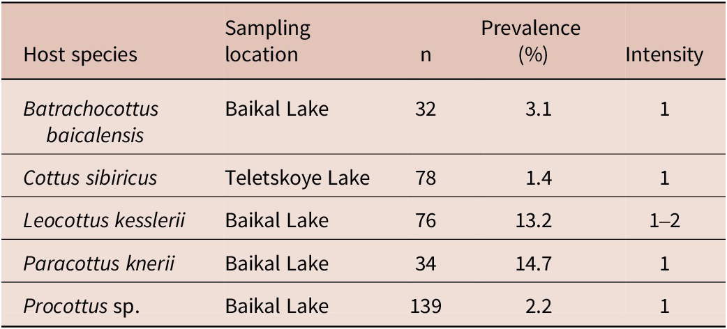
Plerocercoids of Schistocephalus sp. were found in the body cavity of four fish species from Lake Baikal and one species from Lake Teletskoye (Table 1). The intensity of infection ranged from one to two plerocercoids per fish. Additionally, two and one plerocercoids (co-occurring with partially digested cottid fishes) were collected from the stomachs of perch (Perca fluviatilis Linnaeus, 1758) and Amur catfish (Silurus asotus Linnaeus, 1758) in Lake Baikal, respectively. The highest prevalence of infection was recorded in Paracottus knerii (Dybowski, 1874) from Lake Baikal. The lowest prevalence was recorded in C. sibiricus from Lake Teletskoye.
Infestation of the studied fishes by Schistocephalus sp. plerocercoids

Morphological data
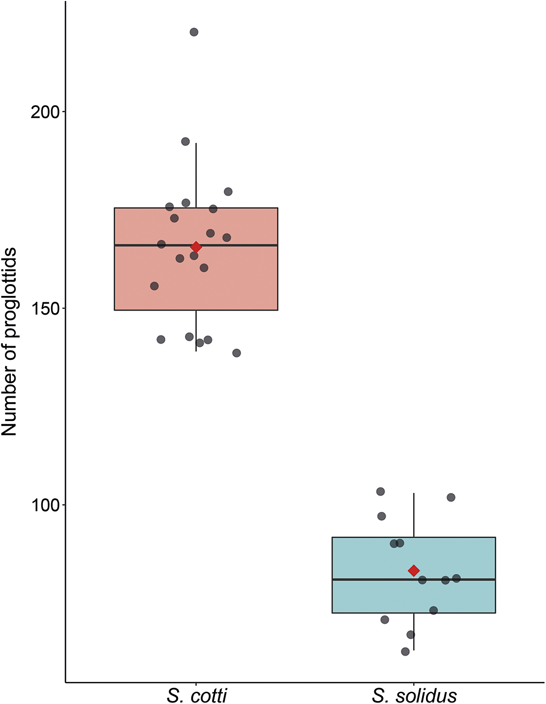
In the examined plerocercoids from sculpins, the number of proglottids ranged from 139 to 220 (Supplementary 2). The first strobilar proglottid of all examined plerocercoids from the body cavity of Cottidae fish possessed a distinctly developed velum, open on both the ventral and dorsal sides, with its margins curved towards the scolex (segmentation incomplete, missing along the mid-line of the strobila) (Figure 2A,B). In the S. solidus specimens from the three-spine stickleback, the first proglottid always possessed a well-defined, continuous velum (Figure 2C,D). The number of proglottids in the examined plerocercoids of this species ranged from 63 to 103 (Figure 3, Supplementary 2).
Scanning electron microscopy (SEM) and general view of Schistocephalus spp. plerocercoids. (A) SEM of the scolex of S. cotti (voucher no. C25105) from Lake Baikal. (B) SEM of the scolex of S. solidus (voucher no. C25122). (C) General view of S. cotti plerocercoids from Baikal sculpins.

Number of proglottids in the examined plerocercoids of S. cotti and S. solidus. Boxplots display median (centre line), interquartile range (box bounds), and range (whiskers). Gray points show individual values; red diamonds indicate means.

Phylogenetic analysis
We obtained five 28S and six cox1 and nad1 sequences from the plerocercoids collected in Lakes Baikal and Teletskoye. In addition, we included two cox1 and three 28S sequences from S. solidus plerocercoids from the three-spine stickleback from Nizhnee Ershovskoye Lake in the analysis.
Phylogenetic analysis of the 28S gene fragments revealed that sequences from plerocercoids in Lakes Baikal and Teletskoye formed a distinct clade, comparable to those of S. solidus and S. pungitii (Figure 4A). The original sequences from G. aculeatus clustered within the S. solidus clade. The nodal support values on the tree were 99% or higher. For the cox1 gene, after including sequences from the slimy sculpin (Cottus cognatus) in the analysis, similar results were obtained. Sequences from sculpins from Lakes Baikal and Teletskoye also formed a separate clade with high support. Distinct clades were formed by S. pungitii and S. solidus, the latter of which included the original sequences from G. aculeatus. Sequences from the slimy sculpin clustered separately (Figure 4B).
Phylogenetic trees of Schistocephalus based on 28S, cox1 gene, and nad1 gene sequences. Original sequences are shown in bold; the S. cotti clade from sculpins of Lakes Baikal and Teletskoye is highlighted in colour. Clades containing more than ten sequences are collapsed.

The phylogenetic tree based on nad1 sequences has a similar topology. Four well-supported, monophyletic clades (with maximum support values) were formed: one each for S. solidus, S. pungitii, and Schistocephalus sp. (from C. cognatus and C. aleuticus in Alaska), and one comprising Schistocephalus sequences from European and Siberian sculpin hosts. As this latter “Eurasian clade” also contained sequences identified as S. cotti from C. gobio, we assign it to the species S. cotti (Figure 4C).
Since the cox1 and nad1 datasets included sequences from cestodes collected in geographically distant localities (Canada and Alaska), an additional analysis of the nad1 dataset was performed, to which the sequences published by Fraser et al. (Reference Fraser, Gray, Dobbs, Andrews, De Reep and Duffy2023) were added. The final alignment comprised only 125 bp due to the short overlapping region between the sequences. As a result, the sequences from Canada (from C. cognatus) and Alaska (from C. cognatus and C. aleuticus) formed two distinct, well-supported clades, in addition to the clades of S. solidus, S. pungitii, and the “Eurasian clade” from European and Siberian sculpin hosts (Figure 5).
Phylogenetic tree of Schistocephalus based on nad1 gene sequences. Original sequences are shown in bold; clades formed by sequences from sculpin hosts (original sequences and those published by Fraser et al. (Reference Fraser, Gray, Dobbs, Andrews, De Reep and Duffy2023) and Heins et al. (Reference Heins, Moody, Arostegui, Harmon, Blum and Quinn2024)) are highlighted in colour. Clades containing more than ten sequences are collapsed.

Discussion
In Siberian water bodies, we recorded Schistocephalus plerocercoids in five fish species. Three ones are endemics of Lake Baikal: Batrachocottus baicalensis (Dybowski, 1874), Leocottus kesslerii (Dybowski, 1874), Procottus sp., and P. knerii which is an endemic of the Yenisei River basin, while C. sibiricus is widely distributed species across the Siberian region. Previous studies in Lake Baikal documented plerocercoids identified as S. solidus in L. kesslerii and P. knerii (Dubinina Reference Dubinina1966, Rusinek Reference Rusinek2024; Rusinek Reference Rusinek2007). Additionally, both species were found to be hosts of S. nemachili (Rusinek Reference Rusinek2024; Rusinek Reference Rusinek2007). Furthermore, a plerocercoid of S. solidus was recorded from the stomach of a lenok (Brachymystax lenok Pallas, 1773) (Matveeva and Matveev Reference Matveeva and Matveev1990).
We recorded Schistocephalus plerocercoids in the stomachs of two piscivorous fish species, P. fluviatilis and S. asotus. We consider these cases of spurious parasitism, as there is no evidence for the successful establishment of Schistocephalus plerocercoids in these fish hosts. Previously in Siberia, Schistocephalus spp. have also been reported from the stomachs of piscivorous fishes (Pugachev, Reference Pugachev2002).
In Lake Teletskoye, we detected Schistocephalus infection in 1 of 72 examined sculpins (prevalence = 1.4%) (Table 1), which is lower than previous records from the same lake. Titova (Reference Titova1954) reported infections in 2 of 15 sculpins (13.3%). In these studies, the plerocercoids were identified as L. intestinalis (Titova Reference Titova1954) and Ligula sp. (Titova Reference Titova1965). Given the strict cyprinid-specificity of Ligula spp. (Orr Reference Orr1967; Dubinina Reference Dubinina1966), these records likely represent misidentifications of Schistocephalus sp. Based on our data and the small sample size from previous study (Titova Reference Titova1954), we can conclude that the infection prevalence in sculpins in Lake Teletskoye is low.
Outside of Lake Baikal, Schistocephalus plerocercoids have also been previously recorded in cottid fishes across Siberia and the Russian Far East (Figure 1). Within the Yenisei River basin, they have been reported from P. knerii (Pugachev Reference Pugachev2002; Gundrizer Reference Gundrizer1981), in Eastern Siberia, from C. poecilopus (Pugachev Reference Pugachev2002), in the Kolyma River basin and rivers draining into the Sea of Okhotsk, from C. kolymensis (Pospekhov et al. Reference Pospekhov, Atrashkevich, Orlovskaya and Mikhailova2021; Boutorina et al. Reference Boutorina, Aseeva, Koval and Nguyen2017), and in water bodies of Chukotka, from C. cognatus (Pugachev Reference Pugachev1984, Reference Pugachev2002; Dubinina Reference Dubinina1959). Although these findings were originally identified as S. solidus, S. pungitii, or Schistocephalus sp., the fact that they were all found in cottid fishes raises the possibility that they may actually belong to S. cotti.
The difference in the number of proglottids among plerocercoids from different fish host species was discussed by Dubinina (Reference Dubinina1966). In addition to variations in number of proglottids between S. pungitii (62–92), S. solidus (79–148), and S. nemachili (228–235), she also identified Schistocephalus sp. from Baikal sculpins (138–190). It was further demonstrated that Schistocephalus spp. are characterized by the formation of the definitive number of strobila proglottids at the plerocercoid stage (Clarke Reference Clarke1954; Dubinina Reference Dubinina1966), and maturation in the definitive host’s intestine occurs very rapidly without significant morphogenesis. Chubb et al. (Reference Chubb, Valtonen, McGeorge and Helle1995) demonstrated a relationship between the number of proglottids and the length of plerocercoids. Additionally, a dependence of both these traits on host body length was observed (Chubb et al. Reference Chubb, Valtonen, McGeorge and Helle1995), suggesting that host size may be a source of their variability. In the description of S. cotti, the difference in number of proglottids was used as a diagnostic character. For S. solidus, values ranged from 55 to 107 (average 74), while for S. cotti they ranged from 122 to 189 (average 146). The range of proglottid counts in the plerocercoids we collected from sculpins did not overlap with that of S. solidus from the three-spine stickleback (Figure 3). Instead, the counts for the majority of these specimens fell within the range reported for S. cotti.
One of the plerocercoids had 220 proglottids (voucher No. 1462, Figure 2 C), which exceeds the upper limit reported for S. cotti (Chubb et al., Reference Chubb, Valtonen, McGeorge and Helle1995), but does not reach the values typical for S. nemachili (Dubinina Reference Dubinina1966). Previously, Schistocephalus plerocercoids with an extremely high number of proglottids (up to 205–230) were also recorded in C. kolymensis from the lower reaches of the Penzhina and Talovka Rivers in Kamchatka Krai (Figure 1; Boutorina et al. Reference Boutorina, Aseeva, Koval and Nguyen2017). Based on their relatively large body length and high proglottid count, these plerocercoids were identified as Schistocephalus sp., distinct from S. solidus found in the three-spine stickleback (Atrashkevich et al. Reference Atrashkevich, Orlovskaja, Regel, Mihailova, Pospekhov, Chereshnev, Chernyavskiy and Kashin2005; Boutorina et al. Reference Boutorina, Aseeva, Koval and Nguyen2017).
However, the number of proglottids alone is an insufficient diagnostic character, as significant variation in this trait between two geographic populations of S. solidus has been demonstrated (Chubb et al. Reference Chubb, Valtonen, McGeorge and Helle1995). A key morphological characteristic of S. cotti is the structure of the first strobilar proglottid: its well-developed velum has incomplete segmentation, curving towards the scolex. In contrast, the first proglottid of S. solidus features a distinct continuous margin. Thus, all specimens we examined from sculpins in Lake Baikal and Lake Teletskoye possess the complete set of morphological characteristics diagnostic for S. cotti.
Molecular data confirm that the plerocercoids from Lake Baikal and Lake Teletskoye belong to the same species. The results of the phylogenetic analysis also indicate that the studied plerocercoids from sculpins are conspecific with S. cotti. Schistocephalus plerocercoids also infect the slimy sculpin (C. cognatus) and the coastrange sculpin (C. aleuticus) in North American water bodies (Fraser et al. Reference Fraser, Gray, Dobbs, Andrews, De Reep and Duffy2023; French and Muzzall Reference French and Muzzall2008; Harmon et al. Reference Harmon, Hilborn and Quinn2015; Heins et al. Reference Heins, Moody, Arostegui, Harmon, Blum and Quinn2024). Previous phylogenetic analyses by Fraser et al. (Reference Fraser, Gray, Dobbs, Andrews, De Reep and Duffy2023) and Heins et al. (Reference Heins, Moody, Arostegui, Harmon, Blum and Quinn2024) demonstrated that Schistocephalus sp. from New World sculpins represents a distinct genealogical lineage. However, these authors maintained identification only to genus level due to insufficient data on morphological variation and host plasticity within the genus Schistocephalus. A less obvious yet plausible obstacle to precise species identification could be the potential existence of Schistocephalus spp. hybrids. Although in vitro experiments have successfully produced hybrids between S. solidus and S. pungitii (Henrich et al. Reference Henrich, Benesh and Kalbe2013), suggesting that natural hybridization may be possible, no evidence of such hybrids has been found in wild populations to date. The results of our phylogenetic analysis demonstrate that Schistocephalus populations from the New World and plerocercoids from Lakes Baikal and Teletskoye form two separate lineages at the species level.
Additional analysis of the nad1 gene revealed that the sequences from Canada and Alaska published by Fraser et al. (Reference Fraser, Gray, Dobbs, Andrews, De Reep and Duffy2023) and Heins et al. (Reference Heins, Moody, Arostegui, Harmon, Blum and Quinn2024) form two distinct, well-supported clades (Figure 5). However, as these results are based on a short fragment, they require further verification using a more comprehensive dataset. Therefore, based on these findings, we can only hypothesize that these plerocercoids represent two distinct species.
This raises the question of which taxa correspond to the North American lineages. One possible candidate is S. thomasi, described from piscivorous birds of North America based on distinctive features of the male reproductive system (Caroian Reference Caroian1960). However, the original description lacks data on strobila proglottid number, preventing the use of this diagnostic character for Nearctic Schistocephalus species identification. Notably, the illustrated morphology of the first proglottid matches the description of S. cotti (Chubb et al. Reference Chubb, Seppälä, Lüscher, Milinski and Valtonen2006) and corresponds to our examined specimens. Nevertheless, direct comparison of mature specimens with plerocercoids may be problematic due to potential morphological changes during cestode maturation. Consequently, we propose that at least one distinct Schistocephalus species, absent in Eurasia, parasitizes North American freshwater fishes. The taxonomic status of S. thomasi requires verification and redescription.
Records of S. cotti in Southern Siberian lakes confirm and substantially extend the prior hypothesis of its broad distribution across the Northern Palaearctic (Chubb et al. Reference Chubb, Seppälä, Lüscher, Milinski and Valtonen2006; Seppälä et al. Reference Seppälä, Chubb, Niemelä and Valtonen2007). Its host range includes multiple species within the Cottoidei suborder, contrasting with the narrow host specificity observed in S. solidus and S. pungitii. Lake Baikal likely represents the aquatic ecosystem with the highest diversity of potential intermediate hosts for S. cotti.
Supplementary material
The supplementary material for this article can be found at http://doi.org/10.1017/S0022149X26101382.
Acknowledgements
The authors are grateful to Dr. Galina Azarkina for her assistance with scanning electron microscopy.
Financial support
Russian Science Foundation, grant no. 25-24-00262 (sampling material from fishes from Lake Baikal), and Russian Science Foundation grant no. 23-74-10101 (sampling material and sequencing of plerocercoids from fish from Lake Teletskoye).
Competing interests
Authors stated no conflict of interest.
Ethical standard
The result of this work has not been published previously and is not under consideration elsewhere.
Ethical approval
All applicable institutional, national, and international guidelines for the care and use of animals were followed.