Introduction
Assisted reproductive technology (ART) treatments have undergone numerous advances over the past decades. However, clinical pregnancy rates per transfer remain low in in vitro fertilization (IVF) cycles (~33.2%) and in intracytoplasmic sperm injection (ICSI) cycles (~32%) (Kupka et al., Reference Kupka, Ferraretti, de Mouzon, Erb, D’Hooghe, Castilla, Calhaz-Jorge, De Geyter and Goossens2014; Sunderam, Reference Sunderam2018). Among the factors that may affect the ART outcome, is the exposure of the spermatozoa to reactive oxygen species (ROS) during gamete handling in vitro (Agarwal and Said, Reference Agarwal and Said2003). At normal concentrations, ROS are important for sperm physiology (e.g. capacitation and acrosome reaction) (O’Flaherty et al., Reference O’Flaherty, de Lamirande and Gagnon2006; de Lamirande and O’Flaherty, Reference de Lamirande and O’Flaherty2008). However, high ROS levels may alter the sperm plasma membrane due to its high content of polyunsaturated fatty acid (Agarwal et al., Reference Agarwal, Makker and Sharma2008; Yun et al., Reference Yun, Gong, Song and Lee2013). Moreover, it has been shown that elevated ROS concentrations may induce sperm DNA fragmentation (Pereira et al., Reference Pereira, Sá, Barros and Sousa2017). Sperm DNA damage has been associated with impaired fertilization (Lopes et al., Reference Lopes, Sun, Jurisicova, Meriano and Casper1998), altered embryo development (Meseguer et al., Reference Meseguer, Martínez-Conejero, O’Connor, Pellicer, Remohí and Garrido2008), increased risk of miscarriage (Robinson et al., Reference Robinson, Gallos, Conner, Rajkhowa, Miller, Lewis, Kirkman-Brown and Coomarasamy2012), and elevated risk of morbidity in the offspring (Ji et al., Reference Ji, Shu, Linet, Zheng, Wacholder, Gao, Ying and Jin1997). During in vitro manipulation, ROS could be generated by several sources such as the presence of leukocytes and abnormal spermatozoa in the semen (Fraczek et al., Reference Fraczek, Sanocka and Kurpisz2004; Kessopoulou et al., Reference Kessopoulou, Tomlinson, Barratt, Bolton and Cooke1992), the culture medium composition (Anbari et al., Reference Anbari, Halvaei, Nabi, Ghazali, Khalili and Johansson2016; Emiliani et al., Reference Emiliani, Van Den Bergh, Vannin, Biramane, Verdoodt and Englert2001), the exposure of spermatozoa to light (Shahar et al., Reference Shahar, Wiser, Ickowicz, Lubart, Shulman and Breitbart2011), the incubation time in culture medium (Talevi et al., Reference Talevi, Barbato, Fiorentino, Braun, Longobardi and Gualtieri2013), and the sperm preparation technique used (Aitken et al., Reference Aitken, Finnie, Muscio, Whiting, Connaughton, Kuczera, Rothkirch and De Iuliis2014; Li et al., Reference Li, Lin, Liu, Xiao and Liu2010).
The cardinal role of sperm preparation techniques is the separation of the motile competent spermatozoa from the seminal plasma (Li et al., Reference Li, Lin, Liu, Xiao and Liu2010). One of the most commonly used techniques for sperm preparation, in ART, is direct swim-up. It is the method of choice when sperm concentration and motility are within normal range (World Health Organization, 2010; Beydola et al., Reference Beydola, Sharma and Agarwal2014). Direct swim-up consists of adding a layer of culture medium on top of an aliquot of semen. Therefore, motile spermatozoa will swim from the bottom semen layer to the upper layer of culture medium (Beydola et al., Reference Beydola, Sharma and Agarwal2014). Subsequently, motile sperm cells will be separated from seminal fluid, which is rich in antioxidants capable of neutralizing ROS (Yun et al., Reference Yun, Gong, Song and Lee2013).
When seminal fluid has been eliminated, spermatozoa become more vulnerable to ROS. This situation is due to their reduced cytoplasm lacking endogenous defence mechanisms (Donnelly et al., Reference Donnelly, McClure and Lewis1999a). In this context, several researchers have tried to study the effect of supplementing sperm culture medium with antioxidants on sperm parameters (Chi et al., Reference Chi, Kim, Ryu, Lee, Park, Chung, Choi, Kim, Chun and Roh2008; Zini et al., Reference Zini, San Gabriel and Libman2010; Talevi et al., Reference Talevi, Barbato, Fiorentino, Braun, Longobardi and Gualtieri2013; Yun et al., Reference Yun, Gong, Song and Lee2013). Vitamin C (ascorbic acid), a highly concentrated antioxidant within the seminal fluid (Dawson et al., Reference Dawson, Harris, Rankin, Charpentier and McGanity1987; Ko and Sabanegh, Reference Ko and Sabanegh2012), was among the tested antioxidants. However, the effect of vitamin C on spermatozoa in vitro did not reach a firm consensus. The reasons behind the discrepancies in the reports are the sample size, the chosen preparation technique (density gradient or indirect swim-up), the time of adding the antioxidant (before or after sperm processing), and the measured outcomes (e.g. sperm DNA fragmentation, motility, intracellular ROS) (Hughes et al., Reference Hughes, Lewis, McKelvey-Martin and Thompson1998; Donnelly et al., Reference Donnelly, McClure and Lewis1999a, Reference Donnelly, McClure and Lewis1999b; Sierens et al., Reference Sierens, Hartley, Campbell, Leathem and Woodside2002; Fanaei et al., Reference Fanaei, Khayat, Halvaei, Ramezani, Azizi, Kasaeian, Mardaneh, Parvizi and Akrami2014; Ahmad et al., Reference Ahmad, Agarwal, Esteves, Sharma, Almasry, Al-Gonaim, AlHayaza, Singh, Al Kattan, Sannaa and Sabanegh2017).
To the best of our knowledge, the influence of vitamin C on functional properties of direct swim-up-enriched sperm population has neither been fully investigated nor well understood. Therefore, the present prospective study aimed to evaluate the effect of vitamin C, during the direct swim-up procedure, on intracellular ROS levels, acrosome reaction, chromatin integrity, and glucose uptake per spermatozoon.
Materials and methods
Collection of semen samples and semen analysis
The present report describes a prospective study conducted at the Azoury IVF Clinic (Mount Lebanon Hospital, Lebanon). Semen samples were obtained from 22 participants attending the clinic for semen analysis. The participants were asked to abstain from ejaculation from 3 to 7 days before their appointments. Semen was collected on-site by masturbation into a sterile urine cup. The ejaculates were allowed to liquefy at 37ºC. After liquefaction, a conventional semen analysis was performed on the raw semen (raw 0 h) according to the recommendations of the World Health Organization manual 2010 (World Health Organization, 2010). Briefly, sperm concentration was estimated using a Makler counting chamber. Sperm motility was evaluated by pipetting 10 µl onto a glass slide and covered with a cover slip (size 22 × 22 mm). This examination was performed at room temperature under a light microscope at a final magnification of ×40. The percentages of spermatozoa with progressive motility, non-progressive motility, and non-motile spermatozoa were assessed for 200 spermatozoa (World Health Organization, 2010). Regarding sperm morphology, after air-drying, the slides were stained using the ‘Spermoscan Kit’ according to the manufacturer’s instructions (Ajina et al., Reference Ajina, Ammar, Haouas, Sallem, Ezzi, Grissa, Sakly, Jlali and Mehdi2017). After staining, at least 200 spermatozoa were examined under a light microscope at a final magnification of ×100 according to Kruger’s strict criteria (World Health Organization, 2010). All samples were assessed by two embryologists and readings were averaged between the two evaluations after calculation of errors (World Health Organization, 2010).
Swim-up procedure using ascorbic acid-supplemented medium
After the initial assessment of the semen, we included only normozoospermic samples with good sperm concentration (≥15 × 106/ml) and with total motility ≥40% (progressive + non-progressive ≥40%; World Health Organization, 2010). After liquefaction, each selected sample (raw 0 h, n=22) was divided into four fractions. Three portions were used separately for the motile sperm selection using the swim-up technique: One of these aliquots was overlaid with an equal volume of pre-warmed sperm medium without AA (0 µM AA) (Sperm medium, COOK Medical). The two remaining portions were overlaid with pre-warmed sperm medium supplemented with 300 µM and 600 µM AA, respectively (Sperm medium, COOK Medical; Lewis et al., Reference Lewis, Sterling, Young and Thompson1997; Donnelly et al., Reference Donnelly, McClure and Lewis1999b; Li et al., Reference Li, Lin, Liu, Xiao and Liu2010). As the swim-up procedure requires a prolonged exposure of the spermatozoa to the seminal fluid (Bormann et al., Reference Bormann, Alagretti, da Motta, Serafini and Smith2010), a fourth semen fraction was used to study the effect of this exposure on the spermatozoa. After 1 h of incubation, the direct swim-up-enriched sperm population and the incubated raw semen (raw 1 h) were selected for further analysis (Fig. 1).
An illustration showing the study design. Twenty-two semen samples (raw semen 0 h) were collected from 22 participants attending the Azoury IVF clinic. Initial assessment for basic and functional sperm tests were performed on the raw semen directly after liquefaction. Next, each semen sample was divided into four aliquots. During direct swim-up, the spermatozoa are exposed to seminal fluid for a period of time (Bormann et al., Reference Bormann, Alagretti, da Motta, Serafini and Smith2010). One of the semen aliquots was used to study the effect of this prolonged exposure on the spermatozoa (raw semen 1 h). At the same time, three semen portions were overlaid with pre-warmed sperm medium (Sperm medium, COOK medical) with respectively 0 µM ascorbic acid (AA), 300 µM AA, and 600 µM AA. Then, the four semen aliquots were incubated at the same time for 1 h. At the end of this incubation period, sperm concentration, motility, morphology, intracellular ROS levels, acrosome reaction, and chromatin integrity were assessed. In addition, glucose uptake per spermatozoon was evaluated on the supernatants derived from the swim-up (0 µM AA, 300 µM AA, 600 µM AA).

Determination of sperm concentration, motility, and morphology
Sperm concentration and motility were analysed in the four groups after 1 h of incubation (raw 1 h, swim-up 0 µM AA, swim-up 300 µM AA and swim-up 600 µM AA) according to the recommendations of WHO 2010 as mentioned above (World Health Organization, 2010). In order to analyse the effect of different culture conditions on sperm morphology, smears from the five groups (raw 0 h, raw 1 h, swim-up 0 µM AA, swim-up 300 µM AA and 600 µM AA) were prepared and stained using the ‘Spermoscan Kit’ according to the manufacturer’s instructions (Ajina et al., Reference Ajina, Ammar, Haouas, Sallem, Ezzi, Grissa, Sakly, Jlali and Mehdi2017). All these evaluations were performed in the same laboratory, using the same products, and evaluated by two embryologists for calculation of error (World Health Organization, 2010).
Detection of intracellular ROS using Nitroblue Tetrazolium test (NBT)
The NBT test was used to detect the level of intracellular ROS in the different culture conditions. The NBT is a yellow water-soluble molecule that can interact with intracellular ROS to form a dark blue pigment called formazan (Esfandiari et al., Reference Esfandiari, Sharma, Saleh, Thomas and Agarwal2003). Formazan can be quantified and correlated with the intracellular ROS concentration (Esfandiari et al., Reference Esfandiari, Sharma, Saleh, Thomas and Agarwal2003). In this context, NBT staining was performed by adding equal volumes of 0.1% NBT solution to the raw (0 h or 1 h) or washed sperm (post-swim-up). The mixture was incubated for 30 min at 37ºC. Smears were prepared and stained with Wright stain and 200 spermatozoa were scored using a light microscope under ×100 magnification with immersion oil. Spermatozoa in smears were scored as NBT(+) (when the sperm is occupied with formazan) and as NBT(−) (when the sperm was not stained with formazan) (Esfandiari et al., Reference Esfandiari, Sharma, Saleh, Thomas and Agarwal2003). Two embryologists evaluated the slides and the readings were averaged between the two assessments.
Evaluation of acrosome status using the triple-stain technique
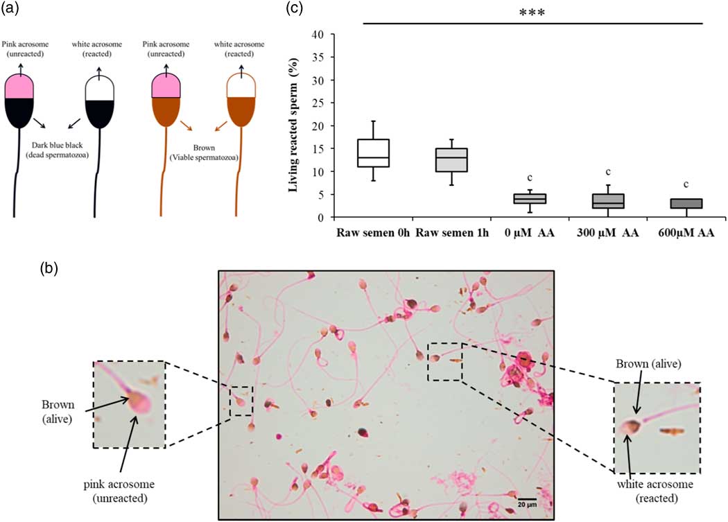
The triple-stain technique permitted the evaluation of the acrosome reaction in viable or dead spermatozoa. Ejaculated or washed (post swim-up) spermatozoa were diluted with an equal volume of trypan blue (2%) and incubated for 15 min at 37ºC. Smears were prepared and the fixation was carried out using 3% glutaraldehyde in 0.1 M cacodylate buffer (pH 7.4) for 30 min. After fixation, slides were stained with 0.8% Bismark brown at 40ºC for 10 min. Next, the smears were stained with Rose Bengal (0.8%) at 24ºC for 45 min. In total, 200 spermatozoa were scored by two embryologists using a light microscope under ×100 magnification. Trypan blue stains dead spermatozoa, Bismark brown stains the post-acrosomal region brown, and Rose Bengal stains the unreacted acrosome pink. As a result, slides treated with this protocol presented four staining patterns (Talbot and Chacon, Reference Talbot and Chacon1981; Tanaka et al., Reference Tanaka, Nagayoshi, Tanaka and Kusunoki2012):
1) dark blue post-acrosomal region and pink acrosome (dead sperm with intact acrosome);
2) dark blue post-acrosomal region and white acrosome (dead sperm lacking acrosome);
3) brown post-acrosomal region and pink acrosome (viable sperm with intact acrosome);
4) brown post-acrosomal region with white acrosome (viable sperm with reacted acrosome).
Assessment of sperm chromatin integrity using toluidine blue assay
Sperm chromatin integrity was assessed in the different culture conditions using the toluidine blue (TB) assay (Tsarev et al., Reference Tsarev, Bungum, Giwercman, Erenpreisa, Ebessen, Ernst and Erenpreiss2009). Toluidine blue is a basic dye that can bind to the phosphate residues of DNA in sperm with altered chromatin and/or fragmented DNA (Erenpreiss et al., Reference Erenpreiss, Bars, Lipatnikova, Erenpreisa and Zalkalns2001). Two hundred spermatozoa per smear were examined under a light microscope by two embryologists at ×100 magnification. The cells were classified into two groups: TB positive cells (TB+) (dark violet cells; altered chromatin) and TB negative cells (TB−) (light blue cells; normal chromatin structure) (Tsarev et al., Reference Tsarev, Bungum, Giwercman, Erenpreisa, Ebessen, Ernst and Erenpreiss2009; Alves et al., Reference Alves, Cancelli, Grassi, Oliveira, Franciscato, Carreira and de Koivisto2018).
Glucose uptake measurement
Vitamin C could be transported to the spermatozoa via glucose transporters (Angulo et al., Reference Angulo, Maldonado, Pulgar, Mancilla, Córdova, Villarroel, Castro and Concha2011). Therefore, at the end of each experiment, the supernatants collected after swim-up (0 µM AA, 300 µM AA and 600 µM AA) were centrifuged for 10 min at 2 rpm. After this centrifugation, the supernatants were used to measure the glucose concentration [Glucose] in the medium. Sperm medium only was used for the control group. The test is based on the enzymatic reaction of hexokinase leading to the production of NADPH. Subsequently, NADPH could be measured photometrically in a C111 Cobas analyzer (Roche, Switzerland) (Akhir et al., Reference Akhir, Chua, Majid and Sarmidi2011). Glucose uptake per spermatozoon (mg/dl) was calculated using the following formula:
[Glucose] in the control group – [Glucose] in the medium after swim-up/total sperm count.
Statistical analysis
Statistical analysis was performed using SPSS Statistics v17.0. Results are expressed as mean±standard deviation (SD) for normally distributed continuous variables and as median (interquartile range) for not normally distributed continuous variables. Differences between groups with more than two categories were compared using the Kruskal–Wallis H test or the one-way analysis of variance (ANOVA) test, according to the data distribution. When P-values were <0.05, the post-hoc Dunn test or Tukey test, respectively, was used to determine which groups differed from each other. Differences were considered statistically significant when P<0.05.
Results
Raw semen characteristics of the selected samples
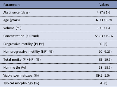
After liquefaction, a semen analysis was performed on raw semen samples (raw semen 0 h, n=22) (Table 1). The participants had a mean±SD abstinence period of 4.87±1.6 days. The mean age was 37.73±6.38 years. In addition, the semen volume mean was 3.71±1.4 ml. Regarding the microscopic semen parameters, the sperm concentration mean was 55.83±19.37×10⁶/ml, the total motile sperm percentage was 62% (±19.5), the percentage of viable spermatozoa was 89.5% (±5.5%), and the percentage of sperm with typical morphology was 4% (±0%) (Table 1).
Table showing the basic semen characteristics of the selected samples

Results are expressed as mean±standard deviation (SD) for normally distributed continuous variables and as median (interquartile range) for not normally distributed continuous variables.
Ascorbic acid supplementation (300 or 600 µM) during swim-up procedure had no effect on sperm concentration, motility and morphology
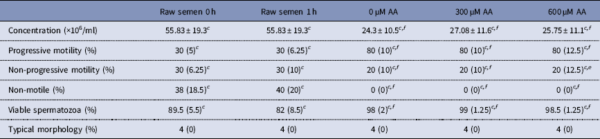
Incubation of the raw semen after liquefaction (raw semen 0 h) for 1 h (raw semen 1 h), did not affect sperm concentration (P>0.05), progressive motility percentage (P>0.05), non-progressive motility percentage (P>0.05), viable spermatozoa percentage (P>0.05), and typical morphology percentage (P>0.05) (Table 2). After swim-up procedure with and without AA (0, 300, 600 µM AA), a significant decrease in sperm concentration was detected when compared with raw semen 0 h (P<0.001) (Table 2). Moreover, after swim-up procedure (0, 300, 600 µM AA), significant differences in progressive motility percentage, non-progressive motility percentage, non-motile sperm, and viable spermatozoa percentage were detected when compared with raw semen 0 h (P<0.001) (Table 2). These results are typical for the direct swim-up procedure, as it consists of separating motile spermatozoa from the semen (World Health Organization, 2010; Beydola et al., Reference Beydola, Sharma and Agarwal2014). Interestingly, there were no statistically significant differences in sperm concentration, motility, vitality and morphology between swim-up in 0 µM AA, 300 µM AA or 600 µM AA (Table 2).
Table comparing the basic sperm parameters in the different culture conditions
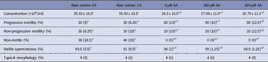
Results are expressed as mean±standard deviation (SD) for normally distributed continuous variables and as median (interquartile range) for not normally distributed continuous variables.
AA, ascorbic acid.
Statistically significant differences between the studied groups: a (P<0.05), b(P<0.01), c(P<0.001).
Statistically significant differences when the swim-up outcome was compared with the raw semen 0 h: d (P<0.05), e (P<0.01), f (P<0.001).
Supplementation of 300 µM ascorbic acid during swim-up procedure helped to select a low percentage of NBT(+) spermatozoa
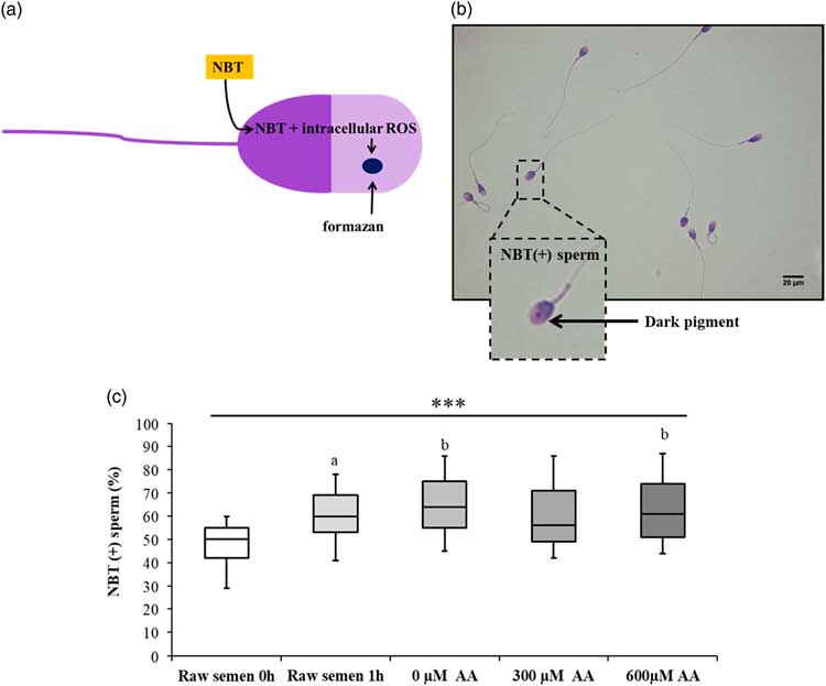
The NBT test was used to detect ROS within the spermatozoa (Agarwal and Majzoub, Reference Agarwal and Majzoub2017) (Figure 2a, b). Compared with the raw semen 0 h, there was a significant increase in the percentage of NBT(+) cells in the raw semen at 1 h (P<0.05), swim-up 0 µM AA (P<0.01), and swim-up 600 µM AA (P<0.01). However, there was no statistically significant increase in the percentage of NBT(+) cells in the swim-up with 300 µM AA compared with raw semen at 0 h (P>0.05) (Figure 2c). Particularly, the increase in the percentage of NBT(+) sperm in the raw semen 1 h was positively correlated with the level of NBT(+) cells in the raw semen 0 h (R=0.829, P <0.001) (Table S1) . Additionally, the increase in the percentage of NBT(+) sperm in the swim-up without AA was positively correlated with level of NBT(+) cells in the raw semen 0 h (R=0.542, P=0.009) and raw semen 1 h (R=0.66, P<0.001) (Table S1).
Figure showing the nitroblue tetrazolium (NBT) test in human spermatozoa. (a) An illustration of the mechanism of NBT action. In the spermatozoon, NBT molecule interacts with intracellular reactive oxygen species (ROS) to form a dark blue pigment called formazan. Spermatozoa incubated with NBT, fixed, and stained with Wright stain. (b) A dark pigment was identified in the sperm acrosome. Scale bar represents 20 µm. Magnification ×100. (c) Boxplots presenting the percentage of NBT(+) sperm (%) in the raw semen 0 h, raw semen 1 h, and post swim-up with 0 µM AA, 300 µM AA, and 600 µM AA. Boxes indicate the interquartile range. Horizontal bars within boxes indicate the median. Whiskers indicate the range of data. Asterisks (***P<0.001) indicate statistically significant differences between the analysed groups. Superscript letters ( a P<0.05) and ( b P<0.01) indicate a statistically significant difference when a study group was compared with the raw semen 0 h group.

Supplementation of 300 µM and 600 µM ascorbic acid during swim-up procedure had no effect on the acrosome reaction
The triple-staining technique allows the identification of viable (brown)/dead (blue) sperm with the reacted (white)/unreacted (pink) acrosome (Fig. 3a, b) (Talbot and Chacon, Reference Talbot and Chacon1981). In the present study, there was no statistically significant difference in the percentage of viable reacted sperm after 1 h of incubation in the raw semen compared with raw semen at 0 h (Fig. 3c). However, swim-up with 0 µM AA, 300 µM AA or 600 µM AA presented lower percentages of viable reacted spermatozoa compared with raw semen 0 h (P<0.001) (Fig. 3c). Of particular interest, there was no statistically significant difference in the percentage of viable spermatozoa with the reacted acrosome between 0 µM AA, 300 µM AA and 600 µM AA (Fig. 3c).
Figure showing the evaluation of acrosome reaction in human spermatozoa. (a) An illustration of the four types of spermatozoa that could be detected after the triple-staining technique. (b) Dead spermatozoa (light blue) with reacted (white) or unreacted (pink) acrosome. Viable spermatozoa (brown) with reacted (white) or unreacted (pink) acrosome. Human spermatozoa stained with the triple-stain technique. Scale bar represents 20 µm. Magnification ×100. (c) Boxplots showing the percentage of viable reacted spermatozoa (%) in the raw semen 0 h, raw semen 1 h, and post swim-up with 0 µM AA, 300 µM AA, and 600 µM AA. Boxes indicate the interquartile range. Horizontal bars within boxes indicate the median. Whiskers indicate the range of data. Stars denote statistically significant difference between the analysed groups (***P<0.001). Superscript ‘c’ indicates a statistically significant difference between the analysed group and the raw semen 0 h ( c P<0.001).
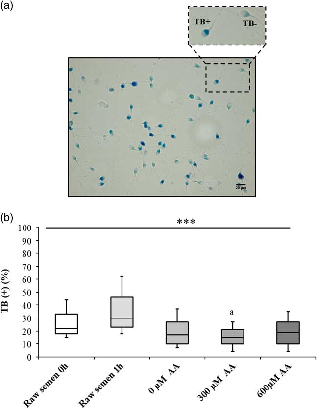
Supplementation of 300 µM ascorbic acid during the swim-up procedure allowed the selection of a low percentage of sperm with altered chromatin integrity
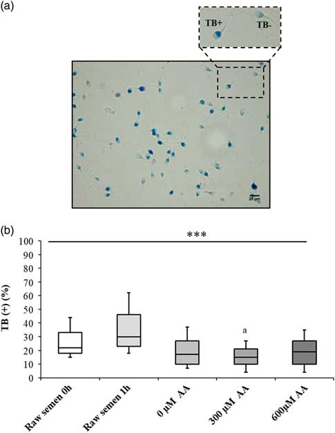
Toluidine blue staining was used to assess sperm chromatin integrity. This dye can bind to phosphate residues of fragmented DNA (Erenpreiss et al., Reference Erenpreiss, Bars, Lipatnikova, Erenpreisa and Zalkalns2001) (Fig. 4a). The increase in the percentage of TB(+) spermatozoa in the raw semen 1 h was statistically insignificant when compared with raw semen 0 h (Fig. 4b). In parallel, swim-up with 300 µM AA selected a significant lower percentage of TB(+) spermatozoa compared with raw semen 0 h (P<0.05) (Fig. 4b). Strikingly, there was a significant positive correlation between the percentage of NBT(+) and TB(+) spermatozoa (R=0.32, P<0.007) (Table S2).
Figure showing the evaluation of chromatin integrity in human spermatozoa. (a) Human spermatozoa stained with toluidine blue (TB). Sperm heads with a light blue colour show normal chromatin integrity (TB−) and sperm heads with dark purple colour show abnormal chromatin integrity (TB+). Scale bar represents 20 µm. Magnification ×100. (b) Boxplots showing the percentage of TB(+) sperm (%) in the raw semen 0 h, raw semen 1 h, and post swim-up with 0 µM AA, 300 µM AA or 600 µM AA. Boxes indicate the interquartile range. Horizontal bars within boxes indicate the median. Whiskers indicate the range of data. Stars denote statistically significant differences between the analysed groups (***P<0.001). Superscript ‘a’ indicates a statistically significant difference between the analysed group and the raw semen 0 h ( a P<0.05).
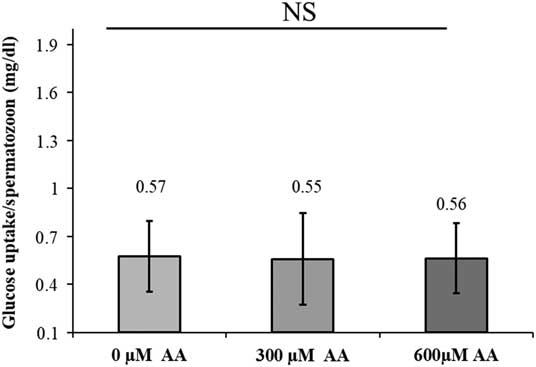
Supplementing sperm medium with vitamin C did not affect glucose uptake per spermatozoon
Vitamin C could be transported to spermatozoa via glucose transporters (Angulo et al., Reference Angulo, Maldonado, Pulgar, Mancilla, Córdova, Villarroel, Castro and Concha2011). We aimed to assess the effect of supplementing sperm medium with vitamin C on glucose uptake by the spermatozoa. There was no statistically significant difference in the glucose uptake per spermatozoon, after swim-up, at the different vitamin C concentrations (Fig. 5).
Figure showing sperm glucose uptake per spermatozoa after exposure to ascorbic acid. Graph presenting the level of glucose uptake/spermatozoon in the different swim-up conditions. Data expressed as mean±standard deviation. NS= not significant.

Discussion
The aim of this prospective study was to evaluate the effect of supplementing the culture medium with vitamin C, during direct swim-up, on sperm functional properties. We showed that adding 300 µM of AA to sperm medium, during direct swim-up, had beneficial effects on intracellular ROS levels and sperm chromatin integrity.
At first, one aliquot of each raw semen sample was used to assess the effect of prolonged incubation in the seminal fluid on basic and functional sperm parameters. A significant increase in the percentage of NBT(+) cells was detected in the raw semen after 1 h of incubation. The reason behind this finding could be that normal and pathological sperm forms are carried by human seminal fluid (Menkveld, Reference Menkveld2010). Spermatozoa with abnormal morphology could be a potential endogenous source of ROS (Aitken et al., Reference Aitken, Fisher, Fulton, Gomez, Knox, Lewis and Irvine1997; Rengan et al., Reference Rengan, Agarwal, van der Linde and du Plessis2012; Agarwal et al., Reference Agarwal, Durairajanayagam and du Plessis2014). High seminal ROS levels or low antioxidants levels may induce oxidative stress (Rengan et al., Reference Rengan, Agarwal, van der Linde and du Plessis2012; Agarwal et al., Reference Agarwal, Roychoudhury, Bjugstad and Cho2016) leading to sperm damage (Pereira et al., Reference Pereira, Sá, Barros and Sousa2017) and impaired embryo development (Burruel et al., Reference Burruel, Klooster, Barker, Pera and Meyers2014; Gualtieri et al., Reference Gualtieri, Barbato, Fiorentino, Braun, Rizos, Longobardi and Talevi2014; Castro et al., Reference Castro D, Assis D, Siqueira, Hamilton, Mendes, Losano, Nichi, Visintin and Assumpção2016).
In parallel, three aliquots of each semen sample, directly taken from raw semen after liquefaction and before any incubation, were used to perform direct swim-up with or without AA (300 µM and 600 µM). There were no statistically significant differences in sperm motility, morphology and viability in the different swim-up conditions. The effect of AA on sperm motility and vitality in vitro was addressed in several reports with contradicting results. For instance, Fananei and colleagues found higher percentages of progressive motility and vitality when 600 µM of AA were added to the medium compared with a medium without AA (Fanaei et al., Reference Fanaei, Khayat, Halvaei, Ramezani, Azizi, Kasaeian, Mardaneh, Parvizi and Akrami2014). By contrast, Donelly et al. (Reference Donnelly, McClure and Lewis1999a) found that AA was not beneficial to sperm motility in vitro. These dissimilarities could be due to the sperm preparation technique used (indirect swim-up versus density gradient), to the population selected (teratozoospermic samples versus normozoospermic and asthenozoospermic samples) or to the protocol applied to evaluate sperm motility.
Furthermore, we showed that the percentage of ROS(+) cells did not increase in the swim-up with 300 µM AA compared with the raw semen. Similar findings were reported by other scientists when they supplemented sperm cryopreservation medium with 300 µM AA (Li et al., Reference Li, Lin, Liu, Xiao and Liu2010). However, Donnelly and colleagues demonstrated that 300 µM and 600 µM AA had no effect on baseline ROS generation. Compared with our report, the authors used the density gradient technique (the AA was present during the procedure) and the ROS were not measured using NBT (Donnelly et al., Reference Donnelly, McClure and Lewis1999b).
Of particular interest, we found that supplementing sperm medium with 600 µM AA had no effect on ROS generation. This finding is in line with the report of Li et al. (Reference Li, Lin, Liu, Xiao and Liu2010). It is noteworthy to mention that AA at a suitable dose acts as ROS scavenger. However, inadequate concentrations of AA might counteract their antioxidant effects and produce sperm damage (Li et al., Reference Li, Lin, Liu, Xiao and Liu2010).
In addition, we detected a significant decrease in the percentage of spermatozoa with altered chromatin integrity, in the 300 µM AA group compared with the raw semen. This finding could be explained by the positive correlation found between NBT(+) cells and TB(+) cells. In fact, the effect of AA on sperm DNA, in vitro, has been investigated in several studies. The majority of these reports highlighted the protective role of vitamin C on sperm DNA during or after sperm processing (Hughes et al., Reference Hughes, Lewis, McKelvey-Martin and Thompson1998; Donnelly et al., Reference Donnelly, McClure and Lewis1999b; Sierens et al., Reference Sierens, Hartley, Campbell, Leathem and Woodside2002; Fanaei et al., Reference Fanaei, Khayat, Halvaei, Ramezani, Azizi, Kasaeian, Mardaneh, Parvizi and Akrami2014). Moreover, the direct swim-up technique, itself, was shown to select a population of spermatozoa with a low percentage of sperm with altered chromatin integrity compared with the unprocessed semen (Marchesi et al., Reference Marchesi, Biederman, Ferrara, Hershlag and Feng2010). Therefore, the presence of 300 µM vitamin C during direct swim-up procedure was beneficial to sperm chromatin integrity. Conversely, vitamin C could have the potential to reduce disulphide bridges of sperm nuclear protamines leading to a chromatin decondensation (Giustarini et al., Reference Giustarini, Dalle-Donne, Colombo, Milzani and Rossi2008; Ménézo et al., Reference Ménézo, Hazout, Panteix, Robert, Rollet, Cohen-Bacrie, Chapuis, Clément and Benkhalifa2007). This could affect preimplantation embryo development and pregnancy outcomes (Ellington et al., Reference Ellington, Evenson, Wright, Jones, Schneider, Hiss and Brisbois1999; Ménézo et al., Reference Ménézo, Hazout, Panteix, Robert, Rollet, Cohen-Bacrie, Chapuis, Clément and Benkhalifa2007; Zidi-Jrah et al., Reference Zidi-Jrah, Hajlaoui, Mougou-Zerelli, Kammoun, Meniaoui, Sallem, Brahem, Fekih, Bibi, Saad and Ibala-Romdhane2016). Therefore, it will be important in the future to study the effect on sperm chromatin decondensation of supplementing the culture medium with vitamin C during direct swim-up.
The acrosome reaction is an additional variable that affects the sperm function. For a successful fertilization, the presence of motile spermatozoa with intact acrosome able to undergo acrosome reaction is of paramount importance (Esteves et al., Reference Esteves, Sharma, Thomas and Agarwal2000). Here we showed that 300 µM and 600 µM of AA did not induce significant changes in acrosome reaction when compared with swim-up without AA. Fanaei et al. (Reference Fanaei, Khayat, Halvaei, Ramezani, Azizi, Kasaeian, Mardaneh, Parvizi and Akrami2014) showed that the addition of 600 µM AA had no effect on acrosome status after the indirect swim-up procedure.
The results of the present prospective autocontrolled study are meaningful but limited by the small sample size. A new study should be conducted in the future on a larger population and with additional objective tests to assess sperm functions.
Conclusion
The direct swim-up procedure is widely used in assisted reproductive therapy (ART). It consists of separating the motile spermatozoa from the antioxidant-rich semen. Therefore, motile spermatozoa will be more vulnerable to ROS due to their plasma membrane characteristics and the lack of endogenous defence mechanisms. Here we showed that supplementing the sperm culture medium used during direct swim-up with 300 µM vitamin C had beneficial effects on intracellular ROS levels and sperm chromatin integrity. This could ameliorate the quality of the spermatozoa derived from the direct swim-up for ART treatments.
Supplementary material
To view supplementary material for this article, please visit https://doi.org/10.1017/S0967199419000030
Acknowledgements
This study was supported primarily by the Azoury IVF Clinic. Glucose measurements were performed by the Al-Hadi Laboratory and Medical Center. An aliquot of AA (Sigma-Aldrich) was provided by the Reproductive Biology Laboratory, Lebanese University, Faculty of Sciences, Section II, Fanar, Lebanon.
Financial support
This research received no specific grant from any funding agency, commercial or not-for-profit sectors.
Ethical standards
The study was performed according to a protocol approved by the Ethical Committee of the Mount Lebanon Hospital. Written informed consent was obtained from each patient before enrolment.
Statement of interest
Authors have reported no conflicts of interest.