Bilateral ducts are not usually seen with confluent pulmonary arteries. We report, however, the case of a fetus with confluent pulmonary arteries and an exceptional merged bilateral duct associated with a retro-esophageal circumflex right aortic arch. The aortic arch encircles the trachea and oesophagus from the right with a left descending aorta.Reference Li, Wen and Liang1 We successively describe the case, discuss the findings, and suggest a developmental hypothesis to this aortic arch and its association with VACTERL syndrome.
Clinical case
A woman aged 30 years old, who has had a normal child born from a first pregnancy, then an extra-uterine pregnancy, had a screening ultrasound scan at 23 weeks gestation that showed anamnios and bilateral renal agenesis, while the first trimester scan was considered normal. The parents chose to terminate pregnancy at 24 weeks.
The male fetus weighed 559 g, and pathological examination showed extra-cardiac malformations. These included facial dysmorphy, limbs and extremities malposition with anamnios sequence, single umbilical artery, thoracic vertebral abnormalities, anal imperforation, right kidney agenesis, left kidney multicystic dysplasia and hypoplasia, and pulmonary hypoplasia. These abnormalities suggested VACTERL syndrome.
Results
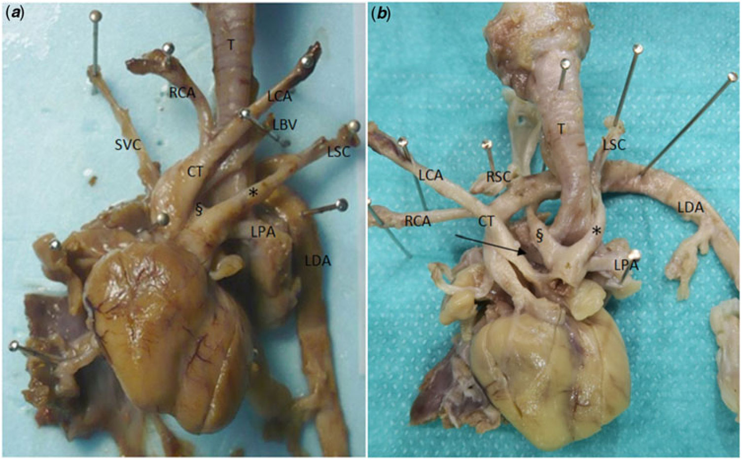
Fetal cardiac examination showed a circumflex right aortic arch with a retro-esophageal segment and a left descending aorta (Fig 1). The right aortic arch gave successively a common carotid trunk dividing into a left and a right carotid artery, a right subclavian artery, and a distal aberrant left subclavian artery.
Anterior views of the heart and the great vessels before (a) and after (b) having reflected the aorta to show the right pulmonary artery (arrow) and the posterior insertion of the right arterial duct (§). Note the left arterial duct (*) joining the right one. CT = carotid trunk; LBV = left brachiocephalic vein; LCA = left carotid artery; LDA = left descending aorta; LPA = left pulmonary artery; LSC = left subclavian artery; RCA = right carotid artery; RSC = right subclavian artery; SVC = superior caval vein; T = trachea.
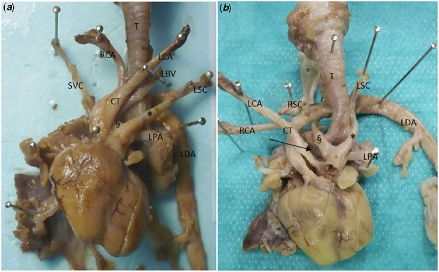
A bilateral arterial duct had a right branch originating from the posterior part of the transverse retro-esophageal aortic arch and a left branch originating from the left subclavian artery. These two branches merged to form a unique vessel connecting to the pulmonary trunk. This trunk was normally divided into confluent right and left pulmonary arteries.
In addition, there was an anomalous course of the left brachiocephalic vein, passing below the aortic arch (retro-aortic) instead of antero-superiorly.
The heart had a normal inner architecture and a normal volume.
Discussion
This is a very rare description of a “Y-shaped” merged bilateral arterial duct, associated here with a circumflex right aortic arch.
Bilateral arterial ducts are usually associated with non-confluent pulmonary arteries, sometimes isolated or most often in the setting of tetralogy of Fallot with pulmonary atresia.Reference Li, Wen and Liang1, Reference Freedom, Moes and Pelech2 These bilateral arterial ducts can be associated with left, right, or double aortic arch with or without isolated or aberrant brachiocephalic arterial trunk or subclavian artery. The aortic arch can also be interrupted.
Circumflex retro-esophageal right aortic arch is characterised by the right-sided arch crossing the midline behind the esophagus, with a left-sided descending aorta. Rarely, the opposite, or mirror-image, is found: left-sided aortic arch with a retro-esophageal segment and right descending aorta.Reference McLeary, Frye and Young3 Usually in circumflex right aortic arch, the most proximal vessel arising from the aorta is the left carotid artery, then the right carotid artery, followed by the right subclavian artery. The most distal vessel is usually the left subclavian artery.Reference Robotin, Bruniaux and Serraf4 A particularity in our case is that the left and right carotid arteries originate from a common carotid trunk.
This malformation has been recently added to the original Stewart classification of congenital aortic arch abnormalities.Reference Li, Wen and Liang1 It is relatively rare among cases of right aortic arch particularly in those discovered during childhood.Reference Hastreiter, D’Cruz, Cantez, Namin and Licata5–Reference Felson and Palayew7 Indeed, a circumflex aortic arch is frequently isolated, hence poorly diagnosed early in age, whereas a right aortic arch with mirror-imaged branching of the brachiocephalic vessels and without retro-esophageal segment is often associated with so-called “conotruncal defects” like tetralogy of Fallot with or without pulmonary atresia and common arterial trunk, with or without microdeletion 22q1.1.Reference Knight and Edwards6, Reference McElhinney, Clark and Weinberg8 Association between a circumflex aortic arch and an intra-cardiac lesion is rare but not exceptional: ventricular septal defect has been reported.Reference Robotin, Bruniaux and Serraf4, Reference Knight and Edwards6
A circumflex aortic arch can sometimes be discovered upon symptoms of esophageal and tracheo-bronchial compression, especially in adults.
Of note, a retro-aortic left brachiocephalic vein, as found in our case, is frequently associated with a right aortic arch.Reference Kahkouee, Sadr and Pedarzadeh9
Developmental hypothesis
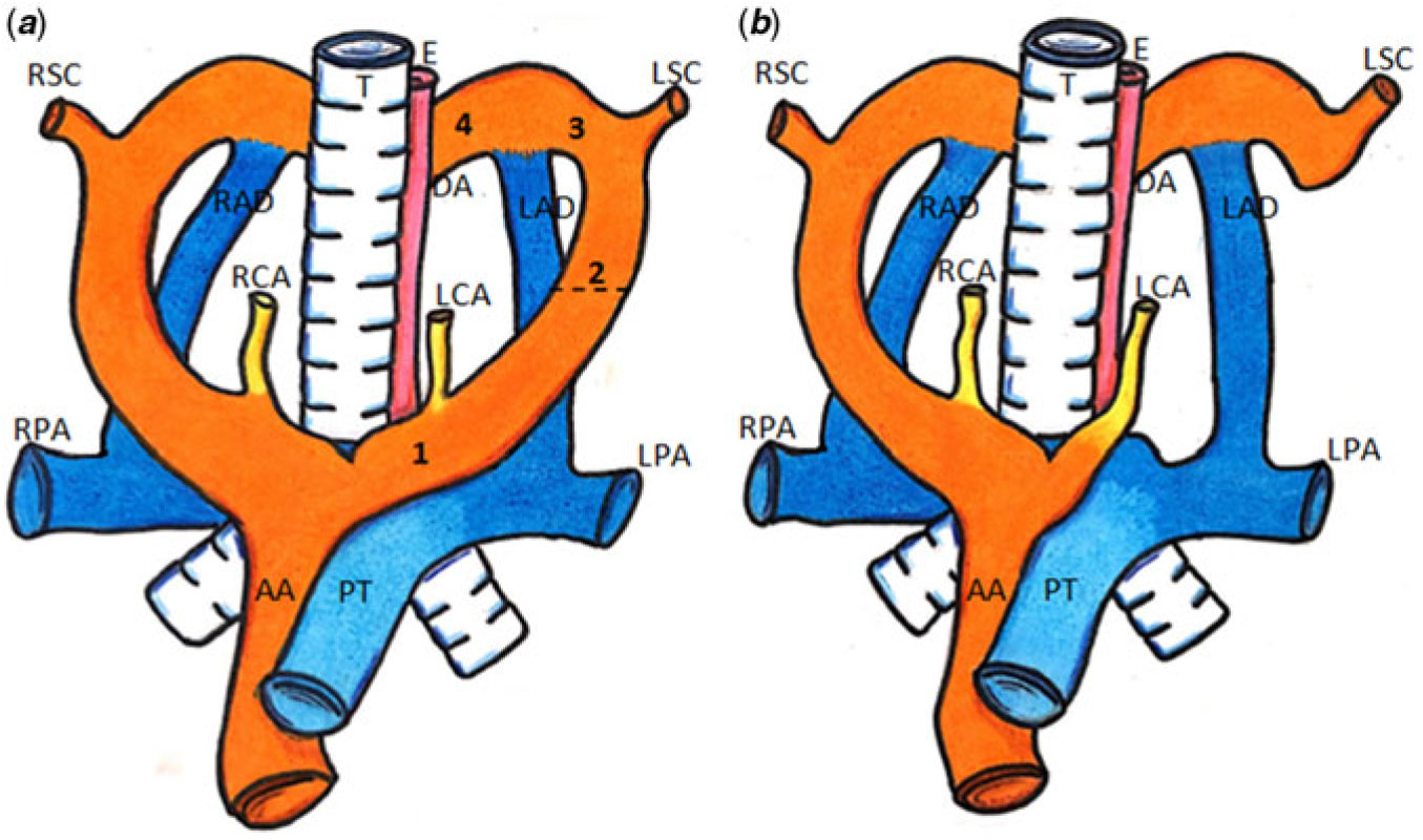
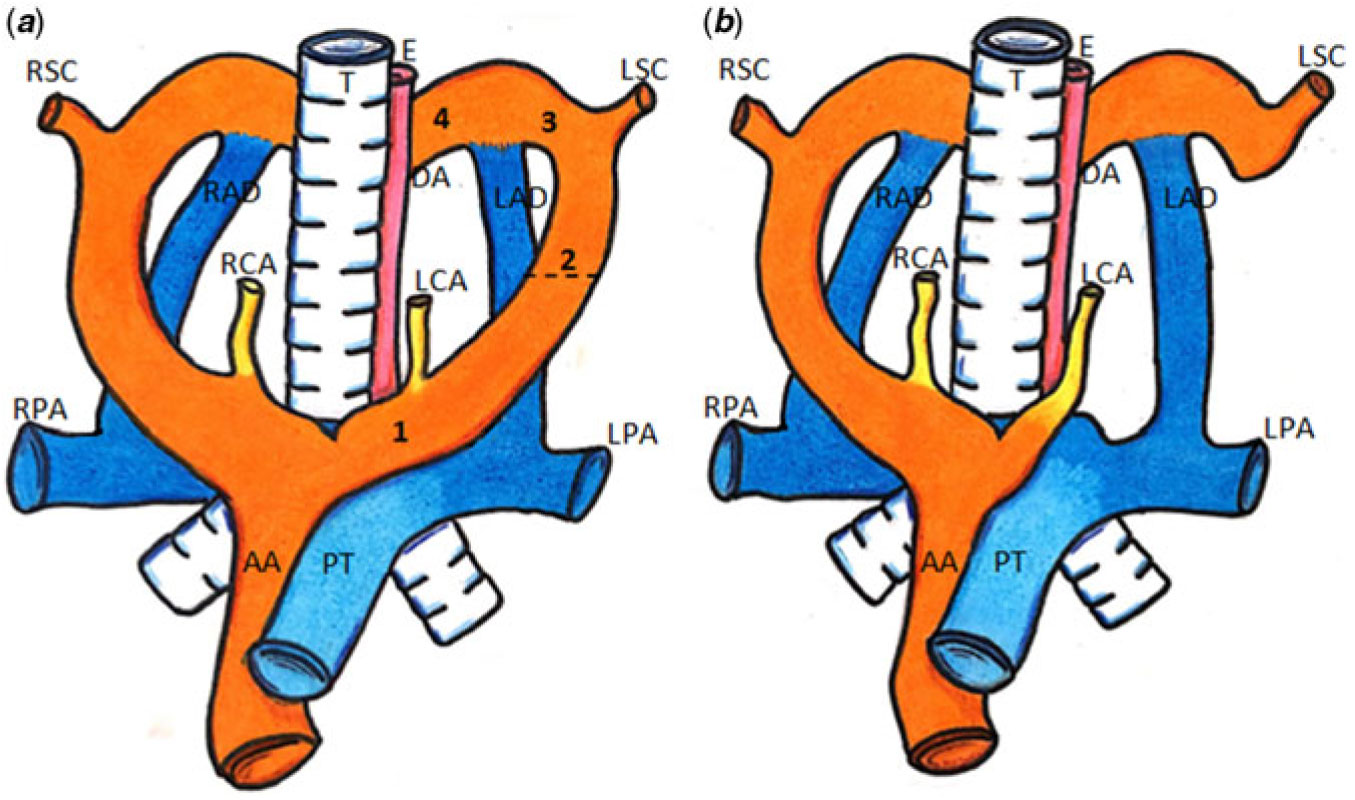
Embryologically, the aortic arches arise from the aortic sac.Reference Gupta, Bamforth and Anderson10 The most cranial is the 3rd aortic arch (which forms the two carotid arteries), then the 4th aortic arch (the right one usually disappears, and the left one forms the segment of the aortic arch between the left carotid artery and the left subclavian artery), then the 6th aortic arch. The 6th aortic arch forms the two arterial ducts. The right duct or right 6th arch artery normally disappears, while the right-sided dorsal aorta involutes.Reference Gupta, Bamforth and Anderson10 The two pulmonary arteries originate from the caudal margin of the 6th aortic arch and then connect to the primitive intrapulmonary arteries and the pulmonary trunk. The pulmonary trunk and the ascending aorta derive from the distal part of the outflow tract and fuse with the aortic sac at the margins of the pericardial cavity. The rare anomaly found in our case could thus be explained on the basis of abnormal remodeling of the aortic sac: the caudal part of the aortic sac has retained its connection with the pulmonary trunk which then feeds directly the persisting 6th arch arteries and the right and left pulmonary arteries. This preserved connection provides a common origin for the 6th arch arteries themselves, the origins of the 6th and 4th arch arteries being separated. This symmetrical state, associated with the fusion of the two 3rd aortic arches in a common carotid trunk, is explained by the preservation of a stage of normal development, the right-sided structures retaining their size compared to the left-sided components. In addition, the left 4th aortic arch disappears (region 2 of the Edwards hypothetical double arch, Fig 2) and regions 3 and 4 persist, leading to aberrant left subclavian artery. Further abnormal remodelling is needed to explain the retroesophagal course of the aortic arch and the position of the descending aorta to the left.
Diagram showing the arterial tree on the basis of the Edwards hypothetical double arch before (a) and after (b) partial disappearance of the left 4th aortic arch. The double arch is broken between the left carotid artery and the left subclavian artery (region 2, see dashed line), whereas regions 1, 3 and 4 persist. AA = ascending aorta; DA = descending aorta; E = esophagus; LAD = left arterial duct; LCA = left carotid artery; LPA = left pulmonary artery; LSC = left subclavian artery; PT = pulmonary trunk; RAD = right arterial duct; RCA = right carotid artery; RPA = right pulmonary artery; RSC = right subclavian artery; T = trachea.
In our case, the existence of at least vertebral, anal, and renal abnormalities confirmed VACTERL diagnosis. The etiology of VACTERL syndrome is heterogenous, and most often sporadic in humans, rare mutations, and mitochondrial dysfunction are described.Reference Solomon, Pineda-Alvarez and Raam11 Ventricular septal defect is the most frequent cardiac anomaly in patients with VACTERL syndrome and also in patients with circumflex aortic arch.Reference Cunningham, Hadley and Hannoush12 In animal models, the association between trachea-esophagus and aortic arch abnormalities, including right aortic arch, has been published and the developmental sonic hedgehog pathway has been involved in 6th aortic arch formation.Reference Kolesová, Roelink and Grim13 This case is to our knowledge the first human description of the association between VACTERL syndrome and circumflex right aortic arch.
Acknowledgements
None.
Financial Support
This research received no specific grant from any funding agency, commercial or not-for-profit sectors.
Conflicts of Interest
None.
Ethical Standards
The authors assert that all procedures contributing to this work comply with the ethical standards of the relevant national guidelines on human experimentation and with the Helsinki Declaration of 1975, as revised in 2008, and has been approved by the institutional committees.
Author Contributions
S.B. wrote the manuscript. R.C.G., M.L., and F.D. took part to data collection. C.O. realised the drawing. D.B. and L.H. reviewed intellectual content.