Introduction
Growth and development of newborn mammals depend on the availability of a sufficient amount of milk from the mother, its secretion by the mammary gland being initiated at about the time of parturition. Milk is a complex fluid comprising of an aqueous phase that contains lactose and other carbohydrates, many minerals, numerous whey proteins, membrane-bound lipid droplets and soluble aggregates of proteins: casein micelles. These various components of milk arise from different secretory pathways that develop in the mammary alveolar cells in response to lactogenic hormones during pregnancy. Milk secretion is obviously of interest due to its physiological and economic importance and also from the point of view of the study of high-efficiency intracellular transport pathways.
Caseins (cas), a family of acidic phosphoproteins (αs1-cas, αs2-cas, β-cas and κ-cas), are the most abundant proteins in milk, except in primates (for review see Swaisgood, Reference Swaisgood, PF and PLH2003). κ-cas is O-glycosylated and forms dimers via highly conserved cysteine residues (Bouguyon et al., Reference Bouguyon, Beauvallet, Huet and Chanat2006). The aggregation of the caseins into micelles involves interaction with calcium phosphate clusters, a phenomenon believed to be initiated in the Golgi apparatus after phosphorylation of the calcium-sensitive caseins (αs1-cas, αs2-cas and β-cas) in this compartment. Co-aggregation of caseins and calcium phosphate clusters proceeds in secretory vesicles during their transport to the cell surface of the mammary epithelial cells (MECs). However, the specific arrangement of caseins in the micelle has not yet been established, and much is still to be learned about the role of individual caseins in the aggregation process.
During full lactation, MECs synthesise and secrete large quantities of milk-specific proteins, lipids and lactose, in a polarised fashion: from their apical surface into the alveolar lumen that they surround. Moreover, these cells transfer milk proteins derived from blood plasma that are internalised at their basolateral surface, transported through the cell and released apically. Depending on their origin, newly synthesised or blood plasma-derived milk proteins travel to the apical side of MECs either via the secretory or the transcytotic route (for review see Burgoyne and Duncan, Reference Burgoyne and Duncan1998; Boisgard et al., Reference Boisgard, Chanat, Lavialle, Pauloin and Ollivier-Bousquet2000). Indeed, the intense transport of milk components across the various membrane-bound compartments of the trafficking pathways of MECs correlates with the generation of a large flow of vesicular and tubular transport carriers. Budding, fission and fusion of these transport carriers involve a concerted action of numerous factors including cytosolic proteins that transiently and reversibly associate with the surface of intracellular organelles and intrinsic factors (specific transmembrane proteins and lipids) of the membrane bilayer (for review see Farsad and De Camilli, Reference Farsad and De Camilli2003). The participation of specific lipid species such as phosphoinositides, phosphatidic or lysophosphatidic acid in these transport processes has been emphasised in the last decade (for review see De Matteis and Godi, Reference De Matteis and Godi2004; Van Meer and Sprong, Reference Van Meer and Sprong2004). In this context, we have shown that both phospholipase D and calcium-independent phospholipase A2 play a fundamental role in the transport of milk proteins in the secretory pathway of MECs (Boisgard and Chanat, Reference Boisgard and Chanat2000; Pechoux et al., Reference Pechoux, Boisgard, Chanat and Lavialle2005).
On the other hand, we highlighted the fact that the fatty acid composition of the membrane phospholipids of mammary cells, and more specifically the content of polyunsaturated fatty acids of the n-6 series, was a key determinant in the optimal synthesis and secretion of milk proteins (Ollivier-Bousquet et al., Reference Ollivier-Bousquet, Lavialle, Guesnet, Rainteau and Durand1997). Indeed, in female rats chronically fed with a lipid-deprived diet, the content of n-6 polyunsaturated fatty acids in cellular phospholipids decreased by 40%. In parallel, synthesis and secretion of milk proteins were reduced by ≈60% and the transport of prolactin in the transcytotic pathway of MECs was substantially slowed down. In this paper, we further investigated the impact of this lipid-deprived diet on the intracellular events that sustain milk secretion, with special attention to post-translational modifications of milk-secretory proteins.
Material and methods
Animals, experimental diets and milk collection
Two groups of Wistar rats, grown in our institute for several consecutive generations, received over their lifespan semi-synthetic experimental diets as detailed in Ollivier-Bousquet et al. (Reference Ollivier-Bousquet, Lavialle, Guesnet, Rainteau and Durand1997). In brief, the control diet contained 6% to 6.5% (w/w) dietary lipids in the form of peanut and rapeseed oils, supplying recommended amounts of essential polyunsaturated fatty acids of both series (n-6 and n-3). Lipid-deprived animals received a diet without added oils and therefore ate only trace amounts of essential polyunsaturated fatty acids. Traces of lipid were originating from the casein and cornstarch preparations entering the diet mix. The gross energy content was 9% lower in the lipid-restricted diet (16.90 v. 15.58 MJ/kg). Milk samples were collected from animals at mid-lactation. Rats were injected intraperitoneally with oxytocin a few minutes before anaesthesia and manually milked. Animal welfare and conditions for animal handling were in accordance with French guidelines (May 2001).
Unless otherwise indicated, chemicals were from Sigma Aldrich.
Milk processing and analysis
Freshly collected milk was kept at room temperature for ≈1 h and aliquots of the aqueous phase were collected below the cream. Samples were either analysed immediately or stored at −20°C. In the latter case, milk samples were thawed and re-warmed to room temperature at least 1 h before sampling.
To prepare casein and whey fractions, aliquots of milk were diluted 50 times in 10 mmol/l N-[2-hydroxyethyl]piperazine-N′-[2-ethanesulphanic acid] (HEPES) KOH pH 6.8, 10 mmol/l CaCl2 and centrifuged for 40 min at 16 000 × g at room temperature. The supernatant was collected and centrifuged as above for 20 min. The resulting supernatant, referred to as whey fraction, was collected and frozen. The first pellet was resuspended in the same buffer and subjected to a second round of centrifugation, as above. The resulting casein fraction was analysed by two-dimensional electrophoresis (2D-PAGE, see below).
Sialidase treatment of caseins was performed essentially as previously described (Chanat, Reference Chanat2006). To prepare caseins, skimmed milk was diluted 50 times in 50 mmol/l 2-[N-morpholino]ethane sulfonic acid (MES) pH 6.0, 1 mmol/l dithiothreitol (DTT) and centrifuged as above. The casein pellet was resuspended in the same buffer supplemented with 1 mmol/l ethylene diamine tetra-acetic acid (EDTA) to help casein solubilisation and CaCl2 was then added to reach a final concentration of 2 mmol/l. Resulting samples were analysed by 2D-PAGE.
Protein assay was according to the Peterson method. Milk osmolarity was measured with an automatic osmometer (freezing point depression, Roebling osmometer, LH Roebling, Berlin, Germany). Lactose concentration was determined enzymatically (Bergmeyer, Reference Bergmeyer and HO1984).
Metabolic labelling
Metabolic labelling of mammary fragments was performed essentially as previously described (Pechoux et al., Reference Pechoux, Boisgard, Chanat and Lavialle2005). Briefly, dams at mid-lactation were killed by decapitation, and the mammary gland was excised bilaterally. Samples were dissected free from connective tissue and muscles, and cut manually into fragments of about 1 mm3 on an ice-cold plastic pad. Fragments were pre-incubated for 30 min in methionine/cysteine-free DMEM under an atmosphere of 95% O2/5% CO2, and then pulse-labelled for 3 min in methionine/cysteine-free DMEM containing 1.85 MBq/ml (50 μCi/ml) [35S] methionine/cysteine mix (>37 TBq/mmol, Life Technologies, Les Ulis, France), all at 37°C. To stop the labelling, fragments were quickly diluted in a 10-fold volume of pre-warmed complete DMEM and filtered on a cell strainer (100 μm; Becton Dickinson France, SA, Le Pont-de-Claix, France). For secretion analysis or to monitor the transport of labelled secretory proteins out of the endoplasmic reticulum (ER), fragments were distributed into cell strainers (100 mg per experimental condition), placed in six-well cluster plates containing complete DMEM and chased under gentle rotary shaking at 37°C in 10% CO2 in air, for the indicated times.
At the end of either the pulse or the chase, tissue fragments in cell strainers were transferred into six-well cluster plates containing ice-cold Tris-buffered saline (TBS, 25 mmol/l Tris-HCl pH 7.4, 4.5 mmol/l KCl, 137 mmol/l NaCl, 0.7 mmol/l Na2HPO4). Mammary fragments were extensively washed with ice-cold TBS and homogenised at 4°C in 1 ml 10 mmol/l HEPES buffer pH 6.7 containing 250 mmol/l sucrose, 1 mmol/l EDTA, 1 mmol/l magnesium acetate and an aliquot (10 μl/ml) of a protease inhibitor cocktail (Sigma Aldrich), with three strokes of a tissue grinder (AA2 Teflon/glass, Thomas Scientific, Swedesboro, NJ, USA). The homogenate was centrifuged for 10 min at 1000 × g, at 4°C, and the resulting supernatant, referred to as post-nuclear supernatant (PNS), was collected. Proteins from the chase media were precipitated with 10% trichloracetic acid (TCA, final concentration). Proteins from the medium and from an aliquot of the PNS (one-fifth) were analysed by 2D-PAGE.
Gel electrophoresis and protein identification
2D-PAGE was as previously described (Chanat, Reference Chanat2006). Gels were stained, destained and dried. For quantification of Coomassie Blue-stained proteins, gels were dried between sheets of cellophane, scanned at 600 d.p.i. (Epson Expression 1600/Pro) and the volume of the spots was measured using the public domain NIH image program ImageJ software (developed at the National Institutes of Health, USA, and available at http://rsb.info.nih.gov/ij/). When appropriate, gels were exposed to a storage phosphor screen (Molecular Dynamics, Sunnyvale, CA, USA) and/or to Kodak Biomax MR-1 films. The phosphor screens were scanned and the radioactivity associated with caseins and whey proteins was quantified using the software Image quaNT (Molecular Dynamics, Sunnyvale, CA, USA).
Protein identification by either peptide mass fingerprint or N-terminal sequencing was as previously described (Bouguyon et al., Reference Bouguyon, Beauvallet, Huet and Chanat2006 and Chanat, Reference Chanat2006, respectively).
Statistical analysis
Paired Student’s t-test was used to compare treated samples to controls. P values are given in figure legends.
Results
Glycosylation of a subset of milk proteins is altered in lipid-deprived rats
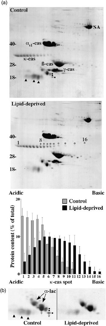
The protein composition of milk from lipid-deprived animals and rats fed with a control diet was compared using 2D-PAGE. The electrophoretic patterns shown in Figure 1a indicate that the four main caseins (see Bouguyon et al., Reference Bouguyon, Beauvallet, Huet and Chanat2006 for casein identification), αs1-cas, β-cas, γ-cas and κ-cas, were present in the milk from the two groups of rats. At least for the three calcium-sensitive caseins (αs1-cas, β-cas and γ-cas), comparison of the Coomassie Blue staining suggests that they were present in similar, if not identical, concentrations. Analysis of whey resulted in an analogous protein pattern for each type of milk. Only the relative amount of a few whey proteins was reduced in the milk from lipid-deprived animals, as will be described below. In line with these observations, milk protein concentration was not significantly affected by the diet (Table 1). Quite interestingly, however, comparison of the 2D-PAGE patterns revealed an obvious effect of the diet on the number and distribution of spots with Mr ≈ 32 000. In milk from lipid-deprived rats, additional spots were resolved in the basic pH range moiety of the gel (Figure 1a, lipid-deprived, spots 12 to 16). These spots, as well as the more acidic spots numbered 8 to 11 (Figure 1a, lipid-deprived, asterisks), were identified as κ-cas by MALDI-MS peptide mass fingerprinting (data not shown). Clearly, the relative proportion of the various molecular forms of κ-cas was modified by the diet. In the milk of lipid-deprived animals, quantification of the κ-cas molecules contained in the various spots showed a decrease in the amount of material focusing in the acidic part of the gel (Figure 1a, lower panel, spots 1 to 5) and an increase of material in the most basic spots. These variations resulted in a bell-shaped distribution of the κ-cas molecules from the milk of lipid-deprived animals along the pH axis.
Milk proteins from control and lipid-deprived rats. Proteins contained in milk fractions from control or lipid-deprived rats were resolved using 2D-PAGE and stained. (a) Two microliter aliquots of skim milk were analysed and the spots identified by symbols were subjected either to N-terminal sequencing (control, dots and triangles) or MS (lipid-deprived, asterisks: some κ-cas forms). The various κ-cas forms present in the milk from control (11 forms) or lipid-deprived (16 forms) rats were quantified from the gels and the amount in each form was expressed as per cent of the total measured in all the present forms. For each type of animal, the mean ± s.d. of three experiments is shown. Mr (kDa) are indicated on the left. (b) Whey proteins prepared from 10 μl of milk were analysed. Identical areas of the 2D-PAGE gels are shown. Dots: GlyCAM-1; triangles: WAP; open diamonds: basic forms of α-lactalbumin and GlyCAM-1. The various forms of α-lactalbumin and GlyCAM-1 were quantified (see Table 1). SA: serum albumin; αs1-cas: αs1-casein; κ-cas: κ-casein; β-cas: β-casein; γ-cas: γ-casein; α-lac: α-lactalbumin.

Milk parameters

The mean ± s.d. of n individual milks is shown.
*P < 0.05.
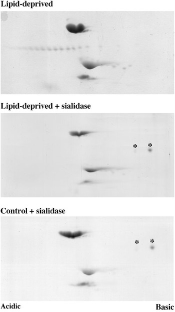
Most of the κ-cas spots essentially differed by a constant interval of pI. Unlike the calcium-sensitive caseins, κ-cas is poorly phosphorylated. Instead, this casein is known to be O-glycosylated on multiple sites (for review see Swaisgood, Reference Swaisgood, PF and PLH2003). Hence, one can speculate that the multiplicity of the κ-cas spots could reflect different levels of glycosylation of the molecule, each form containing different amounts of sialic acid and thus bearing distinct charges. To validate this hypothesis, caseins prepared from the milk of rats fed with either control or lipid-depleted diets were treated with sialidase. In both cases, only two spots with Mr ≈ 32 000 remained (Figure 2, asterisks). The presence of κ-cas in these spots was proven by mass spectroscopy (data not shown). The most basic molecule migrated as spot 16 in Figure 1a and most probably corresponds to the desialylated, non-phosphorylated form of κ-cas, since this spot was still present after sialidase plus phosphatase treatment (data not shown). Accordingly, the second spot might correspond to a phosphorylated form of κ-cas.
The various forms of κ-cas resolved by 2D-PAGE essentially differ by their sialic acid content. Caseins prepared from the milk of control or lipid-deprived rats were incubated in the absence or the presence of sialidase and analysed by 2D-PAGE, followed by protein staining. Representative gels from two independent experiments are displayed. Only the area of the gels containing the caseins is shown. Asterisks: spots analysed by mass spectrometry.

Likewise, whey proteins obtained from the two groups of animals were analysed after selective removal of the caseins from milk by differential centrifugation (Figure 1b). This comparison revealed a significant modification of the pattern of a subset of whey proteins prepared from the milk of the rats fed with the lipid-deprived diet. Spots marked by dots and triangles were processed for N-terminal sequencing and identified as glycosylation-dependent cell adhesion molecule (GlyCAM-1) and whey acidic protein (WAP), respectively. The identification of rat α-lactalbumin in our 2D-PAGE system was recently published (Chanat, Reference Chanat2006). To quantitatively estimate the obvious decrease in GlyCAM-1 content in the milk from lipid-deprived rats, the amount of protein in the various forms of GlyCAM-1 and α-lactalbumin was determined from Coomassie-Blue-stained gels and the ratio of GlyCAM-1 to the total amount of the two whey proteins was calculated. Our results, summarised in Table 2, indicate that the lipid restriction provoked a three-fold reduction in the relative proportion of total GlyCAM-1 (Table 2, column 1). Although not significant, the most basic form of GlyCAM-1 tended to be reduced in the milk of lipid-deprived rats (Table 2, column 2). As for α-lactalbumin, the ratio of the two visible spots was not modified by the diet (Table 2, column 3). Due to animal variability, no clear conclusion could be drawn concerning the possible impact of the lipid-deprived diet on the expression/secretion of WAP, although in several milk samples a decrease of the various molecular forms of the protein was apparent.
Relative proportion of whey proteins in the milk of control and lipid-deprived rats

Proteins present in milk fractions from control or lipid-deprived rats were resolved using 2D-PAGE and stained. The various forms of α-lactalbumin (2 spots, see Figure 1) and GlyCAM-1 (3 spots, see dots in Figure 1) were quantified from Coomassie Blue stained gels. The relative proportion of GlyCAM-1, and of the basic forms of GlyCAM-1 or α-lactalbumin, were calculated and expressed as percent of total GlyCAM-1 plus total α-lactalbumin, total GlyCAM-1 or total α-lactalbumin, respectively. The mean ± s.d. of 5 (control) and 7 (lipid-deprived) individual milks is shown. α-lac: α-lactalbumin; asee open diamonds in Figure 1b.
**P < 0.10.
In conclusion, these variations in the relative proportion of the κ-cas forms in the milk from lipid-deprived rats were attributed to an effect of the diet on the level of glycosylation of the protein. Noteworthy, the molecular forms of GlyCAM-1, which is both O- and N-glycosylated, decreased in the milk of lipid-deprived rats, whereas the relative proportion of the two main spots of α-lactalbumin, which is N-glycosylated only, was not modified by lipid restriction. Similarly, no modification of the electrophoretic mobility of transferrin (N-glycosylated) was noticed in lipid-deprived animals (data not shown). These data strongly suggest that protein O-glycosylation is selectively affected in rats fed with the lipid-depleted diet.
The machinery for O-glycosylation of secretory proteins is affected in lipid-deprived rats
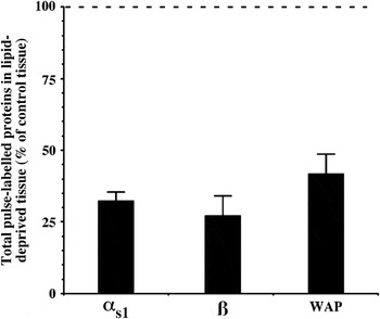
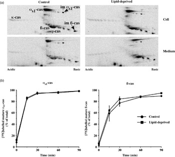
Having established that the level of O-glycosylation of κ-cas was lower in the milk of lipid-deprived animals, it was important to determine whether this proceeds from the degradation of the sugar moieties after protein secretion in milk or from a direct effect of the diet on the post-translational modifications of the protein during its biosynthesis. With this aim, we decided to compare the 2D-PAGE distribution of the newly synthesised κ-cas molecules prepared from MECs obtained from each group of rats. Mammary fragments from control or lipid-deprived rats were pulse-labelled for 3 min with [35S]methionine/cysteine and either collected or chased for 60 min. This length of chase was chosen because it allows the maturation of the newly synthesised secretory proteins, i.e. the post-translational modifications, to occur, while the large majority of the molecules is still intracellular. Tissue proteins were resolved by 2D-PAGE and labelled immature and/or mature forms of αs1-, β-cas and WAP were quantified (κ-cas was excluded from the quantification because we have not identified its immature form in our gels, see also below). We noticed that the total amount of radioactivity incorporated in αs1-, β-cas and in WAP after the pulse was decreased by ≈60% to 65% in lipid-deprived animals as compared with control rats (Figure 3), confirming previous observations from our laboratory (Ollivier-Bousquet et al., Reference Ollivier-Bousquet, Lavialle, Guesnet, Rainteau and Durand1997). On a more interesting note, this procedure revealed that, after a 60-min chase period, the relative proportion of the various molecular forms of newly synthesised κ-cas was different in MECs from lipid-deprived rats, most of the labelled κ-cas forms being less acidic than in control animals (Figure 4a). The κ-cas molecules released in the media during the 60-min chase period were resolved in patterns analogous to those observed in the parent tissues (Figure 4a, medium) and reminiscent of what we observed in the corresponding milk samples (compare Figure 4a with Figure 1a). This indicates that differential glycosylation of κ-cas in lipid-deprived rats occurred during its biosynthesis within MECs and would be due to an effect of the lipid-depleted diet on the glycosylation machinery of these cells.
Protein synthesis is decreased in lipid-deprived animals. Mammary gland explants prepared from control or lipid-deprived rats at mid-lactation were pulse-labelled for 3 min with [35S]methionine/cysteine and aliquots of the tissue proteins were analysed by 2D-PAGE followed by autoradiography, and the indicated proteins were quantified. The amount of pulse-labelled αs1-, β-cas and WAP found in tissue from lipid-deprived rats is expressed as per cent of that found in control animals. The means of two independent experiments are shown; bars indicate the variation of the single values from the mean.

Lipid-deprived diet does not affect the rate of casein secretion. Mammary gland explants prepared from control or lipid-deprived rats at mid-lactation were pulse-labelled for 3 min with [35S]methionine/cysteine and either collected or chase for the indicated times. Aliquots of the tissue proteins (one-fifth as compared with medium, Cell) and of the chase medium (Medium) were analysed by 2D-PAGE, followed by autoradiography. (a) Chase was for 60 min. Only areas of the gels containing caseins are shown. (b) The amount of immature and mature forms of [35S]methionine/cysteine-labelled αs1- and β-cas present at the indicated times of chase were quantified from 2D-PAGE gels. The percentages of mature αs1- and β-caseins were calculated as per cent of the total amount of each radioactive protein (mature + immature). The means of two independent experiments are shown; bars indicate the variation of the single values from the mean. For 90 min chase, the results of a single experiment are shown.

Since in MECs galactose is involved in the biosynthesis of lactose, we wondered whether lactose synthesis, like protein O-glycosylation, would be affected in lipid-deprived animals. With this aim, we compared the amount of lactose in the milk of rats fed with control or lipid-depleted diets. This revealed a large and unexpected increase of lactose concentration in the milk of lipid-deprived animals (Table 1). Although lactose is considered an important osmotic regulator of lactation, this variation in the concentration of the milk sugar was not accompanied by a significant increase in milk osmolarity (Table 1), suggesting that some yet unidentified compound counterbalances the effect of lactose in the milk of lipid-deprived rats.
Finally, we tested whether the kinetics of secretory protein transport in the exocytic pathway of lipid-deprived animals was affected by the diet. Since phosphorylation occurs in the Golgi apparatus, this post-translational modification can be used to monitor the transport of calcium-sensitive caseins (αs1-cas, αs2-cas and β-cas) from the ER to the Golgi stacks. Ideally, we would have liked to also study the transport of κ-cas in the secretory pathway of MECs by analysing its kinetics of O-glycosylation, a post-translational modification, which also occurs in the Golgi apparatus. These experiments were hampered by the fact that the immature κ-cas precursor, i.e. the non-glycosylated and non-phosphorylated ER form of the molecule, was not identified in our 2D-PAGE system. As a matter of fact, the κ-cas molecules contained in spot number 16 (see Figure 1) are most likely not phosphorylated and lack sialic acid. We are confident, however, that this κ-cas form is indeed partly glycosylated and does not correspond to the immature ER form of the protein. This conclusion was supported by the observation that no radioactivity was associated with this spot after a 3-min pulse with [35S]amino acids (data not shown); a length of labelling after which most, if not all, newly synthesised proteins must still be in the ER. Mammary fragments from control or lipid-deprived rats were pulse-labelled for 3 min with [35S]methionine/cysteine and chased for various times, tissue proteins were resolved by 2D-PAGE and labelled immature and mature forms of αs1-, β-cas and WAP were quantified. For both groups of rats the kinetics of conversion of the newly synthesised form to the mature form of each of the caseins were almost identical (Figure 4b). Confirming previous observations (Turner et al., Reference Turner, Handel, Wilde and Burgoyne1993; Boisgard and Chanat, Reference Boisgard and Chanat2000; Pechoux et al., Reference Pechoux, Boisgard, Chanat and Lavialle2005), the maturation of β-cas was slightly delayed as compared with that of αs1-cas. Altogether, these data indicated that, in rats fed with the lipid-depleted diet, despite a marked deficiency in essential polyunsaturated fatty acids in their membranes, the transport of secretory proteins in the early secretory pathway was not significantly modified.
Discussion
Several studies involving our group have led to the conclusion that a lipid-depleted diet induces a large decrease in the level of polyunsaturated fatty acids of membrane phospholipids of mammary cells, notably the level of n-6 polyunsaturated fatty acids including arachidonic acid (20:4 n-6). In parallel, we observed a significant reduction in the intracellular transport of prolactin in the transcytotic pathway of MECs. It was also shown that milk-protein synthesis drops and that the secretagogue effect of prolactin on their secretion disappears in lipid-restricted animals (Ollivier-Bousquet et al., Reference Ollivier-Bousquet, Kann and Durand1993 and Reference Ollivier-Bousquet, Lavialle, Guesnet, Rainteau and Durand1997; Seddiki et al., Reference Seddiki, Delpal, Aubourg, Durand and Ollivier-Bousquet2002). These negative effects are reversed, at least in vitro, by the addition of 20:4 n-6 to mammary fragments (Ollivier-Bousquet et al., Reference Ollivier-Bousquet, Lavialle, Guesnet, Rainteau and Durand1997). In the present work, we demonstrated that the machinery for O-glycosylation of secretory proteins is affected in lipid-deprived rats. Correlatively, we observed a substantial increase in the lactose concentration in the milk of animals receiving the diet without added oil, whereas protein concentration was essentially unchanged.
Effects of lipid-deprived diet on the production of certain milk components
The analysis of the transport of newly synthesised proteins in the mammary tissue from rats receiving a lipid-deprived diet revealed two aspects of the fate of milk proteins in the secretory pathway. First, quantification of various well-identified milk proteins allowed us to conclude that both caseins and the whey protein WAP are produced at a reduced rate in lipid-deprived animals. Second, we demonstrated that despite this large reduction in the biosynthesis of secretory proteins, the kinetics of αs1- and β-cas transport from the ER to the Golgi apparatus are not modified. Therefore, the diet-induced modifications of the lipid composition of cellular membranes seem to have little effect on the membranous events involved in the secretory traffic in the early secretory pathway. This is consistent with previously published data from our laboratory, including the observation that the efficiency of milk protein secretion estimated in vitro was not affected by the lipid-depleted diet (Ollivier-Bousquet et al., Reference Ollivier-Bousquet, Lavialle, Guesnet, Rainteau and Durand1997). Moreover, this is not inconsistent with previous results, which strongly suggest that the secretagogue effect of prolactin, which is likely mediated by arachidonic acid, might be the result of stimulation by the hormone of distal transport steps of the exocytic pathway such as the fusion of carrier vesicles with the plasma membrane (Lkhider et al., Reference Lkhider, Petridou, Aubourg and Ollivier-Bousquet2001).
Since the concentration of milk proteins was not significantly modified by the diet, our data imply that, subsequent to the decrease in milk-protein synthesis, MECs from lipid-deprived animals probably produce less milk fluid. In line with these results, the growth of pups and dams from lipid-deprived dams is affected (Ollivier-Bousquet et al., Reference Ollivier-Bousquet, Lavialle, Guesnet, Rainteau and Durand1997). This conclusion agrees well with the hypothesis that the volume of milk and the volume of water driven to secretory vesicles might be directly related to the flow of material travelling in the secretory pathway of MECs. In this context, one should note that lactose is one of the key osmotic regulators of milk, participating in the determination of the volume of milk produced (Holt, Reference Holt1983), and is critical for milk secretion (Stinnakre et al., Reference Stinnakre, Vilotte, Soulier and Mercier1994). On the other hand, direct relationship between lactose concentration, protein content and osmolarity does not seem obvious in milk (Jost et al., Reference Jost, Vilotte, Duluc, Rodeau and Freund1999). Lactose is synthesised in the trans-most cisternae of the Golgi apparatus and most likely proceeds in secretory vesicles before their fusion with the plasma membrane (Sasaki et al., Reference Sasaki, Eigel and Keenan1978). As lactose cannot diffuse through membranes, its accumulation in the trans Golgi cisternae and in secretory vesicles generates a flow of cellular water into these compartments to maintain the osmotic equilibrium. Obviously, the trans Golgi cisternae and secretory vesicles are swollen and distended in lactating MECs (e.g. see Clermont et al., Reference Clermont, Xia, Rambourg, Turner and Hermo1993). The substantial increase in lactose concentration in the milk from rats receiving the lipid-deprived diet could therefore result from the decrease in the volume of milk produced by these animals, coupled with the maintenance of lactose synthesis at high level. Unfortunately, correct evaluation of the volume of milk produced by small mammals including rats is difficult to achieve. If one speculates that lactose synthesis is not affected by the diet, one can calculate that the volume of milk produced by lipid-deprived rats should be reduced to 60% of that produced by control animals. Yet, given the decrease in milk-protein synthesis in lipid-deprived animals (see above), reduction in milk volume might be up to 30% of control. Therefore, some reduction in lactose synthesis could occur in rats fed the lipid-depleted diet.
Effects of lipid-deprived diet on the O-glycosylation of secretory proteins
The sugar motifs covalently bound to κ-cas were defined in the early 1980s but the composition of the O-glycans in bovine and sheep κ-cas has recently been re-visited using mass spectrometry (Mamone et al., Reference Mamone, Caira, Garro, Nicolai, Ferranti, Picariello, Malorni, Chianese and Addeo2003; Holland et al., Reference Holland, Deeth and Alewood2005). The saccharides found in the antennae were essentially galactose, N-acetylgalactosamine and N-acetylneuraminic acid (in bovine: NeuAcα(2–3)Galb(1–3)[NeuAcα(2–6)]GalNAc).
We interpreted the modification of the relative proportions of the various molecular forms of κ-cas in the milk of lipid-deprived rats as a decrease in the number of neuraminic acid residues linked to the protein. Theoretically, this diminution could result from either a lack of modification of certain O-glycosylation sites or from reduced levels of glycosylation of the various threonine residues by N-acetylgalactosamine and/or galactose. Seemingly, the kinetics of milk protein transport in the secretory pathway of MECs from lipid-deprived animals are not modified, at least between the ER and the Golgi apparatus. On the other hand, it seems that the accumulation of newly synthesised proteins in the media of pulse-labelled mammary fragments prepared from lipid-deprived and control rats is comparable (data not shown). Therefore, one might exclude the possibility that reduced O-glycosylation of secretory proteins in the exocytic pathway of MECs from lipid-deprived animals would be due to variation in the residence time of the substrate proteins in the Golgi apparatus where this post-translational modification is achieved.
Alternative hypotheses concerning the mechanism by which secretory protein O-glycosylation is affected by the diet are as follows: first, glycosyltransferases involved in the O-glycosylation process could be expressed at a lower level in MECs from rats fed the lipid-deprived diet. Second, the necessary nucleotide-sugars (UDP-galactose, UDP-N-acetylgalactosamine and CMP-N-acetylneuraminic acid for κ-cas) could be in reduced amounts in the lumen of the ER/Golgi cisternae in lipid-deprived animals following a defect in either their transport from the cytosol or their biosynthesis. Theoretically, the latter may occur if glucose uptake by MECs is reduced. We do not favour these two hypotheses, however, because we found that the milk of lipid-deprived rats contains more lactose (galactose β1,4-glucose). This strongly suggests that glucose uptake by the cell, as well as its transport into the Golgi compartment, are not affected. In turn, this implies that the synthesis of UDP-galactose and its transport to the lumen of the compartments of the exocytic pathway as well as the activity of the lactose synthase are not rate-limiting in MECs from lipid-deprived rats (for review on lactose synthesis see Brew and Hill, Reference Brew and Hill1975; Kuhn et al., Reference Kuhn, Carrick and Wilde1980). The enzymatic subunit of lactose synthase is the β1,4 galactosyltransferase, an ubiquitous trans-Golgi enzyme, the usual function of which is to transfer galactose to the terminal N-acetylglucosamine acceptor residues in the oligosaccharide complex during either glycoprotein or glycolipid biosynthesis. Therefore, one cannot exclude the possibility that reduced O-glycosylation in lipid-deprived animals would be due to a shift of β1,4 galactosyltransferase activity towards lactose synthesis, hence depleting the cell of the precursor sugar UDP-galactose for post-translational modification of glycoproteins. This may be the case if α-lactalbumin, the non-catalytic subunit of lactose synthase, is expressed at higher levels in MECs from lipid-deprived animals.
Possible effects of lipid-deprived diet on the hexosamine biosynthetic pathway
Another observation that might be relevant to explain our data is the fact that in certain tissues insulin-stimulated glucose uptake by cells increases in calorie-restricted animals (Wetter et al., Reference Wetter, Gazdag, Dean and Cartee1999 and references therein). Indeed, if this is true for MECs, it would be compatible with higher lactose in the milk of lipid-deprived rats. Supporting this, it has recently been shown that expression of both hexokinase and glucose phosphate isomerase is down-regulated in mouse at the initiation of lactation (Rudolph et al., Reference Rudolph, McManaman, Phang, Russell, Kominsky, Serkova, Stein, Anderson and Neville2007). This would favour the partitioning of glucose towards lactose production by decreasing its consumption for glycolysis, in the pentose phosphate cycle and the hexosamine biosynthetic pathways.
With regard to the hexosamine biosynthetic pathway, it has been demonstrated that caloric restriction leads to a reduction in the concentration of UDP-N-acetylhexosamines, namely UDP-N-acetylglucosamine and UDP-N-acetylgalactosamine, the end products of this biosynthetic pathway (Gazdag et al., Reference Gazdag, Wetter, Davidson, Robinson, Buse, Yee, Turcotte and Cartee2000). Worth noting is the fact that a role for N-acetylglucosamine as a nutrient sensor and mediator of insulin resistance has been proposed (for review see Wells et al., Reference Wells, Vosseller and Hart2003; Zachara and Hart, Reference Zachara and Hart2004). On the other hand, these molecules serve as the donor sugar nucleotides for glycosylation reactions, N-acetylgalactosamine being usually the first sugar added to serine or threonine during secretory protein O-glycosylation. Our observation that the level of O-glycosylation of κ-cas is lower in the milk of lipid-deprived animals might therefore be due to reduced glucose flux through the hexosamine biosynthetic pathway and subsequent reduction in UDP-N-acetylhexosamines in lipid-deprived rats. In sum, our data support the hypothesis that, in lipid-restricted MECs, glucose uptake increases and glucose flux through the hexosamine biosynthetic pathway decreases.
Acknowledgements
We are grateful to P. Dahirel (Nutrition et Régulation Lipidique des Fonctions Cérébrales, INRA Jouy-en-Josas, France) for supervision of rat care and C. Henry (Plateau d’Analyse Protéomique par Séquençage et Spectrométrie de Masse, INRA Jouy-en-Josas, France) for expert technical assistance for the MS analysis. We are indebted to Drs J. Wantyghem (Laboratory of Immune Regulations and Development, J Monod Institute, Paris, France) and P. Guesnet (Nutrition et Régulation Lipidique des Fonctions Cérébrales, INRA Jouy-en-Josas, France) for helpful comments on the manuscript and to Ms F. McAlpine for editorial assistance. We wish to thank I. Gicquel for determination of milk lactose (UMR-Production du Lait INRA Rennes, France). This work was partly supported by the ‘Groupe Lipides et Nutrition’, Neuilly-sur-Seine, France.