Introduction
The rat is a reference animal model for physiological studies and for the analysis of multigenic human diseases such as hypertension, diabetes and neurological disorders. As embryonic stem (ES) cells are not yet available for rats, cloning that uses somatic cell nuclear transfer (SCNT) technology with gene targeting could be a potential alternative approach to obtain valuable rat models (Phelps et al., Reference Phelps, Koike, Vaught, Boone, Wells, Chen, Ball, Specht, Polejaeva, Monahan, Jobst, Sharma, Lamborn, Garst, Moore, Demetris, Rudert, Bottino, Bertera, Trucco, Starzl, Dai and Ayares2003; Sendai et al., Reference Sendai, Sawada, Urakawa, Shinkai, Kubota, Hoshi and Aoyagi2006).
The only example of successful production of cloned rats involved injecting metaphase-arrested fetal fibroblasts into oocytes treated with MG132, a proteasome inhibitor, followed by rapid removal of the recipient metaphase plate (Zhou et al., Reference Zhou, Renard, Le Friec, Brochard, Beaujean, Cherifi, Fraichard and Cozzi2003). However, this result has not been reproduced to date. In addition, SCNT embryos produced using metaphase II (MII) oocytes treated with MG132 did not develop to the blastocyst stage in vitro (Popova et al., Reference Popova, Bader and Krivokharchenko2008). Several studies have been conducted to determine the cause of developmental arrest of rat SCNT embryos at the preimplantation stage (Hirabayashi et al., Reference Hirabayashi, Kato, Ishikawa and Hochi2003; Ito et al., Reference Ito, Hirabayashi, Kato, Takeuchi, Ito, Shimada and Hochi2005; Tomioka et al., Reference Tomioka, Mizutani, Yoshida, Sugawara, Inai, Sasada and Sato2007; Yoo et al., Reference Yoo, Demers, Lian and Smith2007; Popova et al., Reference Popova, Bader and Krivokharchenko2008). The failure of nuclear envelope breakdown (NEBD), premature chromosome condensation (PCC) and spindle formation that may be caused by spontaneous activation (Keefer & Schuetz, Reference Keefer and Schuetz1982) have all been described as the main reasons for low in vitro developmental potency (Kato et al., Reference Kato, Hirabayashi, Aoto, Ito, Ueda and Hochi2001; Hirabayashi et al., Reference Hirabayashi, Kato, Ishikawa and Hochi2003). For successful cloning, at least in rodents, it may be necessary for somatic cell nuclei injected into enucleated oocyte cytoplasm to be directly exposed to a high level of maturation-promoting factor (MPF) activity, which results in NEBD, PCC and subsequent pseudo-pronuclei formation following activation (Wakayama et al., Reference Wakayama, Perry, Zuccotti, Johnson and Yanagimachi1998). However, it has not been determined whether failure of PCC and cytoskeletal anomalies are the underlying causes of developmental arrest at the preimplantation stage because of the current lack of an in vitro culture system.
Recently, interspecies SCNT (iSCNT) has emerged as an important tool for the study of nucleus–cytoplasmic interactions, infertility treatments and production of stem cells, cloning of endangered animals whose oocytes are difficult to obtain and for the examination of the in vitro developmental ability of donor nuclei (Beyhan et al., Reference Beyhan, Iager and Cibelli2007). In vitro development to the blastocyst stage was not observed in a previous study, in which PCC of rat cumulus cell nuclei injected into murine MII oocytes occurred (Hirabayashi et al., Reference Hirabayashi, Kato, Ishikawa and Hochi2003). In addition, rat iSCNT embryos made with bovine MII oocytes were shown to be arrested at the 2-cell stage in vitro (Dominko et al., Reference Dominko, Mitalipova, Haley, Beyhan, Memili, McKusick and First1999). On the other hand, bovine and tiger iSCNT embryos made with porcine MII oocytes developed to the blastocyst stage (Hashem et al., Reference Hashem, Bhandari, Kang and Lee2007; Uhm et al., Reference Uhm, Gupta, Kim and Lee2007). These studies indicate that porcine MII oocytes could support the early development of somatic cell nuclei of other species. In addition, our studies suggested that porcine recipient oocytes in the early MII stage retain a high level of MPF activity during the SCNT procedure and thus effectively induce PCC with a normal cytoskeletal structure, followed by a resultant improvement in the subsequent development (Wakai et al., Reference Wakai, Sugimura, Yamanaka, Kawahara, Sasada, Tanaka, Ando, Kobayashi and Sato2008).
In this study, we evaluated the developmental competence of rat iSCNT embryos constructed using murine and porcine MII oocytes. Moreover, to determine the factors that influence this developmental ability, we also evaluated whether donor cell types, such as fetal fibroblasts and cumulus cells, and PCC with normal spindles affected the developmental ability.
Materials and Methods
Animal preparation
Wistar Imamichi rats and C57BL/6 and DBA/2 mice were purchased from Japan SLC Inc. and bred in our laboratory. Green fluorescent protein (GFP)-transgenic rats with a CAG (cytomegalovirus enhancer, chicken actin enhancer–promoter and rabbit globin poly[A] signal) promoter were developed from Wistar rats (Hakamata et al., Reference Hakamata, Tahara, Uchida, Sakuma, Nakamura, Kume, Murakami, Takahashi, Takahashi, Hirabayashi, Ueda, Miyoshi, Kasai and Kobayashi2001; Takeuchi et al., Reference Takeuchi, Sereemaspun, Inagaki, Hakamata, Kaneko, Murakami, Takahashi, Kobayashi and Ookawara2003). Lights were switched on between 08.00 and 20.00 daily. All procedures adhered to the Guide for the Care and Use of Laboratory Animals published by Tohoku University.
Donor cell preparation
GFP-transgenic rat fetal fibroblasts were prepared from 13.5- to 14.5-day post coitus fetuses derived from a female GFP-transgenic Wistar rat as reported previously (Hayes et al., Reference Hayes, Galea, Verkuylen, Pera, Morrison, Lacham-Kaplan and Trounson2001). Briefly, the heads and all internal organs of the fetuses were removed and each carcass was minced individually in Petri dishes. The minced carcass was digested with trypsin–EDTA (Invitrogen) for 1–2 h at 37°C. Cells were then collected by centrifugation at 200 g for 5 min, resuspended and washed in Dulbecco's modified Eagle's medium (DMEM) supplemented with 10% (v/v) fetal bovine serum (FBS; Gemini-Bio Products). Finally, cells from each fetus were placed on two 30 mm dishes that contained DMEM with 10% FBS and incubated overnight in 5% CO2 at 37°C. Once the cells reached confluence, they were frozen as ‘zero passage’ cells (1 vial per 30 mm dish) in a cell conservation medium (Cell banker, Nihon Zenyaku Co.) and thawed as needed. For experiments, donor cells between passages 3 and 7 were used for SCNT. The cells were thawed and cultured for 2 to 5 days after reaching confluence, a single cell suspension was prepared prior to SCNT. By flow cytometry, most of these cells were identified as being in the G0/G1 phase of the cell cycle.
Cumulus cells were also isolated from ovulated oocytes from the oviducts of mature Wistar Imamichi rats. The cells were rinsed with modified rat 1-cell embryo culture medium (mR1ECM) medium (Miyoshi et al., Reference Miyoshi, Kono and Niwa1997) after dispersion with 0.1% (w/v) hyaluronidase (Sigma) and used as the donor cells in nuclear transfer.
Murine oocyte collection
Intraperitoneal injection of sexually mature B6D2F1 (C57BL/6 × DBA/2) female mice, (8 to 10 weeks old) with 5 IU of pregnant mare serum gonadotropin (PMSG, Teikoku Zoki) was followed by an intraperitoneal injection of 5 IU of human chorionic gonadotropin (hCG, Sankyo) 48–49 h later. Animals were sacrificed by cervical dislocation 14–16 h after administration of hCG and their oviducts were removed. The oviducts were placed in bicarbonate-buffered (CZB) medium (Chatot et al., Reference Chatot, Lewis, Torres and Ziomek1990) and treated with 0.1% (w/v) hyaluronidase. Cumulus–oocyte complexes (COCs) were collected by sectioning the oviducts in the medium. After washing three times with CZB medium, oocytes were transferred to 100 μl droplets of CZB medium overlaid with liquid paraffin (Nacalai Tesque).
In vitro maturation of porcine oocytes
Ovaries from prepubertal gilts were collected at a local slaughterhouse and transported to the laboratory in a warm chamber at 37°C. COCs were aspirated from ovarian follicles of 3–5 mm diameter. COCs were washed with modified Dulbecco's PBS medium containing 5.56 mM glucose (Wako), 0.33 mM sodium pyruvate (Wako), 0.01 mg/ml antibiotic antimycotic solution (Sigma) and 4 mg/ml fatty acid-free BSA (Sigma) (PBI medium; Quinn et al., Reference Quinn, Barros and Whittingham1982) (Quinn et al., Reference Quinn, Barros and Whittingham1982) and only those with a uniform cytoplasm and compact cumulus cells were selected for maturation. About 10 COCs were cultured in each 100 μl drop of North Carolina State University (NCSU)-23 medium (Petters & Wells, Reference Petters and Wells1993) supplemented with 50 μM prepubertal β-mecaptoethanol (Sigma), 0.6 mM cysteine (Sigma), 5 mg/ml insulin (Sigma), 10% (v/v) porcine follicular fluid, 1 mM dbcAMP (Sigma), and hormonal supplements of 10 IU/ml hCG (Sankyo) and of 10 IU/ml PMSG (Asuka Seiyaku Co.) for 22 h at 38.5°C under an atmosphere of 5% CO2 in air. The COCs were then cultured in the same medium without dbcAMP and hormones for an additional 18 h. After the maturational step cultivation, oocytes were denuded from cumulus cells by pipetting in PB1 medium that contained 0.1% hyaluronidase and those oocytes with the first polar body were selected as mature.
Nuclear transfer and activation
Production of iSCNT embryos using murine MII oocytes
Oocytes were enucleated by a previously reported method (Wakayama et al., Reference Wakayama, Perry, Zuccotti, Johnson and Yanagimachi1998). In brief, the oocytes were treated with 5 μg/ml cytochalasin B (Sigma) in HEPES-buffered CZB medium (Chatot et al., Reference Chatot, Lewis, Torres and Ziomek1990) for 10 min at room temperature. Enucleation was performed in the same medium under an inverted microscope equipped with Nomarski or Hoffmann optics using a piezo-driven micromanipulator (Prime Tech Ltd). A GFP-expressing fetal fibroblast was inserted into the perivitelline space of the enucleated oocyte. Donor–recipient pairs were placed between the electrodes of a fusion chamber filled with a solution of 0.3 M mannitol and 0.1 mM MgSO4, 0.05 mM CaCl2 and 0.1 mg/ml polyvinyl alcohol (Sigma) at 37°C. The fusion of donor–recipient pairs was induced by two direct current pulses of 1.20 V/cm for 70 μs with an inter-pulse interval of 1 s (Shinozawa et al., Reference Shinozawa, Mizutani, Tomioka, Kawahara, Sasada, Matsumoto and Sato2004). The embryos were then placed in a culture droplet and checked every 10 min. Embryos usually fused within 20 min. iSCNT embryos were incubated in CZB medium for approximately 1 h prior to activation treatment. At 1 h after cell fusion, iSCNT embryos were activated by treatment with Ca2+-free CZB medium plus 10 mM Sr2+ and 5 μg/ml cytochalasin B for 1 h. After activation, the embryos were treated in Ca2+-free CZB medium plus 5 μg/ml cytochalasin B for 5 h. The embryos were then transferred to CZB medium and culture was continued under 5% CO2 in air at 37°C.
Production of iSCNT embryos using porcine MII oocytes
Oocytes were enucleated by a previously reported method (Kawahara et al., Reference Kawahara, Wakai, Yamanaka, Kobayashi, Sugimura, Shimizu, Matsumoto, Kim, Sasada and Sato2005). Mature porcine oocytes were transferred to PB1 medium supplemented with 2.5 μg/ml cytochalasin D (Sigma) for 10 min at room temperature. Enucleation was performed by aspiratation of the first polar body and adjacent cytoplasm. The oocytes had been previously treated for 5 to 10 min in PB1 medium that contained 5 μg/ml Hoechst 33342 (Sigma). Successful enucleation was confirmed by visualization under ultraviolet light. For microinjection of the donor nucleus, the cell membrane was ruptured by gentle pipetting and the nucleus was directly injected into the enucleated oocyte. iSCNT embryos were incubated in PZM-3 medium (Yoshioka et al., Reference Yoshioka, Suzuki, Tanaka, Anas and Iwamura2002) for approximately 3 h prior to activation treatment. At 3 h after microinjection, SCNT embryos were activated by ionomycin (Sigma) and cycloheximide (Sigma) (Yamanaka et al., Reference Yamanaka, Sugimura, Wakai, Shoji, Kobayashi, Sasada and Sato2007); embryos were treated with 15 μM ionomycin in PZM-3 medium for 20 min at 38.5°C in 5% CO2 in air. After ionomycin treatment, the embryos were cultured in PZM-3 medium plus 5 μg/ml cycloheximide and 2.5 μg/ml cytochalasin D for 5 h at 38.5°C in 5% CO2 in air (Sugimura et al., Reference Sugimura, Kawahara, Wakai, Yamanaka, Sasada and Sato2008). After activation, the embryos were cultured in porcine zygote medium (PZM)-8 medium at 38.5°C in 5% CO2 in air.
Immunocytochemical examination of chromosomes and microtubules
Immunocytochemical staining was performed by a previously reported method (Wakai et al., Reference Wakai, Sugimura, Yamanaka, Kawahara, Sasada, Tanaka, Ando, Kobayashi and Sato2008). In brief, after washing three times in PBS plus 0.1% (w/v) polyvinyl alcohol (PBS–PVA), SCNT embryos were fixed in PBS–PVA that contained 2% paraformaldehyde (Sigma) and 0.2% (v/v) Triton-X (Wako) for 40 min. The fixed embryos were washed three times for 15 min each in PBS–PVA and then stored in PBS–PVA supplemented with 1% (w/v) BSA (PBS–PVA–BSA) overnight. On the following day, the embryos were treated by blocking with 10% fetal bovine serum (Gemini Bio-Products) in PBS–PVA–BSA plus a monoclonal anti-α-tubulin antibody (1:250 dilution; Sigma) at 4°C overnight, washed in PBS–PVA–BSA three times for 15 min each and incubated with fluorescein isothiocyanate (FITC)-conjugated goat anti-mouse IgG (1:50 dilution; Oregon Teknika) for 40 min at room temperature. After the embryos were washed in PBS–PVA three times for 15 min each, they were stained with propidium iodide (Sigma) for 1 h and then mounted onto glass slides. Immunocytochemical examination was carried out using a confocal microscope (MRC-1204, Bio-Rad). Laser Sharp Processing software (Bio-Rad) was used to analyse the confocal images.
Experimental design
Experiment 1: Developmental ability of iSCNT embryos
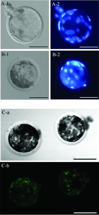
First, to investigate the effect of recipient cytoplasts, oocytes from two different species – mouse and pig – were evaluated. Next, to determine the difference in developmental ability in donor cell type, iSCNT embryos were constructed with GFP-expressing fetal fibroblasts or cumulus cells. After hCG injection for 14 to 16 h or IVM for 40 h, matured murine or porcine oocytes were selected and subjected to iSCNT, respectively. The rates of cleavage and blastocyst formation were evaluated on days 2 or 7 of in vitro culture, respectively. All blastocysts on day 7 of in vitro cultivation were fixed in PBS–PVA that contained paraformaldehyde and Triton-X and stained with 5 μg/ml Hoechst 33342. The number of nuclei was counted under ultraviolet light using a phase-contrast microscope (Fig. 1). In addition, cytoplasmic expression of GFP in iSCNT blastocysts with GFP-expressing fetal fibroblasts was examined by confocal microscopy using a FITC filter set (Fig. 1).
Morphology (A-1, B-1, C-a), embryonic cells (A-2, B-2) and GFP expression (C-b) of blastocysts derived from iSCNT embryos reconstructed with GFP-expressing fetal fibroblasts (A, C) and cumulus cells (B). Bar = 100 μm.

Experiment 2: Spindle formation of iSCNT embryos using porcine MII oocytes
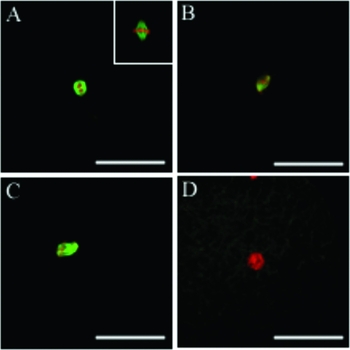
To evaluate the effect of donor cell type on nuclear remodelling, we examined the morphological changes in transferred nuclei in recipient cytoplasts. The formation of NEBD, PCC and microtubules was observed at 3 h after nuclear injection (Figs. 2 and 3). As shown in Fig. 3, four types of PCC spindle morphologies were identified: (A) normal bipolar spindle with aligned chromosome assembly; (B) normal bipolar spindle but with misaligned chromosome assembly; (C) abnormal spindle structure; and (D) disarrayed microtubules in the oocyte cytoplasm.
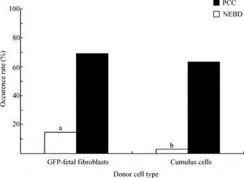
The occurrence of NEBD and PCC in iSCNT embryos using porcine MII oocytes. At 3 h after cell injection, the formation of NEBD and PCC was observed. The number of iSCNT embryos examined for each donor cell type was 68. Values are expressed as mean ± S.E. a,bValues with different superscripts are significantly different (p < 0.05).

Spindle morphologies after injection of donor nuclei. In the chromosomal configuration of iSCNT embryos, various types of PCC spindle were observed at 3 h after cell injection (A–D). All images were double labelled for microtubules (green) and DNA (red). (A) Normal bipolar spindle with aligned chromosome assembly, inset: meiotic spindle in metaphase II oocytes; (B) normal bipolar spindle but with misaligned chromosome assembly; (C) abnormal spindle structure; and (D) disarrayed microtubules in the oocyte cytoplasm. Bar = 50 μm.

Statistical analysis
The total cell number of iSCNT blastocysts was analysed with ANOVA followed by Tukey's test for determining significant difference (p < 0.05). Other data were analysed by the chi-squared test (p < 0.05).
Results
Experiment 1: Developmental ability of iSCNT embryos
As shown in Table 1, we investigated the developmental competence of iSCNT embryos using murine or porcine oocytes as recipient. While the iSCNT embryos using porcine oocytes developed to the blastocyst stage, iSCNT embryos using murine oocytes did not. The cleavage rate was approximately equal between iSCNT embryos using porcine MII oocytes reconstructed with GFP-expressing fetal fibroblasts and cumulus cells. In addition, the rate of blastocyst formation in iSCNT embryos using GFP-expressing fetal fibroblasts tended to increase compared with those embryos using cumulus cells (5.6% vs. 2.7%). The total cell number of iSCNT blastocysts derived from GFP-expressing fetal fibroblasts was significantly higher than for those blastocysts derived from cumulus cells (32.3 ± 11.2 vs. 12.7 ± 1.5). Moreover, as shown in Fig. 1C-b, iSCNT embryos made using GFP-expressing fetal fibroblasts exhibited cytoplasmic expression of GFP. In addition, the embryos showed 100% cytoplasmic expression of GFP at the 2-cell, 4-cell, 8-cell and morula stages (data not shown).
In vitro developmental competence of iSCNT embryos using murine or porcine oocytes.

ND, no data. *Mean ± S.D. a , bValues with different superscripts are significantly different for each donor cell type (p < 0.05).
Experiment 2: Spindle formation in iSCNT embryos using porcine MII oocytes
iSCNT embryos constructed using GFP-expressing fetal fibroblasts as donor cells increased the occurrence of NEBD and PCC (14.7% and 69.1%) in comparison with cumulus cells (2.9% and 63.2%) (Fig. 2). As shown in Table 2, the number of iSCNT embryos with normal spindles was higher in those embryos made with GFP-expressing fetal fibroblasts than in those embryos made with cumulus cells (48.9% [Fig. 3A: 19.1%; Fig. 3B: 29.8%] vs. 25.6% [Fig. 3A: 16.3%; Fig. 3B: 9.3%]). Moreover, the number of iSCNT embryos that had PCC with abnormal spindle was higher in those embryos that were constructed using cumulus cells than in those embryos made using GFP-expressing fetal fibroblasts (51.2% vs. 31.9%) (Fig. 3C). In addition, iSCNT embryos with disarrayed microtubules in the oocyte cytoplasm were higher in those embryos that were constructed using cumulus cells than in those embryos made using GFP-expressing fetal fibroblasts (19.1% vs. 23.3%) [Fig. 3D].
Spindle formation of iSCNT embryos using porcine MII oocytes.

a , bValues with different superscripts are significantly different for each donor cell type (p < 0.05).
Discussion
In the present study, to determine the factors that affect developmental ability of rat SCNT embryos, we evaluated PCC spindle formation and developmental ability of rat iSCNT embryos made using porcine MII oocytes. We demonstrated that rat iSCNT embryos made using porcine MII oocytes have many PCC with normal bipolar spindle and developmental potential up to the blastocyst stage in vitro. In addition, the embryos reconstructed by GFP-expressing fetal fibroblasts exhibited cytoplasmic expression of GFP at the blastocyst stage (Fig. 1C). This observation suggested that the developmental arrest of rat SCNT embryos in the preimplantation stage (Hirabayashi et al., Reference Hirabayashi, Kato, Ishikawa and Hochi2003; Tomioka et al., Reference Tomioka, Mizutani, Yoshida, Sugawara, Inai, Sasada and Sato2007; Nakajima et al., Reference Nakajima, Inomata, Ito and Kashiwazaki2008; Popova et al., Reference Popova, Bader and Krivokharchenko2008) was attributed to recipient oocytes rather than to the donor nucleus.
Donor nuclei in the G0/G1 phase exhibit a high occurrence of NEBD and PCC when they are transferred into recipient cytoplasts with high levels of MPF activity before activation. These changes, defined as chromatin remodelling, are considered necessary for complete functional reprogramming of the transferred nucleus (Wakayama et al., Reference Wakayama, Perry, Zuccotti, Johnson and Yanagimachi1998). In contrast, cytoplasts derived from activated oocytes with low levels of MPF activity cannot induce NEBD and PCC in transferred nuclei. SCNT embryos made by using these oocytes underwent irregular cleavages and only a small percentage of the embryos developed into blastocysts (Wakayama & Yanagimachi, Reference Wakayama and Yanagimachi2001). In the present study, we produced rat iSCNT embryos by using porcine oocytes at the early MII stage, which retains a high level of MPF activity (Wakai et al., Reference Wakai, Sugimura, Yamanaka, Kawahara, Sasada, Tanaka, Ando, Kobayashi and Sato2008), this approach resulted in the occurrence of PCC with normal spindle. Hence, the development of rat iSCNT embryos to the blastocyst stage may depend on beneficial factors of porcine early MII oocytes such as high MPF activity.
In contrast, we demonstrated that when using murine MII oocytes to produce rat iSCNT embryos, the embryos were arrested at the 2-cell stage. This result is consistent with findings by Hirabayashi et al. (Reference Hirabayashi, Kato, Ishikawa and Hochi2003). It is hypothesized that developmental arrest at the 2-cell stage might arise from aberrant nucleus–cytoplasmic interactions, notably a leading cause for anomalous spindle formation (Wakayama et al., Reference Wakayama, Perry, Zuccotti, Johnson and Yanagimachi1998; Miyara et al., Reference Miyara, Han, Gao, Vassena and Latham2006; Balbach et al., Reference Balbach, Jauch, Bohm-Steuer, Cavaleri, Han and Boiani2007). Such abnormality may lead to chromosome destabilization and to an increased risk of segregation errors at each cleavage division (Bean et al., Reference Bean, Hunt, Millie and Hassold2001, Reference Bean, Hassold, Judis and Hunt2002). Thus, rat iSCNT embryos that were produced using murine MII oocytes may have abnormal spindle structures and this situation may result in developmental arrest at the 2-cell stage.
Following SCNT, the ooplasm restores a chromosomal spindle structurally different from a normal spindle of MII oocytes. This findiing may reflect a ‘meiosis–mitosis mismatch’ between recipient ooplasm (meiotic) and donor nucleus (mitotic) (Balbach et al., Reference Balbach, Jauch, Bohm-Steuer, Cavaleri, Han and Boiani2007). Balbach et al. stated that the use of ES cells should be less of a ‘mismatch’, because ES cells have been regarded as the ‘earliest germ cells’ (Zwaka & Thomson, Reference Zwaka and Thomson2005). In addition, they indicated that SCNT embryos derived from ES cells have a high occurrence of morphologically normal spindles. In the present study, many rat SCNT embryos that had been constructed using GFP-expressing fetal fibroblasts from passages 3 to 7 induced PCC with normal spindle formation compared with those embryos made using cumulus cells. On the basis of the report by Balbach et al., we speculated that GFP-expressing fetal fibroblasts might contain many stem cell-like cells, which results in the induction of normal PCC spindle formation observed in the present study.
iSCNT technique is an effective tool for the investigation of nucleus–cytoplasmic interaction (Beyhan et al., Reference Beyhan, Iager and Cibelli2007). If this interaction failed, poor quality blastocyst formation may occur (Beyhan et al., Reference Beyhan, Iager and Cibelli2007). Aberrant nucleus–cytoplasm interaction probably causes decreased total cell number of blastocysts and increased apoptosis incidence (Mastromonaco et al., Reference Mastromonaco, Favetta, Smith, Filion and King2007). In the present study, the total cell number of iSCNT blastocyst derived from GFP-expressing fetal fibroblasts was higher than that derived from cumulus cells. This finding may be due to a difference in the nucleus–cytoplasmic interaction status.
In conclusion, this report is the first in which rat SCNT embryos constructed using porcine MII oocytes have been shown to have the potential to develop to the blastocyst stage in vitro. In addition, this study indicated that the iSCNT technique, when using early porcine MII oocytes, provides a new bioassay system for the evaluation of the developmental potential of rat somatic cells.
Acknowledgements
We thank Dr Eiji Kobayashi of Jichi Medical School for providing GFP-transgenic rats and the staff of the Meat Inspection Office, Sendai City, for supplying porcine ovaries. This work was supported by the Japan Society for the Promotion of Science Grant JSPS-16108003 (to E.S.).