Introduction
Kaposi's sarcoma, an acquired immunodeficiency syndrome (AIDS) defining illness, is a leading cause of morbidity and mortality in patients with human immunodeficiency virus (HIV) infection.Reference Lorenzo, Konstantinopoulos, Pantanowitz, Trollo, Placido and Dezube1 Kaposi's sarcoma lesions comprise aberrant vessels and spindle-shaped tumour cells which progress from an early patch stage to a plaque stage and may ultimately form a tumour (nodular stage). While skin lesions constitute the most frequent manifestation of AIDS-related Kaposi's sarcoma, extracutaneous involvement is common, especially in patients not receiving highly active antiretroviral therapy. Kaposi's sarcoma of the oral cavity occurs in approximately one-third of afflicted AIDS patients, most frequently in the areas supplied by the palatine arteries.Reference Reichart, Gelderblom, Becker and Kuntz2, Reference Schiodt and Pindborg3 However, oral Kaposi's sarcoma associated with actual erosion of underlying bone is very rare. We report a case of palatal Kaposi's sarcoma with intraosseous extension and destruction of the adjacent maxillary bone, and we review the literature on this uncommon but devastating complication.
Case report
A 45-year-old, malnourished, homosexual man presented with an oral lesion associated with significant discomfort. He had a history of intravenous drug abuse, HIV infection for over 20 years (currently, cluster of differentiation four glycoprotein (CD4 +) T-lymphocyte count = 64 cells/mm3, HIV viral load = 57 922 copies/ml, and antiretroviral therapy naive), hepatitis B and C co-infections, and prior Pneumocystis carinii pneumonia. Six months prior to presentation, he had had his upper second molar tooth extracted, but this had continued to drain, bleed and cause significant pain.
Examination of the patient's oropharynx revealed an oral lesion that was very tender and friable to touch. It was large, fungating and involved the entire left hard palate and some of the soft palate. Additional findings included oral thrush and purulent drainage from an oral–antral fistula on the right, just behind the upper bicuspid and first molar teeth. Nasal endoscopy detected no turbinate necrosis and apparently normal rostra of the sphenoid sinuses. Direct laryngoscopy revealed a left true vocal fold mass. The rest of the physical examination was unremarkable.
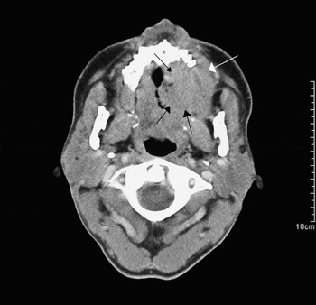
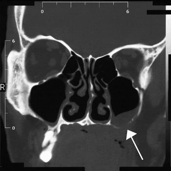
A computed tomography (CT) scan of the neck demonstrated an irregular mass involving the left palate, which invaded the maxillary bone (Figure 1). Coronal maxillofacial CT showed erosion of the floor of the left maxillary sinus and thickening of the left maxillary sinus mucosa (Figure 2). There was no evidence of erosion into the skull.
Axial computed tomography scan of the neck, showing an irregular mass (black arrows) involving the left palate, which has invaded the maxillary bone (white arrow).

Coronal maxillofacial computed tomography scan showing erosion of the floor of the left maxillary sinus (arrow) and thickening of the left maxillary sinus mucosa.

Otorhinolaryngology, haematological oncology, radiation oncology and infectious disease consultations were obtained.
The patient underwent panendoscopy, which showed a 7.5 × 5.5 cm, irregular, friable mass encompassing the entire left hard palate, encroaching onto the soft palate and eroding into the maxilla on the left, with compromise of the tooth roots.
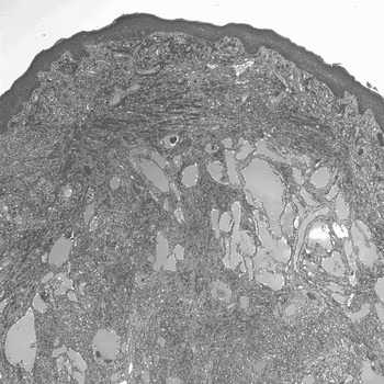
The left lateral maxillary teeth were extracted as they were unstable. Excisional biopsies were performed and demonstrated nodular Kaposi's sarcoma (Figure 3).
Photomicrograph of biopsy of the hard palate lesion, consistent with nodular Kaposi's sarcoma (H&E; ×40).

Immunohistochemical analysis showed the tumour cells to be immunoreactive with CD34 and CD31 and negative for factor VIII, consistent with the diagnosis of Kaposi's sarcoma.
Biopsy of the left true vocal fold demonstrated a vascular-hyaline polyp.
Oesophagoscopy and bronchoscopy were normal.
The patient was subsequently treated with palliative radiation and systemic chemotherapy with liposomal doxorubicin. His treatment course was complicated by radiation mucositis, which was treated supportively. The patient declined further therapy and was discharged to a hospice.
Discussion
Kaposi's sarcoma is a vascular neoplasm which involves mucocutaneous and visceral body sites.Reference Lorenzo, Konstantinopoulos, Pantanowitz, Trollo, Placido and Dezube1 While oral involvement is a common manifestation of AIDS-related Kaposi's sarcoma, it is rarely observed in other types of Kaposi's sarcoma, such as classic, endemic (African) and iatrogenic (transplant-related) Kaposi's sarcoma. The most common sites of intraoral AIDS-related Kaposi's sarcoma include the soft and hard palate, the gingiva and the tongue.Reference Reichart, Gelderblom, Becker and Kuntz2, Reference Schiodt and Pindborg3 Intraoral Kaposi's sarcoma lesions appear as painless, red to blue macules located in the palatal mucosa or gingiva. They may develop into large tumour masses which eventually ulcerate or become superinfected. Most oral lesions tend to be exophytic and either singular or multifocal.
To date, there have been 10 reported cases of Kaposi's sarcoma involving the jaw bones (Table I).Reference Lausten, Ferguson, Barker and Cobb4–Reference Righi, Pierleoni and Ficarra8 All reported cases involved men with AIDS whose age averaged 36 years (range, 27–52 years). Their median CD4+ T-lymphocyte count was 45 cells/mm3 (range, 0–321 cells/mm3). This suggests that the degree of HIV-associated immunosuppression may correlate with the risk of developing intraosseous, maxillofacial Kaposi's sarcoma. The maxilla was involved in six cases and the mandible in four. One patient presented with bilateral maxillary bone involvement. In most of the cases (nine of ten), the jaw bones were secondarily involved, typically by deep extension from an overlying, contiguous, mucosal Kaposi's sarcoma lesion. Hence, it appears that, in this setting, intraosseous Kaposi's sarcoma results mainly from direct extension of long-standing mucocutaneous Kaposi's sarcoma, as observed in our case.Reference Lausten, Ferguson, Barker and Cobb4, Reference Kleinegger and Sarubin5
Published cases of AIDS-related kaposi's sarcoma involving the jaw bones

AIDS = acquired immunodeficiency syndrome; Ref = reference; 1° = primary; 2° = secondary; CD4 = cluster of differentiation four glycoprotein; KS = Kaposi's sarcoma; M = male; CT = computed tomography
Kaposi's sarcoma presenting as an isolated bone defect, without involvement of the overlying mucosa or skin (i.e. primary intraosseous Kaposi's sarcoma), is exceedingly rare. As in ours and two other reported cases, Kaposi's sarcoma of the jaw bones arose six months to one year after tooth extraction.Reference Langford, Pohle and Reichart6 This raises the interesting possibility that tooth extraction may provoke Kaposi's sarcoma development, in the correct setting. Accordingly, Kaposi's sarcoma has been reported to arise in surgical wounds.Reference Webster-Cyriaque9 There has also been one reported case in which the patient presented with bilateral alveolar bone Kaposi's sarcoma following radiation therapy for oral Kaposi's sarcoma.Reference Lausten, Ferguson, Barker and Cobb4 Indeed, post-radiation Kaposi's sarcoma has also been previously reported.Reference De Pasquale, Nasca and Micali10
Clinically, reported patients with Kaposi's sarcoma of the jaw bones have been mainly asymptomatic, but have also presented with increased teeth mobility at the involved site (six cases), a mass effect (two cases) and lip paraesthesia (one case). On examination, palpable intraoral Kaposi's sarcoma tumours are non-tender, with pain only on deep probing. These patients have also had notable periodontitis and associated caries. Intra-operatively, osseous Kaposi's sarcoma lesions have been described as crater-like bony defects lined by granulation-type tissue.Reference Langford, Pohle and Reichart6
The differential diagnosis of maxillary bone destruction in an HIV-seropositive patient includes periodontal abscess, necrotising ulcerative periodontitis, opportunistic infections caused by atypical mycobacteria or fungi, and neoplasia. The development of periapical or periodontal abscess may occur secondary to dental caries or HIV-associated periodontitis. Other nodular mucosal lesions which can directly invade subjacent bone include bacillary angiomatosis, haemangiomas, pyogenic granulomas, and malignancies such as lymphoma, melanoma, plasmacytoma and squamous cell carcinoma. In HIV-positive patients, non-Hodgkin's lymphoma (e.g. plasmablastic lymphoma) is probably the most common malignancy associated with involvement of the maxilla or mandible. The clinical and histopathological similarities between Kaposi's sarcoma and other vascular lesions such as bacillary angiomatosis and pyogenic granuloma may lead to confusion in diagnosis and hence improper treatment. Documentation of bartonella infection, via either special staining of tissue material or microbiological studies (e.g. culture), is usually necessary to confirm a diagnosis of bacillary angiomatosis.Reference Glick and Cleveland11
Intraosseous Kaposi's sarcoma can usually be identified in plain radiographs as a radiolucent area consistent with osteolysis and bone destruction.Reference Restrepo, Martinez, Lemos, Carrillo, Lemos and Ojeda12 However, osseous Kaposi's sarcoma lesions may not be detected on routine X-rays. Computed tomography scanning is helpful in assessing the involvement of deep tissue planes and the extent of nodal disease. Computed tomography findings consistent with Kaposi's sarcoma include nodular or polypoid protrusions, infiltration of deep tissue planes, and lymphadenopathy. Because of its direct multiplanar capability and superior tissue differentiation, magnetic resonance imaging can be very helpful in evaluating neoplasms of the sinonasal region, whereas CT and plain radiographs may be more limited in defining soft tissue planes.Reference Fliss, Parikh and Freeman13
Detailed information regarding the management of AIDS-related intraosseous Kaposi's sarcoma of the jaw bones is very limited. Nichols et al. treated their patient with Kaposi's sarcoma of the maxilla using a combination of systemic chemotherapy (two courses of vinblastine and bleomycin) and local intralesional vinblastine (eight treatments over six months), which resulted in complete clinical and radiographic resolution of the tumour.Reference Nichols, Flaitz and Hicks7 In two other cases, localised, palliative radiotherapy (up to 3000 cGy) was used;Reference Lausten, Ferguson, Barker and Cobb4, Reference Langford, Pohle and Reichart6 one of these patients responded but the other died six weeks later.
The major goals of treatment for Kaposi's sarcoma include: symptom palliation; tumour shrinkage to reduce soft tissue oedema, organ compromise and psychological stress associated with disfigurement; and prevention of disease progression.Reference Lorenzo, Konstantinopoulos, Pantanowitz, Trollo, Placido and Dezube1 Treatment decisions depend on the presence and extent of symptomatic and extracutaneous Kaposi's sarcoma, the HIV-1 viral load, and the host status (i.e. CD4 count and overall medical condition). Accordingly, numerous treatment options are currently available. Although low dose, fractionated radiation therapy has been used for palliative treatment of AIDS-related Kaposi's sarcoma in several patients,Reference Swift14, Reference Gressen, Rosenstock, Xie and Corn15 including our patient, it is important to note that radiation of oral lesions may induce severe radiation mucositis, especially when combined with systemic chemotherapy (particularly anthracyclines). Vinblastine is perhaps the most widely used intralesional agent for treating Kaposi's sarcoma, and has a very good response rate of approximately 70 per cent.Reference Ramirez-Amador, Esquivel-Pedraza, Lozada-Nur, De, Rosa-Garcia, Volkow-Fernandez and Suchil-Bernal16 Treated lesions usually fade and regress, although typically do not resolve completely. While intralesional chemotherapy is relatively easy to administer and may diminish the need for systemic chemotherapy, side effects may include pain, mucosal ulceration and temporary paraesthesia.
Administration of systemic cytotoxic chemotherapy (mainly liposomal anthracyclines and taxanes) is warranted in patients with more advanced or rapidly progressive disease.Reference Gill, Wernz, Scadden, Cohen, Mukwaya and von Roenn17, Reference Northfelt, Dezube, Thommes, Miller, Fischl and Friedman-Kien18 Typical indications for systemic therapy include widespread skin involvement (e.g. more than 20 lesions), extensive Kaposi's sarcoma of the oral cavity (as in our case), symptomatic pedal or scrotal oedema, and symptomatic visceral involvement.
Importantly, all patients with AIDS-related Kaposi's sarcoma should be advised to take highly active antiretroviral therapy in order to achieve an optimal decrease in HIV-1 viral load. Effective antiretroviral regimens are associated with a regression in the size and number of existing lesions.Reference Krown19 This response is clearly related to reconstitution of the immune system, and possibly also due to a direct effect (e.g. anti-angiogenic) on tumour biology.Reference Sgadari, Barillari, Toschi, Carlei, Bacigalupo and Baccarini20
• Oral involvement is a common manifestation of acquired immunodeficiency syndrome (AIDS) related Kaposi's sarcoma, and occurs in approximately one-third of afflicted patients
• This paper describes an unusual case of Kaposi's sarcoma of the hard palate, which invaded the underlying maxillary bone
• This case highlights the need for a multidisciplinary approach, consisting of ENT, oncology and infectious disease consultations, in order to appropriately manage maxillofacial, intraosseous, AIDS-related Kaposi's sarcoma
Surgical excision should be considered for single, expansile, exophytic, intraoral Kaposi's sarcoma lesions.Reference Singh, Har-el and Lucente21 This strategy is often required to achieve shrinkage (i.e. debulking) of the tumour prior to radiation therapy or systemic chemotherapy. Unfortunately, most oral Kaposi's sarcoma lesions present as multifocal, macular or broad-based, exophytic lesions that are not amenable to surgical removal.Reference Epstein22 Moreover, surgical management is best restricted to accessible lesions. In one other case, maxillary edentulation with surgical closure was performed together with dental prosthetics, but was complicated by post-surgical oral–antral perforation.Reference Lausten, Ferguson, Barker and Cobb4
Conclusion
Bone involvement is a relatively rare complication of Kaposi's sarcoma. As exemplified by our case, a multidisciplinary approach which includes ENT, infectious disease and oncology consultations should be employed in order to appropriately manage this dire complication.