




No CrossRef data available.
Article contents
ATP diphosphohydrolase from Schistosoma mansoni egg: characterization and immunocytochemical localization of a new antigen
Published online by Cambridge University Press: 10 June 2004
Abstract
The fact that the Schistosoma mansoni egg has two ATP diphosphohydrolase (EC 3.6.1.5) isoforms with different net charges and an identical molecular weight of 63000, identified by non-denaturing polyacrylamide gel electrophoresis and immunological cross-reactivity with potato apyrase antibodies, is shown. In soluble egg antigen (SEA), only the isoform with the lower net negative charge was detected and seemed to be the predominant species in this preparation. By confocal fluorescence microscopy, using anti-potato apyrase antibodies, the S. mansoni egg ATP diphosphohydrolase was detected on the external surface of miracidium and in von Lichtenberg's envelope. Intense fluorescence was also seen in the outer side of the egg-shell, entrapped by the surface microspines, suggesting that a soluble isoform is secreted. ATP diphosphohydrolase antigenicity was tested using the vegetable protein as antigen. The purified potato apyrase was recognized in Western blots by antibodies present in sera from experimentally S. mansoni-infected mice. In addition, high levels of IgG anti-ATP diphosphohydrolase antibodies were detected by ELISA in the same sera. This work represents the first demonstration of antigenic properties of S. mansoni ATP diphosphohydrolase and immunological cross-reactivity between potato apyrase and sera from infected individuals.
Keywords
Information
- Type
- Research Article
- Information
- Copyright
- © 2004 Cambridge University Press
INTRODUCTION
Despite considerable advances in the understanding of the immunopathological basis of the granulomatous inflammation in schistosomiasis and the fact that more information on schistosomula and adult worm antigens has been obtained, the identity of most of the egg antigens, secreted or released after the egg's death and disintegration, remains unknown (Ashton et al. 2001; Stadecker, Hernandez & Asahi, 2001). The proteins present in soluble egg antigen (SEA) preparations and their role in granuloma formation have been intensively investigated and some of them have been recently identified, such as phosphatases, N-acetyl-B-glucosaminidase, peptidases, thioredoxin peroxidase and phosphoenolpyruvate carboxykinase (Cesari et al. 2000; Stadecker et al. 2001).
In the present work, the presence of active ATP diphosphohydrolase (or apyrase; EC 3.6.1.5) isoforms in S. mansoni egg preparations – homogenized egg 2500 g supernatant (HES) and the standard soluble egg antigen (SEA; Boros & Warren, 1970) that is commonly used in studies of granulomatous immune response (Stadecker et al. 2001) – were characterized. These enzymes, previously detected in adult S. mansoni (Vasconcelos et al. 1996), hydrolyse di- and triphosphate nucleosides and, in mammals, are involved in a variety of physiological responses such as platelet aggregation inhibition and inflammatory or other immune responses (Vasconcelos et al. 1996; Gendron et al. 2002). Their localization and preliminary antigenic properties were also analysed.
MATERIALS AND METHODS
Chemicals
3-(N-morpholino)propanesulfonic acid (MOPS), nucleotides, ouabain, sodium azide, sodium orthovanadate, bafilomycin A, peroxidase-conjugated sheep anti-mouse IgG, o-phenylenediamine (OPD), protein molecular weight markers, and protease inhibitors were obtained from Sigma Chemical Co. (St Louis, MO, USA). All other reagents were also of the highest analytical grade available. Potato apyrase (52 kDa) was purified from a commercial strain of Solanum tuberosum in this laboratory as described by Kettlun et al. (1992). Polyclonal antiserum against potato apyrase was developed in a New Zealand White rabbit that was immunized with 3 injections of 100 μg of the purified potato apyrase at 15-day intervals, the first being emulsified in Freund's complete adjuvant and the others in Freund's incomplete adjuvant (Harlow & Lane, 1988).
Animals
Eight-week-old male Swiss mice were subcutaneously infected with approximately 150 cercariae of S. mansoni obtained from infected Biomphalaria glabrata snails, as described by Smithers & Terry (1965). After 7 weeks these mice were used to obtain eggs and immune sera.
Homogenized egg 2500 g supernatant (HES) and soluble egg antigen (SEA) preparations
S. mansoni eggs were isolated by differential centrifugation from homogenized liver tissue of infected mice (Boros & Warren, 1970). Homogenized egg 2500 g supernatant (HES) from purified eggs was prepared by sonication and grinding in 5 mM Tris–HCl, pH 7·4, 8% sucrose plus leupeptin (0·5 μg/ml), pepstatin (0·07 μg/ml), soybean trypsin inhibitor (50 μg/l) and phenylmethylsulfonyl fluoride (2 μg/ml), using a Potter homogenizer with a Teflon pestle, with subsequent centrifugation at 2500 g. The supernatant obtained was then stored at −20 °C. Protein concentration was determined according to Lowry et al. (1951). Soluble egg antigen (SEA), obtained by centrifugation at 100000 g, was prepared as described by Boros & Warren (1970).
Activity measurements
Activity measurements were performed in standard reaction medium containing 50 mM MOPS buffer, pH 7·4, 5 mM CaCl2, 2 mM MgCl2, 150 mM NaCl, 5 mM KCl, 1 mM ouabain, 10 mM sodium azide, 0·05 mg protein/ml and 3 mM of substrate, unless otherwise specified (Vasconcelos et al. 1996). Inorganic pyrophosphate (Pi) liberated was determined spectrophotometrically according to Taussky & Shorr (1953). Values represent averages from (n) determinations±S.D. (standard deviation), using different preparations. For cation dependence analysis, ATPase and ADPase hydrolysis were measured in medium containing MOPS, pH 7·4, 150 mM NaCl, 5 mM KCl, 1 mM ouabain, 100 μM vanadate and 10 mM sodium azide, with or without 1 mM CaCl2 or MgCl2. The endogenous activity (control) was measured in the absence of added bivalent cations.
Purification with non-denaturing polyacrylamide gel electrophoresis
The purification was performed as modified from Vasconcelos et al. (1996). Aliquots of 200 μg HES were solubilized in standard reaction medium without nucleotides, supplemented with 0·2% Triton X-100 plus 0·4% sodium deoxycholate. These samples were applied to a 6% polyacrylamide gel with 0·1% (v/v) Triton X-100 plus 0·1% (w/v) sodium deoxycholate in the gel and running buffer and subjected to electrophoresis using a Mini-Protean III Cell (Bio-Rad) apparatus. The gels were then incubated overnight in standard reaction medium supplemented by 100 μM vanadate, 1 mM levamisole, plus 5 mM CaCl2 and 5 mM ADP or ATP. The white calcium phosphate precipitate indicated a phosphohydrolytic activity in situ which was photographed against a dark background. Regions of the gels corresponding to the reactive bands were cut out and electroeluted in an Electro-Eluter (Bio-Rad, model 422) according to the manufacturer's instructions. Eluted samples were precipitated with 10% trichloroacetic acid, washed by centrifugation with water and dissolved in gel loading buffer. Two samples were combined (eluted protein originated from 400 μg of HES) and submitted to sodium dodecyl sulfate polyacrylamide gel electrophoresis (SDS–PAGE) on 10% polyacrylamide gels (Laemmli, 1970). Gels were either stained with Coomassie blue or were electroblotted onto nitrocellulose membrane.
Western blotting
Purified potato apyrase or egg ATP diphosphohydrolase isoforms were electroblotted onto nitrocellulose filters, followed by blocking with skim-milk using standard procedures (Harlow & Lane, 1988). Solutions of serum with anti-potato apyrase antibodies (1[ratio ]1000) raised in rabbits, or sera from experimentally S. mansoni-infected Swiss mice (1[ratio ]100) were incubated overnight. Assays were developed by chemiluminescence with secondary antibody coupled to horseradish peroxidase and Luminol as substrate using the ECL kit (Amersham, UK) and exposed to X-ray film.
Immunocytochemistry
Specimens of mouse liver were removed 8 weeks post-infection and frozen in Tissue-Tek OCT Compound by immersion in liquid nitrogen. Cryostat sections (10 μm thick) were adhered to glass slides previously coated with poly-L-lysine and fixed in acetone for 30 min at −20 °C. After drying, the sections were blocked by incubation in PBS containing 2% skim-milk, 2·5% bovine serum albumin plus 8% fetal calf serum for 45 min at room temperature and washed 3 times in PBS. The sections were then incubated overnight at 4 °C in PBS plus anti-potato apyrase, diluted 1[ratio ]250. The slides were washed twice for 15 min each with gentle agitation in PBS and incubated for 60 min at 37 °C with PBS plus TRITC-conjugated donkey anti-rabbit IgG antibody, diluted 1[ratio ]800. The slides were again washed as described above before addition of DABCO. As a control, sections were incubated with secondary antibody alone. To reduce and discriminate the excessive red autofluorescence from the egg-shell, sections were analysed in an inverted confocal laser scanning microscope (LSM 410-Zeiss), applying a narrow long pass filter (LP570) for the green emissions.
Enzyme-linked immunosorbent assays (ELISA)
ELISAs were performed as described by Harlow & Lane (1988). Microtitration plates (Nunc-Immuno Plate PolySorp™) were coated overnight at 4 °C with the purified potato apyrase (5 μg/ml) diluted in carbonate-bicarbonate buffer (pH 9·6). Sera from infected mice (n=15) were tested, in duplicate, diluted from 1[ratio ]500 to 1[ratio ]4000. Soluble egg antigen (SEA), used as a positive reference, was included in each plate. Anti-ATP diphosphohydrolase antibodies present in sera from infected mice, bound to the potato apyrase-plate, were detected using peroxidase-conjugated sheep anti-mouse IgG and OPD/H2O2 as substrate. The reaction was measured at 492 nm (A492) on a microplate reader (Molecular Devices Corp., Menlo Park, CA). The considered values of A492 were the means of 2 determinations with a variation of no more than 10% between them. For the interpretation of seropositivity, a threshold value was calculated for each dilution used by taking the mean A492 value obtained from tested uninfected mice sera (n=4) plus 2 standard deviations. A492 values equal to or greater than the threshold were considered as positive.
RESULTS
Phosphohydrolytic activity detected in the homogenized egg 2500 g supernatant fraction (HES) hydrolysed equally either ATP or ADP, in the range of 58±18 nmol Pi/mg/min (n=10) or 62±17 nmol Pi/mg/min (n=8), respectively (Table 1). In the presence of ADP plus ATP, the phosphohydrolytic activity (60±17 nmol Pi/mg/min; n=6) was similar to the values obtained in the presence of each nucleotide separately, rather than the sum of each activity, suggesting that a single enzyme is responsible for nucleotide hydrolysis. Further evidence for the presence of the ATP diphosphohydrolase activity in HES was obtained by hydrolysis of UTP, UDP, GTP and GDP (Table 1). Using the same experimental conditions described for HES, the phosphohydrolytic activity in SEA was as effective as HES in hydrolysing ADP (59±0·6 nmol Pi/mg/min; n=4). However, a lower range of activity was observed for ATP (20 nmol Pi/mg/min; n=2), UDP (31 nmol Pi/mg/min; n=2) and GDP (47±25 nmol Pi/mg/min; n=4).
Table 1. Substrate specificity (Activity measurements were performed in standard reaction medium in the presence of ouabain and sodium azide, as described in the Materials and Methods section. All substrates were used at 3 mM. Values represent averages±S.D. and the number of determination are shown in parentheses, using homogenized egg 2500 g supernatant (HES) preparations.)

The interference of 5′-nucleotidase, phosphatases, F-, P-, or V-type ATPases activities was discarded since classical inhibitors (see Coimbra et al. 2002) such as ammonium molybdate 100 μM, levamisole 1 mM, ouabain 1 mM, vanadate 100 μM, sodium azide 10 mM or bafilomycin A 10 μM, added to the reaction medium for non-denaturing gel development (Fig. 1) and/or in activity measurements (data not shown), did not significantly affect the ATP diphosphohydrolase activity. Interference of 5′-nucleotidase, inorganic pyrophosphatase and phosphatase activity was also discarded because little Pi was released when 5′-AMP, inorganic pyrophosphate and p-NPP were used as substrate (see Table 1).

Fig. 1. Separation of ATP diphosphohydrolase by non-denaturing gels. Proteins from homogenized egg 2500 g supernatant (HES) or soluble egg antigen (SEA) were separated and lanes cut out from the gels were immersed in medium for activity measurements containing either ATP (A) or ADP (B) as substrate. After 12 h of incubation at 37 °C, white deposits of calcium phosphate appeared as a result of nucleotide hydrolysis catalysed by the enzyme. The gel was photographed against a dark background.
ATPase and ADPase activities from HES were stimulated by calcium, with increases of approximately 91% (control: 23 nmol Pi/mg/min; test: 44 nmol Pi/mg/min; n=2) and 50% (control: 50 nmol Pi/mg/min; test: 75 nmol Pi/mg/min; n=2), respectively. The hydrolysis of ATP was also stimulated (79%) in the presence of Mg2+. However, this ion was much less effective than Ca2+ when ADP was the substrate, causing an increase of only 11% in the hydrolytic activity. The endogenous activity (control) could be removed completely by the addition of 1 mM of EDTA or EGTA, thus confirming dependence of activity on bivalent ions.
Purification of ATP diphosphohydrolase was performed by non-denaturing gel electrophoresis. Figure 1 shows an electrophoretic pattern of homogenized egg 2500 g supernatant (HES) and soluble egg antigen (SEA). HES shows 2 distinct calcium phosphate precipitate bands using ATP (A) or ADP (B) as substrate, corresponding to 2 enzymatic forms with different electrophoretic mobilities (Fig. 1, HES). A single band, matching the upper active band from HES, was obtained when SEA was applied under the same conditions (Fig. 1, SEA).
SDS–PAGE was developed with the material electroeluted from either the upper or the lower active band from HES non-denaturing gel (Fig. 1, HES). As showed in Fig. 2, considerable purification was achieved for the upper band (lane U; 63 kDa), as compared to lane T (total proteins from HES), both stained by Coomassie blue (Fig. 2, HES, lanes U and T). The electro-elution of the lower band (Fig. 1, HES) yielded a non-purified material with 3 major bands of approximately 29, 51 and 63 kDa (Fig. 2, HES, lane L). Anti-potato apyrase polyclonal antibodies reacted in Western blots with the 63 kDa band (Fig. 2, HES-Wb, lanes U and L), which coincides with a single band of 63 kDa in homogenized egg 2500 g supernatant (Fig. 2, HES-Wb, lane T), thus confirming the ATP diphosphohydrolase identity.

Fig. 2. Identification of proteins extracted from active bands on non-denaturing gels. Proteins from homogenized egg 2500 g supernatant (HES) or soluble egg antigen (SEA) were separated in non-denaturing gels (Fig. 1). Proteins (50 μg) from HES or SEA (lanes T) and electro-eluted samples of active upper (U) or lower (L) bands were separated by SDS–PAGE. For HES, the gel was stained with Coomassie blue (lanes T and U) or silver stain (lane L). For HES(Wb) and SEA(Wb), proteins were electroblotted onto nitrocellulose and developed with anti-potato apyrase antibodies (1[ratio ]1000).
Extraction of the SEA upper active band from non-denaturing gel (Fig. 1, SEA), followed by Western blotting developed with anti-potato apyrase antibodies, revealed the same 63 kDa band (Fig. 2, SEA-Wb, lane U), also observed in the total SEA preparation (Fig. 2, SEA-Wb, lane T). An insignificant amount of ATP diphosphohydrolase, also reactive with potato apyrase antibodies, was extracted from the gel slice corresponding to the HES lower active band (Fig. 1, HES) run in parallel (Fig. 2, SEA-Wb, lane L).
In cryostat labelled sections of infected mouse liver, ATP diphosphohydrolase in S. mansoni egg was detected on the external surface of the miracidium (Fig. 3B, arrow 1) and in the region between the miracidium and the egg-shell (arrow 2). Immunoreactive granular material was also seen immediately outside (Fig. 3C and D, arrows) and spreading away from the egg-shell, forming deposits in the central periovular area of granulomatous reaction (Fig. 3B, arrow 3). No reactivity was observed with the secondary antibody alone (Fig. 3A).

Fig. 3. Immunocytochemical localization of ATP diphosphohydrolase from Schistosoma mansoni egg. Fluorescence confocal microscopy images are shown. Anti-potato apyrase antibodies and secondary antibody coupled to TRITC were used for fluorescence detection of ATP diphosphohydrolase on cryostat sections (10 μm thick) of infected mouse liver. (B) General aspect of S. mansoni egg showing fluorescence homogeneously distributed on external surface of miracidium (arrow 1), in the region between the miracidium and the inner side of the egg-shell (arrow 2) and immediately outside and spreading away from the egg-shell (arrow 3). (C) Granular material entrapped by the surface microspines located outside the egg-shell (arrow) and in (D), the enlargement of a region of the egg-shell surface (arrows). In (A), sections were probed with secondary antibody alone.
Because of the immunological cross-reactivity observed between antibodies against potato apyrase and S. mansoni ATP diphosphohydrolase isoforms, and because of the large amount of highly purified potato apyrase available (Fig. 4A–I, lanes 1 and 2), the ATP diphosphohydrolase antigenicity was tested using the vegetable protein as antigen. As shown in Fig. 4A–I, purified potato apyrase (lane 3) was recognized in Western blots by diluted sera (1[ratio ]100) from S. mansoni-infected Swiss mice. Sera from uninfected mice (dilution 1[ratio ]100) did not react with potato apyrase (data not shown). Pooled ATP diphosphohydrolase isoforms from HES (Fig. 4A–II, lane 2) were also efficiently recognized by diluted serum (1[ratio ]100) from S. mansoni-infected Swiss mice.

Fig. 4. Antigenicity analysis of the Schistosoma mansoni ATP diphosphohydrolase. (A) Samples (1 μg) of purified potato apyrase (I) or pooled ATP diphosphohydrolase isoforms from S. mansoni eggs (II) were separated by SDS–PAGE, and stained with Coomassie blue (I, lane 1), or electroblotted onto nitrocellulose and developed with anti-potato apyrase antibodies raised in rabbit (I, lane 2; II, lane 1), or with serum from S. mansoni-infected Swiss mice (I, lane 3; II, lane 2). (B) Reactivity of S. mansoni-infected mice sera (dilution 1[ratio ]2000) against the potato apyrase protein was assayed by ELISA. The absorbance values of the sera minus the absorbance mean of control sera (A492=0·077) plus 2 standard deviations (S.D.=0·005) are represented.
The reactivity of IgG specific antibodies against the ATP diphosphohydrolase was analysed by ELISA, using potato apyrase as coating antigen. Figure 4B shows that the potato apyrase was recognized by 13 out of 15 (87%) of the sera tested, at a 1[ratio ]2000 dilution, with reactivity values higher than 2 S.D. (standard deviation) above the mean of the sera from uninfected mice. At a 1[ratio ]4000 dilution, 67% (10/15) of the sera still showed positive recognition of the potato apyrase (data not shown).
DISCUSSION
In this work, the presence of active ATP diphosphohydrolase isoforms in S. mansoni egg was demonstrated. Definitive proof was obtained from immunological cross-reactivity with antibodies against potato apyrase that identified 2 isoforms possessing different net charges and presenting identical molecular weights of 63000, similar to those observed in the tegument from the adult worm (Vasconcelos et al. 1996). Broad substrate specificity for di- and triphosphate nucleosides, activated by bivalent cations, non-significantly affected by phosphatase, 5′-nucleotidase and several ATPase inhibitors, excluded other phosphohydrolases. This strongly suggested that the ATP diphosphohydrolase isoforms were responsible for the high phosphohydrolytic activity observed in HES, under the experimental conditions utilized.
Comparing SEA preparations and HES developed in non-denaturing gel, the hydrolytic activity of the isoform with higher mobility did not appear in the former. In addition, SEA showed a 30–60% lower hydrolytic activity for ATP, UDP and GDP, than the activity found in HES. The results suggested that the isoform with lower net negative charge (lower mobility) was responsible for most of the phosphohydrolytic activity found in SEA, with an ATPase/ADPase ratio <1. This isoform may represent a soluble species, present in the 100000 g supernatant (SEA), accumulated in the egg envelope, which is considered the source of egg secretions (Ashton et al. 2001). Otherwise, the species with higher net negative charge (higher mobility) observed in HES probably corresponds a non-easily-extractable membrane-bound isoform.
Confirming this hypothesis, immunolabelled cryostat sections of infected mouse liver revealed positive reactions on the external surface from the miracidium, indicating the presence of an isoform of ATP diphosphohydrolase associated with membrane, which probably corresponds to the lower band of HES. Intense fluorescence was also detected in the region between the miracidium and the inner side of the egg-shell and in the outer side of the egg-shell, located in von Lichtenberg's envelope, and entrapped by the surface microspines, respectively. These results suggest that a soluble ATP diphosphohydrolase isoform is also secreted, and may represent the upper band observed in HES and SEA. Recently, while this work was in progress, the cloning of a S. mansoni ATP diphosphohydrolase (SmATPDase gene) from adult worms was reported, and the mRNA coding this gene was detected in all stages of the S. mansoni life-cycle (DeMarco et al. 2003). According to the authors, it is likely that a single-copy of this gene is present in the genome of S. mansoni (DeMarco et al. 2003). It is possible that the ATP diphosphohydrolase isoforms described in this work are the result of post-translational modifications, such as glycosylation, that could modify the protein net charge, determining their cellular localization.
The high IgG antibody reactivity in infected mouse sera suggests that the secreted ATP diphosphohydrolase isoform is antigenic and it could be involved in the host immune response during the course of the parasitic infection, interfering in the immunopathology of the granulomatous inflammation in schistosomiasis in vivo. In addition, the immunological cross-reactivity between potato apyrase and sera from experimentally S. mansoni-infected mice was demonstrated for the first time, suggesting that this vegetable protein may be considered relevant for schistosomiasis immunodiagnostic tests. Interestingly, potato apyrase seems to have predominant proteic epitopes, since it is not a highly glycosylated protein (Handa & Guidotti, 1996), reducing the cross-reactivity with other antigens. ATP diphosphohydrolases have been characterized in other parasites (see Coimbra et al. 2002) and, therefore, the specificity and sensitivity should be evaluated for this purpose. These experiments are being carried out in our laboratory.
Since there is a broad expression of ATP diphosphohydrolase in different stages of the S. mansoni life-cycle (Vasconcelos et al. 1996; DeMarco et al. 2003), with high specific phosphohydrolytic activity (Vasconcelos et al. 1996), we propose that this enzyme expresses possible functions related to nucleotide catabolism. Then, it is hypothesized by us that ATP diphosphohydrolase activity in S. mansoni could regulate the concentration of purine nucleotides around the intravascular worms and eggs, enabling them to escape from the host haemostasis by preventing ADP-induced platelet activation (Vargaftig, Chignard & Beneviste, 1981) and/or ATP-mediated cytolytic T lymphocyte reactivity (Filippini et al. 1990).
This work was supported in part by grants from the Fundação de Amparo à Pesquisa do Estado de Minas Gerais (FAPEMIG) and Conselho Nacional de Desenvolvimento Científico e Tecnológico (CNPq). Faria-Pinto P. is recipient of a fellowship from the Coordenação de Aperfeiçoamento de Pessoal de Nível Superior (CAPES). We would like to acknowledge the Grupo Interdepartamental de Estudos sobre Esquistossomose, GIDE, UFMG, Belo Horizonte, Brazil, for maintaining the S. mansoni biological cycle, and to Aparecida F. S. Oliveira, Alberto G. Santos, for technical assistance, and Nelson D. L. for revising the manuscript. Patrícia A. M. Martinez and Flávia R. M. Lamarão for the superior quality of the images obtained by confocal microscopy.
References
REFERENCES
ASHTON, P. D.,
HARROP, R.,
SHAH, B. &
WILSON, R. A.
(
2001).
The schistosome egg: development and secretions.
Parasitology
122, 329–338.CrossRefGoogle Scholar
BOROS, D. L. &
WARREN, K. S.
(
1970).
Delayed hypersensitivity-type granuloma formation and dermal reaction induced and elicited by a soluble factor isolated from Schistosoma mansoni eggs.
Journal of Experimental Medicine
132, 488–507.CrossRefGoogle Scholar
CESARI, I. M.,
BALLEN, D. E.,
PERRONE, T.,
ORIOL, O.,
HOEBEKE, J. &
BOUT, D.
(
2000).
Enzyme activities in Schistosoma mansoni soluble egg antigen.
Journal of Parasitology
86, 1137–1140.CrossRefGoogle Scholar
COIMBRA, E. S.,
GONÇALVES DA COSTA, S. C.,
CORTE-REAL, S.,
FREITAS, F. G. R.,
DURÃO, A.,
SOUZA, C. S. F.,
SILVA SANTOS, M. I. &
VASCONCELOS, E. G.
(
2002).
Characterization and cytochemical localization of an ATP diphosphohydrolase from Leishmania amazonensis promastigotes.
Parasitology
124, 137–143.CrossRefGoogle Scholar
DeMARCO, R.,
KOWALTOWSKI, A. T.,
MORTARA, R. A. &
VERJOVSKI-ALMEIDA, S.
(
2003).
Molecular and characterization of Schistosoma mansoni ATP diphosphohydrolase.
Biochemical and Biophysical Research Communications
307, 831–838.CrossRefGoogle Scholar
FILIPPINI, A.,
TAFFS, R. E.,
AGUI, T. &
SITKOVSKY, M. V.
(
1990).
Ecto-ATPase activity in cytolytic T-lymphocytes.
Journal of Biological Chemistry
265, 334–340.Google Scholar
GENDRON, F. P.,
BENREZZAK, O.,
KRUGH, B. W.,
KONG, Q.,
WEISMAN, G. A. &
BEAUDOIN, A. R.
(
2002).
Purine signaling and potential new therapeutic approach: possible outcomes of NTPDase inhibition.
Current Drug Targets
3, 229–245.CrossRefGoogle Scholar
HANDA, M. &
GUIDOTTI, G.
(
1996).
Purification and cloning of a soluble ATP diphosphohydrolase (apyrase) from potato tubers (Solanum tuberosum).
Biochemical and Biophysical Research Communications
218, 916–923.CrossRefGoogle Scholar
HARLOW, E. &
LANE, D.
(
1988).
Antibodies: A Laboratory Manual. Cold Spring Harbor Laboratory, Cold Spring Harbor, NY.
KETLLUN, A. M.,
URRA, R.,
LEYTON, M.,
VALENZUELA, M. A.,
MANCILLA, M. &
TRAVERSO-CORI, A.
(
1992).
Purification and characterization of two isoapyrases from Solanum tuberosum var. Ultimus.
Phytochemistry
31, 3691–3696.CrossRefGoogle Scholar
LAEMMLI, U. K.
(
1970).
Cleavage of structural proteins during the assembly of the head of bacteriophages T4.
Nature, London
227, 680–685.CrossRefGoogle Scholar
LOWRY, O. H.,
ROSEBROUGH, N. J.,
FARR, A. L. &
RANDALL, R. J.
(
1951).
Protein measurement with the Folin phenol reagent.
Journal of Biological Chemistry
193, 265–275.Google Scholar
SMITHERS, S. R. &
TERRY, R. J.
(
1965).
The infection of laboratory hosts with cercariae of Schistosoma mansoni and the recovery of adult worms.
Parasitology
55, 695–700.CrossRefGoogle Scholar
STADECKER, M. J.,
HERNANDEZ, H. J. &
ASAHI, H.
(
2001).
The identification and characterization of new immunogenic egg components: implications for evaluation and control of the immunopathogenic T cell response in schistosomiasis.
Memórias do Instituto Oswaldo Cruz
96, 29–33.CrossRefGoogle Scholar
TAUSSKY, H. M. &
SHORR, E.
(
1953).
A microcolorimetric method for the determination of inorganic phosphorus.
Journal of Biological Chemistry
202, 675–685.Google Scholar
VARGAFTIG, B. B.,
CHIGNARD, M. &
BENEVISTE, J.
(
1981).
Present concepts on the mechanism of platelet aggregation.
Biochemical Pharmacology
30, 263–271.CrossRefGoogle Scholar
VASCONCELOS, E. G.,
FERREIRA, S. T.,
CARVALHO, T. M. U.,
SOUZA, W.,
KETTLUN, A. M.,
MANCILLA, M.,
VALENZUELA, M. A. &
VERJOVSKI-ALMEIDA, S.
(
1996).
Partial purification and immunohistochemical localization of ATP diphosphohydrolase from Schistosoma mansoni.
Journal of Biological Chemistry
271, 22139–22145.CrossRefGoogle Scholar
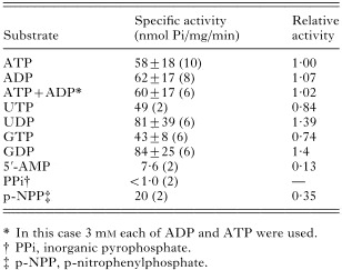
Table 1. Substrate specificity
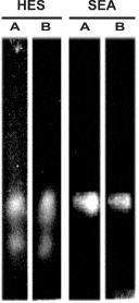
Fig. 1. Separation of ATP diphosphohydrolase by non-denaturing gels. Proteins from homogenized egg 2500 g supernatant (HES) or soluble egg antigen (SEA) were separated and lanes cut out from the gels were immersed in medium for activity measurements containing either ATP (A) or ADP (B) as substrate. After 12 h of incubation at 37 °C, white deposits of calcium phosphate appeared as a result of nucleotide hydrolysis catalysed by the enzyme. The gel was photographed against a dark background.
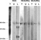
Fig. 2. Identification of proteins extracted from active bands on non-denaturing gels. Proteins from homogenized egg 2500 g supernatant (HES) or soluble egg antigen (SEA) were separated in non-denaturing gels (Fig. 1). Proteins (50 μg) from HES or SEA (lanes T) and electro-eluted samples of active upper (U) or lower (L) bands were separated by SDS–PAGE. For HES, the gel was stained with Coomassie blue (lanes T and U) or silver stain (lane L). For HES(Wb) and SEA(Wb), proteins were electroblotted onto nitrocellulose and developed with anti-potato apyrase antibodies (1[ratio ]1000).
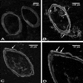
Fig. 3. Immunocytochemical localization of ATP diphosphohydrolase from Schistosoma mansoni egg. Fluorescence confocal microscopy images are shown. Anti-potato apyrase antibodies and secondary antibody coupled to TRITC were used for fluorescence detection of ATP diphosphohydrolase on cryostat sections (10 μm thick) of infected mouse liver. (B) General aspect of S. mansoni egg showing fluorescence homogeneously distributed on external surface of miracidium (arrow 1), in the region between the miracidium and the inner side of the egg-shell (arrow 2) and immediately outside and spreading away from the egg-shell (arrow 3). (C) Granular material entrapped by the surface microspines located outside the egg-shell (arrow) and in (D), the enlargement of a region of the egg-shell surface (arrows). In (A), sections were probed with secondary antibody alone.

Fig. 4. Antigenicity analysis of the Schistosoma mansoni ATP diphosphohydrolase. (A) Samples (1 μg) of purified potato apyrase (I) or pooled ATP diphosphohydrolase isoforms from S. mansoni eggs (II) were separated by SDS–PAGE, and stained with Coomassie blue (I, lane 1), or electroblotted onto nitrocellulose and developed with anti-potato apyrase antibodies raised in rabbit (I, lane 2; II, lane 1), or with serum from S. mansoni-infected Swiss mice (I, lane 3; II, lane 2). (B) Reactivity of S. mansoni-infected mice sera (dilution 1[ratio ]2000) against the potato apyrase protein was assayed by ELISA. The absorbance values of the sera minus the absorbance mean of control sera (A492=0·077) plus 2 standard deviations (S.D.=0·005) are represented.
 You have
Access
- 31
- Cited by