Introduction
Preimplantation embryonic development is a complex process that is regulated by both genetic and epigenetic factors. Gametes acquire specific epigenetic marks during gametogenesis and some of these parental marks are retained faithfully in the zygote, while others undergo reprogramming to make the zygote totipotent. During development, these marks are again reprogrammed to make the embryos fit for further differentiation. This epigenetic reprogramming includes the processes of DNA methylation and histone modifications. Histone modifications are involved in the regulation of gene expression and include methylation, acetylation, phosphorylation, ubiquitination, SUMOylation and citrullination. Methylation and acetylation are known to be involved in preimplantation embryonic development (Henery et al., Reference Henery, Miranda, Wiekowski, Wilmut and DePamphilis1995; Wiekowski et al., Reference Wiekowski, Miranda, Nothias and DePamphilis1997; Reik et al., Reference Reik, Dean and Walter2001; Li Reference Li2002) and are catalyzed by enzymes such as histone methyltransferases (HMTs), histone deacetylases (HDACs), acetylases, and demethylases.
Peptidylarginine deiminases (PADs) are a family of calcium-dependant enzymes that convert protein-bound positively charged arginine residues into neutral citrulline residues (Vossenaar et al., Reference Vossenaar, Zendman, Venrooij and Pruijn2003) in a type of post-translational modification called citrullination. The loss of the positive charge results in increased hydrophobicity of the protein and leads to its unfolding (Tarcsa et al., Reference Tarcsa, Marekov, Mei, Melino, Lee and Steinert1996), making it vulnerable to modifications. This protein modification by peptidylarginine deiminases changes the functional properties of the target proteins and therefore has an important role in development and cell differentiation (Senshu et al., Reference Senshu, Kan, Ogawa, Manabe and Asaga1996; Nakashima et al., Reference Nakashima, Hagiwara, Ishigami, Nagata, Asaga, Kuramoto, Senshu and Yamada1999). So far, five isoforms of human, mouse and rat PAD have been isolated and characterized, namely PAD1, PAD2, PAD3, PAD4/5 and ePAD/PAD6 (Nakashima et al., Reference Nakashima, Hagiwara, Ishigami, Nagata, Asaga, Kuramoto, Senshu and Yamada1999; Kanno et al., Reference Kanno, Kawada, Yamamouchi, Yosida-Noro, Yoshiki, Shiraiwa, Kusakabe, Manabe, Tezuka and Takahara2000; Ishigami et al., Reference Ishigami, Ohsawa, Asaga, Akiyama, Kuramoto and Maruyama2002; Guerrin et al., Reference Guerrin, Ishigami, Méchin, Nachat, Valmary, Sebbag, Simon, Senshu and Serre2003; Chavanas et al., Reference Chavanas, Mechin, Takahara, Kawada, Nachat, Serre and Simon2004). PADs are cytosolic proteins except for PAD4, which is the only nuclear isoform with a nuclear localization sequence (NLS) (56-PPAKKKST-63). PAD4 targets histones (H2, H3 and H4) and nucleophosmin/B23 (Hagiwara et al., Reference Hagiwara, Nakashima, Hirano, Senshu and Yamada2002; Nakashima et al., Reference Nakashima, Hagiwara and Yamada2002, Cuthbert et al., Reference Cuthbert, Daujat, Snowden, Erdjument-Bromage, Hagiwara, Yamada, Schneider, Gregory, Tempst, Bannister and Kouzarides2004; Sarmento et al., Reference Sarmento, Digilio, Wang, Perlin, Herr, Allis and Coonrod2004; Wang et al., Reference Wang, Wysocka, Sayegh, Lee, Perlin, Leonelli, Sonbuchner, McDonald, Cook, Dou, Roeder, Clarke, Stallcup, Allis and Coonrod2004). It has been shown that modifications mediated by PAD4 are involved in regulation of chromatin structure and gene expression in a variety of cell types (Cuthbert et al., Reference Cuthbert, Daujat, Snowden, Erdjument-Bromage, Hagiwara, Yamada, Schneider, Gregory, Tempst, Bannister and Kouzarides2004; Sarmento et al., Reference Sarmento, Digilio, Wang, Perlin, Herr, Allis and Coonrod2004; Wang et al., Reference Wang, Wysocka, Sayegh, Lee, Perlin, Leonelli, Sonbuchner, McDonald, Cook, Dou, Roeder, Clarke, Stallcup, Allis and Coonrod2004; Raijmakers et al., Reference Raijmakers, Zendman, Egberts, Vossenaar, Raats, Soede-Huijbregts, Rutjes, van Veelen, Drijfhout and Pruijn2007).
Thus, revelation of the involvement of PAD4 in the regulation of gene expression, which is an active component of embryonic development, prompted us to study localization and expression of the nuclear PAD isoform (PAD4) and subsequent citrullinated nuclear proteins in porcine and mouse oocytes and parthenogenetic or in vitro fertilized (IVF) embryos, as an initial step in the understanding of PAD4's role in mammalian embryonic development. Parthenogenetic embryos in pig were used as it was technically difficult to obtain the large numbers of IVF/intracytoplasmic sperm injection (ICSI) embryos required for this study, owing to the low success rate of IVF (due to high incidence of polyspermy) and ICSI in the pig. It has also been reported that the development of parthenogenetic embryos is similar to that of in vivo embryos (at least up to the blastocyst stage) in terms of development rate, timing of compaction and number of blastomeres formed (Kure-bayashi et al., Reference Kure-bayashi, Miyake, Katayama, Miyano and Kato1996, Reference Kure-bayashi, Miyake, Okada and Kato2000; Thuan et al., Reference Thuan, Harayama and Miyake2002).
Materials and methods
This study was approved by the Institutional Animal Care and Use Committee (Permission number: 19-5–30, 19-5–31, 22-05–13 and 22-05–15) and carried out in accordance with the Kobe University Animal Experimentation Regulations.
In vitro maturation (IVM) of pig oocytes
Prepubertal gilt (Sus scrofa domestica) ovaries were collected from a local abattoir and transported to the laboratory within 2 h. The ovaries were washed once with cationic detergent, 0.2% (w/v) cetyltrimethyl ammonium bromide (CETAB) followed by thorough washing with Ca2+-free Dulbecco's phosphate-buffered saline (PBS) that contained 0.1% (w/v) polyvinyl alcohol (PVA; Sigma-Aldrich Chemical Co., St. Louis, MO, USA). Healthy antral follicles sized between 4 to 6 mm were collected according to the technique described by Moore and Trounson (Reference Moore and Trounson1977). Oocyte–cumulus–granulosa cell complexes (OCGCs) were separated intact from the inner surface of these follicles and cultured to maturation in 2 ml TCM-199 (Earl's salt; Nissui Pharmaceutical Co. Ltd, Tokyo, Japan) supplemented with 10% (v/v) heat-inactivated fetal calf serum (FCS; ICN Biomedicals, Inc., Aurora, Ohio, USA), 0.1 mg/ml sodium pyruvate, 0.08 mg/ml kanamycin sulphate (Sigma-Aldrich) and 0.1 IU/ml hMG (Pergonal; Teikokuzoki, Tokyo, Japan) along with two thecal shells freed from follicular fluid and granulosa cells (Mattioli et al., Reference Mattioli, Galeati, Bacci and Seren1988). Culture was carried out in a CO2 incubator at 38.5 °C under 5% CO2 in humidified air for 42–48 h with gentle agitation. Oocytes were collected at various stages such as at the germinal vesicle (GV) stage (0 and 21 h), early metaphase I (MI, 27 h), anaphase I (AI, 32 h) and metaphase II (MII, 42–48 h) for epifluorescence microscopy as well as for western blot analysis.
Following denudation of pig MII oocytes, cumulus cells were collected, washed with PBS-PVA and used for both immunostaining and western blot analysis.
Production of parthenogenetic embryos
Mature pig oocytes after 42–48 h maturation culture were briefly exposed to 0.01% (w/v) hyaluronidase (Sigma-Aldrich) and gently pipetted to remove the cumulus cells. Denuded oocytes with first polar bodies in groups of 20 were selected and washed thoroughly in a field solution composed of 0.3 mM mannitol, 0.1 mM MgSO4 and 0.05 mM CaCl2 (Zimmermann and Viemken, Reference Zimmermann and Viemken1982) supplemented with 0.01% (w/v) PVA before placing them in 100 μl of field solution between two parallel stainless steel electrodes in an activation chamber (FTC-03; Shimadzu Co., Ltd., Kyoto, Japan). A single direct current pulse of 1500 V/cm for 100 μs was supplied from an electro cell fusion device (LF101; Bex Co., Tokyo, Japan). The oocytes were then washed thoroughly in equilibrated porcine zygote medium (PZM3; Yoshioka et al., Reference Yoshioka, Suzuki, Tanaka, Anas and Iwamura2002) supplemented with 3 mg/ml bovine serum albumin (BSA; Wako Pure Chemical Industries, Osaka, Japan) before placing them in cytochalasin B (CB; Sigma-Aldrich) supplemented (5 mg/ml) PZM3 medium at 38.5 °C in humidified incubator with 5% CO2 in air for 4 h. Then the activated oocytes were washed and placed in fresh equilibrated PZM3 drops for further culture. In vitro cultured embryos were collected at various stages of development such as 2-cell, early 4-cell, morula and blastocyst stages at 24, 48, 96, and 120 h after electro-stimulation, respectively, for immunostaining and western blot analysis.
Mouse sample collection
ICR mice were superovulated with 5 IU/ml eCG and 5 IU/ml hCG injection at an interval of 48 h. Oviducts were collected 16–18 h post hCG and MII oocytes were released into HEPES-buffered CZB medium (Chatot et al., Reference Chatot, Ziomek, Bavister, Lewis and Torres1989) that contained 0.1% hyaluronidase. Cumulus cells were removed by gentle pipetting. A group of MII oocytes were used for indirect immunocytological study and western blot analysis.
For IVF embryos, MII oocytes were incubated with capacitated sperm (5 × 10⁶ sperm/ml) in human tubal fluid (HTF) medium (Quinn et al., Reference Quinn, Kerin and Warnes1985). After 4 h, extrusion of the second polar body was confirmed, washed thoroughly and in vitro cultured up to the blastocyst stage. IVF embryos were collected at 2-cell (D1), 4-cell (D2), morula (D3) and blastocyst (D4–D5) stages and subjected to indirect immunofluorescence study for both PAD4 and modified citrulline residue detection after removing the zona by treating the embryos with acid Tyrode's solution (pH 2.5) for 30 s.
For immunostaining analysis, cumulus cells were used after denudation of MII oocytes.
The caudae epididymes from retired breed ICR male mice were cut and sperm was released into equilibrated HTF medium and kept in the incubator for capacitation for 1 h.
Indirect immunofluorescence staining
The pig oocytes and embryos collected at various stages of preimplantation development were washed with PBS-PVA followed by fixation in 2% paraformaldehyde supplemented with 0.2% Triton X-100 (Nacalai Tesque, Inc., Kyoto, Japan) for 20 min. The fixed oocytes/embryos were washed thoroughly twice in PBS-PVA for 5 min each and then stored in 1% BSA supplemented PBS-PVA at 4 °C until use. They were blocked with 10% goat serum in 1% BSA-PBS-PVA for 1 h before incubation with primary antibodies at 4 °C overnight. Primary antibodies used were: 10 μg/ml rabbit polyclonal anti-human PAD4 antibody (AP1072C, Abgent Inc, San Diego, CA, USA), mouse anti-B23 (1:100; Sigma-Aldrich), and rabbit anti-H3–9712 (1:50; Cell Signaling Technology Inc, Danvers, MA, USA). The next day the samples were washed thoroughly with PBS-PVA and incubated with the corresponding secondary antibodies for 1 h at room temperature (4 μg/ml: Alexa Fluor 488 donkey anti-rabbit IgG, or Alexa Fluor 488 donkey anti-mouse IgG; Invitrogen, Carlsbad, CA, USA). After washing three times with PBS-PVA for 5 min each, DNA was stained with 200 μg/ml propidium iodide (PI; Sigma-Aldrich) or 2 μg/ml 4′,6-diamidino-2-phenylindole (DAPI) (Molecular Probes Inc, Eugene, OR, USA). The oocytes and embryos were washed again before being mounted on vectashield medium (Vector Laboratories, Burlingame, CA, USA) and observed under a fluorescence microscope (BX51-Fl; Olympus Optical Company Ltd., Tokyo, Japan) at ultraviolet (UV) for DAPI staining and for others with appropriate excitation. Control labelling was done using the same protocol but without the primary antibody. As it was difficult to remove the zonae before immunostaining, the zonae of the pig embryos were made to rupture by pressing down on the coverslips to expose the eggs/embryos to confirm that the signals observed were not from the zonae. To check antibody specificity an appropriate concentration (1 μg/ml) of binding peptide (BP, ap1072c) was mixed with anti-PAD4 antibody (ap1072c) during incubation.
The pig cumulus cells were spread on slides and cultured for appropriate periods. The rest of the procedure was the same as that described above. Mouse MII oocytes, IVF embryos and cumulus cells were treated in the same way as for pigs as described above.
Sodium dodecyl sulphate polyacrylamide gel electrophoresis (SDS-PAGE) and western blotting
Pig oocytes and embryos collected at various stages of development [oocytes: each stage = μ50, 2-cell (n = 50), 4-cell (n = 35), morula (n = 35), blastocyst (n = 20)] were placed in a minimum volume of PBS-PVA and stored at –80 °C until use. Before use, the samples were lysed in an equal amount of 2× sample buffer that contained 10% gycerol, 2% SDS, 80 mM Tris–HCl (pH 6.8) and 0.02% bromophenol blue and boiled for 5 min. Electrophoresis was done on 12.5% minigel SDS-PAGE (AE 6530, Atto Bioscience and Technology, Tokyo, Japan) and the proteins were transferred to polyvinylidene difluoride (PVDF) membranes (Immobilon P, Millipore, Bedford, MA, USA) on a semidry transfer cell (Trans-Blot SD, BioRad Laboratories, Hercules, CA, USA) for 1 h. Membranes were either cut into two, to process two different antibodies or the same membrane was reprobed after probing for the first antibody. The membranes were blocked for 1 h at room temperature with 5% FCS or fat-free milk in 0.1% Tween-20 in PBS (PBST) with gentle agitation and incubated overnight at 4 °C with a dilution of primary antibodies in the blocking solution. The primary antibodies used were: 0.4 μg/ml rabbit polyclonal anti-human PAD4 (ab38772; Abcam, Cambridge, MA, USA) or 1:10000 mouse monoclonal anti-β actin antibody (A2228; Sigma-Aldrich). Subsequently, membranes were washed in PBST and incubated with their respective horseradish peroxidase (HRP)-linked secondary antibodies (1:20,000 anti-rabbit IgG, HRP- linked whole antibody from donkey; GE Healthcare UK Ltd, Buckinghamshire, UK or 1:20,000 goat anti-mouse HRP-linked IgG; Pierce, ThermoFischer Scientific inc., Rockford, IL, USA) for 2 h at room temperature. Membranes were washed three times with PBST and the HRP activity was detected using ECL SuperSignal substrate (Pierce, ThermoFisher Scientific Inc., Rockford, IL, USA). As a positive control for PAD4, human carcinoma cell line lysate (A431) was used. The concentration of cumulus cells used was 400 × 106 cells/ml.
Analysis of citrullinated proteins
Citrullinated nuclear proteins were analyzed by indirect immunofluorescence study using an anti-citrulline (modified) detection kit (Millipore, Billeria, MA, USA). The samples were fixed with 2% paraformaldehyde for 20 min followed by permeabilization with 0.2% Triton X-100 for 5 min. After washing with PBS-PVA twice for 5 min each, the samples were transferred into 1% BSA for 15 min. Blocking was performed with 10% goat serum-supplemented 1% BSA for 1 h. Then the samples were chemically treated in equal amounts of solutions A and B provided in the kit at 37 °C on a heating plate in the dark for 30 min to modify the citrulline residues. Following modification, the samples were washed thoroughly with PBS-PVA and then incubated with anti-citrulline (modified) antibody in 1:500 dilution overnight. The rest of the procedure was the same as described earlier (see Indirect immunofluorescence staining).
For the western blotting analysis, initially a similar number of oocytes/embryos to that used in PAD4 detection (GV/M2 = 50, 2-cell = 25, 4-cell = 25, morula/blastocyst = 15, cumulus = 100 × 106 cells/ml, sperm = 100 × 106 sperm/ml) was run on 12.5% minigel. Later on, higher concentrations of cumulus (400 × 106 cells/ml) and higher numbers of blastocysts (n = 100) were used. The antibodies used were primary antibody – 2μg/ml anti-citrullinated H3 antibody (ab5103, Abcam), and secondary antibody – 1:20,000 anti-rabbit IgG, HRP-linked whole antibody from donkey. For detection of citrullinated proteins using anti-modified citrullinated proteins antibody (1:1000; anti-modified citrulline detection kit, Millipore, Billeria, MA, USA) the membranes were treated with the modification reagent for 3 h in the dark at 37 °C before blocking. The rest of the procedure was the same as that described earlier in sodium dodecyl sulphate polyacrylamide gel electrophoresis (SDS-PAGE) and western blotting.
Each immunofluorescence experiment was repeated at least three times, with a minimum of five samples in each repeat; western blot analysis was carried out at least twice.
Results
PAD4 expression and localization in pig oocytes
To determine the expression of PAD4 in mammalian oocytes, in vitro-matured pig oocytes collected at different stages of maturation were subjected to western blot analysis. PAD4 was recognized as a 67 kDa protein in all the oocytes stages (Fig. 1A) along with the positive control, human epithelial carcinoma cell (A431) lysate (lane1). Indirect immunofluorescence microscopy revealed fluorescent signals in the cytoplasm of GV oocytes (Fig. 1B: a2), whereas in the MI and MII oocytes, anti-PAD4 labelling was detected in the cytoplasm (Fig. 1B: c2, d2) as well as around the metaphase chromosomes (arrowhead in Fig. 1B: c2, d2, inset in d2). Higher magnification revealed that PAD4 was localized along the metaphase spindle (Fig. 1B: inset in d2). No PAD4 signal was detected in the first polar body (white arrow in Fig. 1B: inset in d2). Nuclear PAD4 was not observed in the GV at the initial stage, but was seen localized around the spindle in MI and MII oocytes. To check the time of PAD4 nuclear translocation, GV oocytes were subjected to PAD4 labelling after 18 h and 21 h of maturation culture, before GV breakdown (GVBD). GV oocytes that were nearing GVBD (21 h) expressed PAD4 in the nucleoplasm excluding the nucleoli (Fig. 1B: b2). Negative control oocytes treated with only the secondary antibody showed faint background signals (Fig. 1B: e2) and when the antibody specificity was checked using blocking peptide along with the anti-PAD4 antibody there was reduction in the PAD4 labelling (Fig. 1B: f2) and the disappearance of the spindle-localized signal (arrowhead in Fig. 1B: f2).
Localization and expression of peptidylarginine deiminase 4 (PAD4) in pig oocytes. (A) Western blot analysis of PAD4 in pig oocytes. A431: positive control, AI: anaphase I, GV: germinal vesicle, MI: metaphase I, MII: metaphase II (n = 50). PAD4 was expressed in all oocytes from GV to MII. (B) Immunofluorescent localization of PAD4 in pig oocytes. a: GV (0 h), b: GV (21 h), c: MI (27 h), d: MII (44 h), e: MII oocyte as negative control treated only with secondary antibody, f: MII oocyte treated with anti-PAD4 antibody and its binding peptide. PI stained chromatin (a1–f1) and Alexa fluor 488 labelled PAD4 (a2–f2). Uniform cytoplasmic PAD4 labelling was seen in all the stages and nuclear signal was seen in the late GV (b2) stage and spindle-localized signals were seen in the metaphase oocytes, arrowhead in c2,d2 and inset in d2. Spindle-specific signal disappeared in binding peptide-treated MII oocytes in f2 (arrowhead). Zona pellucida was ruptured (V shaped) in the samples to expose the oocytes. Scale bar: 20 μm. A colour version of this figure is available online.

PAD4 detection in pig parthenogenetic preimplantation embryos
Embryos collected at different stages of development such as 2-cell at 24 h, early 4-cell at 48 h, morula at 98 h and blastocyst at 120 h after electro-stimulation were subjected to indirect immunofluorescence microscopy and western blotting. In western blot analysis, PAD4 was detected as a single band of 67 kDa throughout the preimplantation embryonic stages (Fig. 2A).
Localization and expression of peptidylarginine deiminase 4 (PAD4) in pig parthenogenetic embryos. (A) Western blot analysis showing PAD4 expression in parthenogenetic embryos. 2C: 2-cell (n = 50), 4C: early 4-cell (n = 35), Mor: morula (n = 35), Bl: blastocyst (n = 20), Cum: cumulus (400 × 106 cells/ml). (B) Immunofluorescence localization of PAD4 in pig parthenogenetic diploid embryos. a: 2-cell (24 h), b: early 4-cell (48 h), c: morula (96 h), d: blastocyst (120 h), e: negative control (blastocyst) treated with only secondary antibody, f: morula treated with anti-PAD4 and binding peptide. Note the partial nuclear translocation of PAD4 in blastocysts (d2). PAD4 signal from the cytoplasm decreased in the binding peptide treated control (f2). Zona pellucida was ruptured (V shaped) in the samples to show that the signals are not from the zona. (C) Immunofluorescence localization of PAD4 in pig cumulus cells (g) localizing in the cytoplasm and nucleus. DAPI (a1–c1, e1, f1) or PI (d1, g1) stained the chromatin and Alexa fluor 488 labelled PAD4 (a2–g2). Scale bar: 20 μm. A colour version of this figure is available online.
In the indirect immunological study it was observed that PAD4 was found predominantly in the cytoplasm until the morula stage (Fig. 2B: a2–c2). However, at the blastocyst stage, PAD4 was again found to be partially translocated into or around the nucleus (Fig. 2B: d2). Control blastocyst embryo treated with only the secondary antibody showed weak cytoplasmic background (Fig. 2B: e2) and when the antibody specificity was checked using blocking peptide along with the anti-PAD4 antibody, there was significant reduction in the green fluorescence for PAD4 labelling (Fig. 2B: f2). Cumulus cells showed PAD4 present in the cytoplasm and the nucleus (Fig. 2C: g2) and expression was confirmed in western blot analysis (Fig. 2A).
Localization of nucleophosmin/B23 and histone H3 in pig oocytes and parthenogenetic embryos
B23 signals varied in their localization from stage to stage. In GV oocytes, B23 was found in the nucleoplasm with a clear ring-shaped expression on and around the chromatin (Fig. 3: a2, a3). After GVBD, B23 was found primarily in the cytoplasm in MII oocytes (Fig. 3: b2, b3). B23 was mainly found in the cytoplasm at the 2-cell stage (Fig. 3: c2, c3), but at later stages such as early 4-cell (Fig. 3: d2, d3), morula (Fig. 3: e2, e3) and blastocyst (Fig. 3: f2, f3) it was localized in the nuclei, and the nucleolar localization was observed in the morula and in the blastocyst (Fig. 3: e, f), but not in 4-cell embryos. The B23 signal was absent in metaphase cells (arrowhead in Fig. 3: e2).
Localization of nucleophosmin (B23) in pig oocytes and parthenogenetic embryos. a: Germinal vesicle (GV) oocyte, b: metaphase II, c: 2-cell (24 h), d: early 4-cell (48 h), e: morula (96 h), f: blastocyst (120 h after electro-stimulation). B23 was localized mainly in the cytoplasm and the nucleolar translocation was noticed only after the 4-cell stage, when there is formation of nucleoli. Arrowhead in e2 indicates absence of B23 signal in the metaphase cell. (a3–f3) Enlarged version of boxed area in (a2–f2). DAPI stained chromatin (a1–f1) and Alexa fluor 488 labelled B23 a2,a3–f2,f3). Scale bar: 20 μm. A colour version of this figure is available online.
Histone H3 signals were found universally in granular form both on the condensed chromatin of oocytes (Fig. 4: a, b) and on decondensed chromatin of the interphase embryonic cells at morula and blastocyst stages (Fig. 4: e, f). However, H3 detection varied in 2-cell and 4-cell embryos. In fact 84% (21/25) of the 2-cell embryos and 65% (13/20) of the 4-cell embryos had H3 signals. Among the H3 detected embryos, 28% (7/21) of the 2-cell embryos and 20% (4/20) of the 4-cell embryos showed partial and asymmetrical localization. Thus some cells were devoid of H3 signals (arrowhead in Fig. 4: c2). Compared with morula, several blastomeres did not show the presence of the H3 signals. In all other stages, such as GV, MII and blastocysts, signals were observed in all the cells.
Localization of histone H3 in pig oocytes and embryos. a: Germinal vesicle (GV) oocyte, b: metaphase II (MII) oocyte, c: 2-cell (24 h), d: early 4-cell (48 h), e: morula (96 h), f: blastocyst (120 h after electro-stimulation). Histone H3 signals were seen in granular form in the chromatin of all the stages except the 2-cell and early 4-cell where the localization was asymmetric. Arrowhead in c2 indicates absence of the signal. (a3–f3) Enlarged version of boxed area in (a2–f2). DAPI stained chromatin (a1–f1) and Alexa fluor 488 labelled Histone H3 (a2,a3–f2,f3). Scale bar: 20 μm. A colour version of this figure is available online.
Localization of citrullinated proteins in pig oocytes and parthenogenetic embryos
Following confirmation of the expression of PAD4, the oocytes and embryos were checked for citrullinated proteins. As anti-citrullinated histone H3 antibody did not work for immunocytology in the pig, the anti-modified citrulline antibody that would detect all the citrullinated proteins was used for the detection of citrullination in pig oocytes and parthenogenetic embryos. Samples were modified chemically before incubation with the anti-modified citrulline antibody. Modified citrulline labelling was detected faintly in the cytoplasm in all the oocytes and embryos (Fig. 5). However, on the chromatin, right from the GV stage oocyte (Fig. 5: a) up to the blastocyst stage (Fig. 5: f) signals were in strong granular form, similar to histone H3 localization kinetics. Thus, cells of some 2-cell and 4-cell embryos showed varied localization and strength pattern, and showed asymmetrical signals in the 2-cell embryos (arrowhead in Fig. 5: c2 indicates a signal-negative cell). Condensed chromatin that surrounded the nucleolus of GV (Fig. 5: a2), MII chromosomes (Fig. 5: b2) and polar body chromatin (PB in Fig. 5: b2, c2, d2) exhibited intense signals. The negative control, in which morulae were treated with only secondary antibody after chemical modification, did not show any labelling (Fig. 5: g2). Pig cumulus cells showed similar granular citrullinated signals on their chromatin (Fig. 5: h2) with anti-modified citrulline antibody treatment after chemical modification.
Localization of citrullinated proteins in pig oocytes and parthenogenetic embryos. Samples were chemically modified before incubation with anti-modified citrulline antibody. a: Germinal vesicle (GV) oocyte, b: metaphase II (MII) oocyte, c: 2-cell (24 h), d: early 4-cell (48 h), e: morula (96 h), f: blastocyst (120 h after electro-stimulation), g: negative control, where morulae were chemically modified and treated with only secondary antibody, h: cumulus cell labelled with anti-modified citrullinated protein antibody after modification. PI stained chromatin (a1–h1) and Alexa fluor 488-labelled modified citrullinated proteins (a2–h2, a3–f3). Citrullinated signals were granular, similar to that of histone H3 signals. It followed the localization pattern of histone H3 with asymmetric signals in 2-cell and 4-cell stages. Arrowhead in c2 indicates absence of citrullinated protein signal. (a3–f3) Enlarged version of boxed area in (a2–f2). PB, polar body. Scale bar: 20 μm. A colour version of this figure is available online.
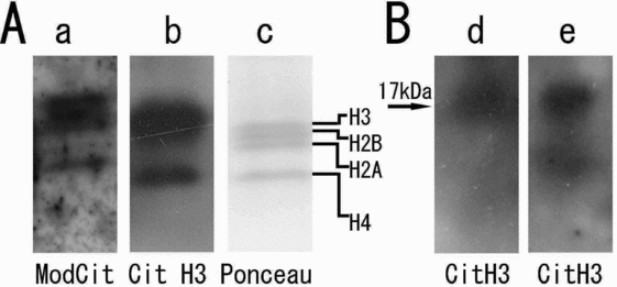
Cumulus cells subjected to western blotting with anti-modified citrulline antibody detected bands for histone H2A, H2B, H3 and H4 (Fig. 6A: a), which were later substantiated by re-probing the same membrane with anti-citrullinated H3 antibody (Fig. 6A: b). Ponceau staining of the membrane (Fig. 6A: c) was used to show protein blotting. Blastocysts that exhibited the strongest signals for modified citrulline in their nuclei were subjected to western blot analysis using anti-citrullinated histone H3 antibody; a moderate band for histone H3 at 17 kDa was detected (Fig. 6B: d) in parallel with the cumulus sample (Fig. 6B: e).
Western blot analysis of citrullinated proteins in the pig. (A) Expression of citrullinated histones in cumulus cells detected by modified citrulline antibody (a); by anti-citrullinated histone H3 antibody (citrullinated at R2, R8 and R17) (b); and Ponceau stain (c). Detection of citrullinated histones was confirmed in this section. (B) Detection of citrullinated proteins by anti-citrullinated histone H3 antibody in blastocysts (d: n = 100) and in cumulus cells (e). The citrullinated histone residue was compared with the citrullinated histones of cumulus cells in this section.
Localization of PAD4 and citrullinated residues in mouse oocytes and IVF preimplantation embryos
To confirm the commonality of the PAD4 localization in mammals, mouse MII oocytes and IVF embryos were checked for PAD4 localization by immunocytological study. MII oocytes showed cytoplasmic and the spindle colocalization (Fig. 7A: a2).
Localization of peptidylarginine deiminase 4 (PAD4) and citrullinated residues in mouse oocytes and IVF embryos. Immunolocalization of PAD4 (A) in mouse MII oocytes and IVF embryos. a: metaphase II (MII), b: 2-cell (Day 1), c: 4-cell (Day 2), d: morula (Day 3), e: blastocyst (Day 4–5). The PAD4 localization was fundamentally similar to that of pig. PAD4 was cytoplasmic and spindle colocalized in MII oocytes (a2). In in vitro fertilized (IVF) embryos PAD4 was gradually translocated into the nuclei from 2-cell onwards, concentrating around the nucleoli. (B) Localization of modified citrulline residues in mouse MII oocytes and IVF embryos detected using anti-modified citrulline antibody. a: MII, b: 2-cell (Day 1), c: 4-cell (Day 2), d: morula (Day 3), e: blastocyst (Day 4–5). Chromatin was stained with PI (a1–e1). Modified citrulline residues were stained by Alexa Fluor 488 (a2–e2). Citrullinated protein signals were granular and steadily increased from early stages to the late. (C) Two-cell embryo showing reduced citrullinated protein signal. PI stained chromatin (a1) and Alexa fluor 488 stained modified citrulline residues (a2, a3). Scale bar: 20 μm. A colour version of this figure is available online.
Along with the cytoplasmic PAD4, mouse IVF embryos showed gradual translocation of PAD4 into the nuclei from the 2-cell to the blastocyst (Fig. 7A: b2-e2), with the blastocyst showing the strongest nuclear localization (Fig. 7B: e2). Nuclear localization was found to be concentrated around the nucleoli. Oocytes and IVF preimplantation embryos subjected to detection of modified citrulline residues showed a granular pattern similar to that of pig. Mouse MII oocytes had similar modified citrulline signals on their metaphase chromosome (Fig. 7B: a2). At the 2-cell stage, the signals varied as in the case of pig. Some embryos showed strong signals (Fig. 7B: b2) whereas some showed reduced signals (Fig. 7C: a2, a3). Overall the signals increased in intensity from early (Fig. 7B: b2) to the late (Fig. 7B: e2) stages, with the blastocyst exhibiting the strongest signals, findings similar to that of pig.
Discussion
The objective of this study was to analyze the expression and localization of the enzyme PAD4, that plays a role in gene regulation in mammalian oocytes and embryos, as an initial step in understanding its role, if any, in mammalian preimplantation embryonic development. PADs are known to be mostly localised in the cytoplasm. But PAD4, which has a nuclear localization signal, is found in the nucleus (Vossenaar et al., Reference Vossenaar, Zendman, Venrooij and Pruijn2003). Studies have shown that in oligodendrocytes, PAD4 is primarily cytosolic and that tumour necrosis factor (TNF) induced its nuclear translocation (Mastronardi et al., Reference Mastronardi, Wood, Mei, Raijmakers, Tseveleki, Dosch, Probert, Casaccia-Bonnefil and Moscarello2006). In concurrence with this report, the present study revealed that, in mammalian oocytes and embryos, PAD4 is predominantly cytoplasmic and that translocation happens only during specific stages of development, such as prior to GVBD during oocyte maturation when there is re-initiation of meiosis – although the involvement of any specific factor in inducing this translocation is unknown. After GVBD, PAD4 was found to be translocated into the nuclei and localized around both the MI and MII chromosomes.
The natural substrates of PADs include structural proteins, while the targets of PAD4 activity are histones (H3, H2A and H4) and nucleophosmin (Cuthbert et al., Reference Cuthbert, Daujat, Snowden, Erdjument-Bromage, Hagiwara, Yamada, Schneider, Gregory, Tempst, Bannister and Kouzarides2004; Hagiwara et al., Reference Hagiwara, Nakashima, Hirano, Senshu and Yamada2002; Nakashima et al., Reference Nakashima, Hagiwara and Yamada2002; Sarmento et al., Reference Sarmento, Digilio, Wang, Perlin, Herr, Allis and Coonrod2004; Wang et al., Reference Wang, Wysocka, Sayegh, Lee, Perlin, Leonelli, Sonbuchner, McDonald, Cook, Dou, Roeder, Clarke, Stallcup, Allis and Coonrod2004). Nucleophosmin is a multifunctional nucleolar phosphoprotein that acts as a chaperone and is involved in ribosome biogenesis and centrosome duplication (Spector et al., Reference Spector, Ochs and Busch1984; Szebeni & Olson, Reference Szebeni and Olson1999). It is known to shuttle between the cytoplasm and the nucleus (Borer et al., Reference Borer, Lehner, Eppenberger and Nigg1989). B23 is known to be in cytoplasm when the nucleoli disassemble at late prophase and is translocated back into the nucleoli at the reassembly of the nucleoli, after telophase (Scheer et al., Reference Scheer, Thiry and Goessens1993). In contrast, histones are the nuclear proteins around which DNA wraps to form nucleosomes in the chromatin (Hansen. Reference Hansen2002). In mouse it has been shown that there is variation in the synthesis of histones during the early embryonic developmental stages (Wiekowski et al., Reference Wiekowski, Miranda, Nothias and DePamphilis1997).
Before focusing on the substrates of PAD4, the localization of B23 and histone H3 were examined in pig oocytes and parthenogenetic embryos by indirect immunological analysis. Svarcova et al. (Reference Svarcova, Dinnyes, Polgar, Bodo, Adorjan, Meng and Maddox-Hyttel2009) have shown the localization of B23 in the nuclei and nucleolar entities from late 2-cell stage (after zygotic genome activation (ZGA)) in the mouse. Similar to this finding, the present results revealed that nucleophosmin localization varied depending on the embryo cell cycle stage and the nucleolar translocation was seen after ZGA in the pig, that is after the 4-cell stage. In the GV it was primarily nuclear, whereas at the metaphase stage it was only cytoplasmic. In the early cleaving stage embryos, it was seen to be both cytoplasmic and nuclear, but in the later preimplantation stages, it was predominantly in the nuclei with nucleolar translocation in those cells that are probably transcriptionally active. This finding is in agreement with an earlier report that the nucleolar translocation of B23 is dependent on the active rRNA transcription, localizing to nucleoli during interphase when ribosomes are actively synthesized (Yung et al., Reference Yung, Busch and Chan1985) and the study of B23 in porcine in vivo embryos by Hyttel et al. (Reference Hyttel, Laurincik, Rosenkranz, Rath, Niemann, Ochs and Schellander2000). In the present study histone H3 signals were found in a homogenously granular form on the chromatin of all the oocytes and embryos, except in earlier stages of embryos in which the histone H3 signals and distribution varied, being asymmetric and partial. This factor may be explained on the basis of the work of Wiekowski et al. (Reference Wiekowski, Miranda, Nothias and DePamphilis1997) who reported the changes in the synthesis of histones at the beginning of mouse development. They showed that at the 1-cell stage, the amount of nascent histone H3 and H4 was greater than the H1 and H2, whereas at the 2-cell stage, it was vice versa. Therefore, the absence or asymmetric detection of histone H3 signals in some of the 2-cell and 4-cell embryos may be due to an immunocytologically undetectable amount of H3 prior to or around zygotic gene expression in pig embryos. Our immunological signals of modified citrullinated residues were comparable with the histone H3 localization that were seen as granules on the condensed chromatin of oocytes and decondensed chromatin of embryonic nuclei. Discrepancies noticed in the modified citrulline signals of early embryos such as 2-cell or 4-cell embryos were in concurrence with the asymmetric histone H3 localization pattern that was observed in the 2-cell and 4-cell blastomeres and/or embryos in this study. These results suggest that histone is the target of PAD4, which was later substantiated with the western blot analysis results that detected both citrullinated H3 residues (R2, R8 and R17) and modified citrulline residues in this study. This situation leads to the assumption that PAD4 mainly targets histones in the pig oocytes and embryos. As the anti-citrullinated H3 antibody (R2, R8, R17) did not work for immunocytological analysis in the pig, anti-modified citrulline antibody that would detect all the citrullinated peptides was used. The results of immunofluorescence microscopy revealed modified citrulline signals both in the cytoplasm as well as in the nuclei and including oocyte chromatin. Results of western blot analysis revealed a band of around 55 kDa in all the oocyte and embryo stages except in somatic cells (cumulus cells) and in sperm (data not shown). We presumed that this band as well as the cytoplasmic signals seen in immunocytological analysis could be that of the citrullinated cytoplasmic sheets that are unique to mammalian oocytes and embryos. PAD6 is an oocyte and early embryo-abundant PAD isoform that localizes in the cortical granules and targets egg cytoplasmic sheets (Wright et al., Reference Wright, Bolling, Calvert, Sarmento, Berkeley, Shea, Hao, Jayes, Bush, Shetty, Shore, Reddi, Tung, Samy, Allietta, Sherman, Herr and Coonrod2003). These cytoplasmic sheets are composed of keratins K5, 6, 8, 16 and Z (Gallicano et al., Reference Gallicano, Larabell, McGaughey and Capco1994) that vary from 52 to 67 kDa in size.
It has been shown that citrullination results in protein unfolding (Tarcsa et al., Reference Tarcsa, Marekov, Mei, Melino, Lee and Steinert1996) and that hypercitrullination of histone H3 by PAD4 mediates chromatin decondensation in neutrophils (Wang et al., Reference Wang, Li, Stadler, Correll, Li, Wang, Hayama, Leonelli, Han, Grigoryev, Allis and Coonrod2009). Mammalian blastocyst formation involves processes such as cell proliferation, differentiation, as well as cell death. Trophectoderm differentiation is related directly to compaction, as this cell layer is derived from the polarized outer cells of the compacted morula (Watson et al., Reference Watson, Kidder and Schultz1992; Reference Watson, Westhusin, De Sousa, Betts and Barcroft1999). Initiation and regulation of the process of cavitation is brought about by the gene products. This factor illustrates the immense transcriptional activity that happens around the blastocyst stage. This situation may account for the nuclear translocation of PAD4 at the blastocyst stage showing strongest citrullinated histone signals. Thus, the present results of citrullinated protein signals on the chromatin of oocytes and embryos suggest PAD4 involvement in fertilization and development.
The comparative study in mouse MII oocytes and embryos showed PAD4 localization and expression similar to that of pig. In MII oocytes, PAD4 was found to be cytoplasmic and colocalizing with the spindle. It was expressed in oocytes and all preimplantation parthenogenetic embryos (data not shown). To check the PAD4 localization pattern in fertilized embryos, we examined mouse IVF embryos as to obtain in vitro or in vivo pig embryos was difficult. PAD4 seems to be translocating into the nuclei gradually from the early to late stages. All these results suggest that PAD4 localization and expression may be fundamentally similar in mammals. However, the PAD4 expression pattern may be different in fertilized embryos in which there is presence of both maternal and paternal genomes. In addition, detection of PAD4 and citrullinated histone H2, H3 and H4 residues in pig cumulus cells reveals PAD4 expression and concurrent citrullination in somatic cells.
Thus the expression of PAD4 and its target proteins in the gametes and preimplantation embryos indicates a possible role of PAD4 in embryonic development that needs to be investigated.
Acknowledgements
We are grateful to Prof. Takashi Miyano and Prof. Hiroshi Harayama for their advice and help in discussion.