Introduction
Schwannomas, also termed neurilemmomas, are benign tumours arising from the Schwann cells of nerve sheaths. They usually occur in the head and neck region, and have various complicated growth patterns.Reference Sharma, Ahmad, Chandra and Mahapatra1, Reference MacNally, Rutherford, Ramsden, Evans and King2
Trigeminal schwannomas may arise from the trigeminal nerve root, the gasserian ganglion and/or the three peripheral branches of the trigeminal nerve, and may involve multiple anatomical compartments.Reference Kouyialis, Stranjalis, Papadogiorgakis, Papavlassopoulos, Ziaka and Petsinis3–Reference Yoshida and Kawase5 Intraosseous schwannomas within the mandible may mimic the appearance of benign odontogenic tumours.Reference Gallego, Junquera, Rodriguez-Recio and Fresno6 Complex cases of intraosseous schwannoma involving extraosseous and intracranial extension have not been fully described in previous reports.
This article documents two cases of schwannoma within the mandible, originating from the trigeminal nerve, with intracranial extension along the mandibular nerve. Clinical, radiological and histopathological characteristics are summarised, previous literature is reviewed, and helpful information for pre-operative diagnosis is discussed.
Case reports
Case one
A 22-year-old man presented complaining of pain and loosening of the left mandibular molars over the previous six months. Facial swelling had been prominent for two months.
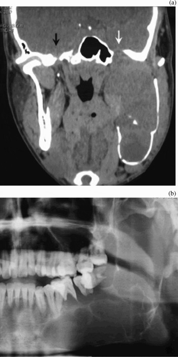
A panoramic X-ray demonstrated radiolucent areas within the bony structure of the left mandibular body and ramus, having clear margins and containing several bony septae (Figure 1). The cortex of the mandible was obviously thinned and the mandibular teeth appeared to ‘float’. The roots of the affected molars were short and thinned.
(a) Coronal computed tomography image of case one, showing an expanded left mandible and displaced medial and lateral pterygoid muscles. An intraosseous mass extends into the pterygomandibular and infratemporal space. Note the expanded left foramen ovale (white arrow), compared with the normal right foramen ovale (black arrow). (b) Segmental panoramic X-ray of case one showing a multilocular radiolucency in the left mandibular body and ramus. The molars are ‘floating’ and their roots are thinned and resorbed.

Computed tomography (CT) showed that the mass extended from the mandible into the cranium via an enlarged foramen ovale. The tumour was of heterogeneous radiological density and contained multiple low-density areas.
The patient underwent segmental resection of the mandible, and reconstruction with a vascularised fibular flap.
Recovery was uneventful, and no recurrence was found at two-year follow up.
Case two
A 65-year-old woman presented complaining mainly of facial swelling. She also reported intermittent toothache of the right mandibular molars over the previous two years.
Physical examination showed diffuse swelling of the buccal and lingual side of the right mandible, with a shallowed buccogingival sulcus.
A panoramic X-ray revealed a multilocular, radiolucent lesion with clearly defined margins extending from the molar region to the sigmoid notch.
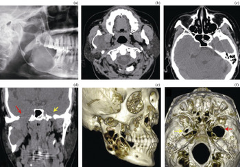
Computed tomography showed that the lesion extended into the middle cranial fossa via an obviously expanded right foramen ovale (Figure 2). The neighbouring medial and lateral pterygoid muscles were displaced.
(a) Segmental panoramic X-ray of case two, showing a well defined, multilocular radiolucency with a notch-like margin in the right mandibular body and ramus. (b) Axial computed tomography (CT) image showing the mass extending outside the mandible into the pterygomandibular space. (c) Axial CT scan showing an intracranial mass located in the middle cranial fossa and partially extending into the posterior cranial fossa, with a dumbbell appearance. The pontine cistern is compressed and the sphenoid sinus deformed. (d) Coronal CT scan showing a significantly expanded right foramen ovale (red arrow). The lesion extends outside the mandible and into the middle cranial fossa via the expanded foramen ovale. Note the normal appearance of the left foramen ovale (yellow arrow). (e) Volumetric rendering of craniofacial bone structure, showing severe destruction of the mandibular body and ramus. (f) Volumetric rendering of the skull base, superior view, showing a grossly expanded right foramen ovale (red arrow) and a normal left foramen ovale (yellow arrow).

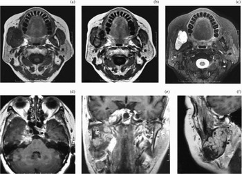
Magnetic resonance imaging (MRI) revealed a heterogeneous, intermediate to low density signal on T1-weighted and proton density images, and hyperintensity on T2-weighted images (Figure 3). The lesion enhanced heterogeneously when gadolinium was administered. Solid and cystic components of the lesion could be clearly distinguished. The intracranial lesion straddled the middle and posterior cranial fossae via Meckel's cave. The cavernous sinus and the internal carotid artery were also involved.
(a) Axial magnetic resonance imaging (MRI) scan of case two (T1 flair sequence; Repetition Time (TR) = 2706, Echo Time (TE) = 10.9) showing a mixed intermediate signal intensity and low signal intensity area inside the right mandible which extends into the pterygomandibular space. (b) Axial MRI scan (T1 flair sequence; TR = 2706, TE = 10.9) showing inhomogeneous enhancement after gadolinium administration. (c) Axial, fat-suppressed, T2-weighted MRI scan showing a lesion of inhomogeneously increased signal intensity. The round area with hyperintensity indicates a cystic part of the lesion. (d) Axial, gadolinium-enhanced MRI scan (T1 flair sequence; TR = 2706, TE = 10.9) showing an intracranial mass located in the middle cranial fossa and extending to the posterior cranial fossa, with a dumbbell appearance. Note the compressed pontine cistern and deformed sphenoid sinus due to expansion of the lesion. The cavernous sinus and internal carotid artery are also involved. (e) Coronal, enhanced, proton density MRI scan (T1 flair sequence; TR = 2706, TE = 10.9) showing connection between the intracranial and mandibular lesions via the infratemporal fossa and expanded foramen ovale. (f) Sagittal, gadolinium-enhanced MRI scan (T1 flair sequence; TR = 2962, TE = 25.4) showing the bead-like appearance of the lesion within the mandible.

The patient underwent segmental resection of the mandible and reconstruction using a vascularised fibular flap.
Histopathology
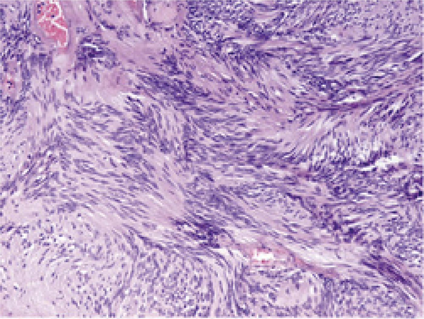
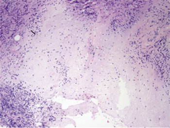
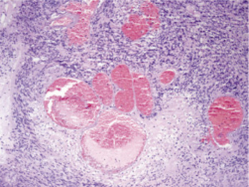
Microscopic examination of the two tumours revealed a similar histological pattern. Both tumours were well encapsulated and composed predominantly of an Antoni A component with scattered Antoni B areas. The Antoni A areas were composed of dense, spindle-shaped Schwann cells arranged in short bundles or interlacing fascicles. Nuclear palisading and Verocay bodies were observed (Figure 4). The Antoni B areas showed a haphazard arrangement of Schwann cells in a loose meshwork of myxoid and microcystic tissue containing small vacuoles. Pleomorphism and mitotic activity were unremarkable, but isolated cells with bizarre hyperchromatic nuclei were occasionally encountered (Figure 5). Large, irregularly spaced blood vessels were most prominent in case one, with gaping and tortuous lumina, thickened and hyalinised vessel walls, and thrombus (Figure 6). Haemorrhage, substantial haemosiderin deposition, focal necrosis and foamy macrophage infiltration were also noted. In the case one specimen, we noted the presence of cysts lined by Schwann cells.
Photomicrograph showing Verocay body and hyaline thickening of vessel walls. (H&E; original magnification ×200)
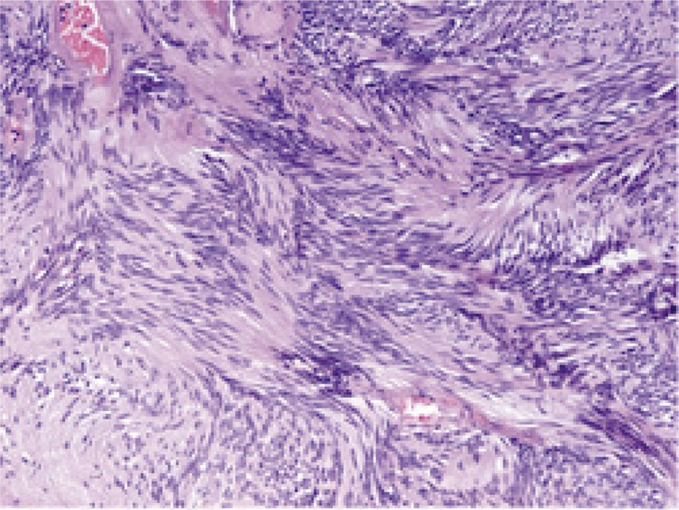
Photomicrograph showing Antoni B area of lesion. (H&E; original magnification ×100)

Photomicrograph showing dilated vascular lumina with thickened, hyalinised walls and thrombus. (H&E; original magnification × 100)

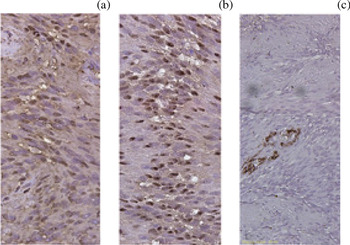
Immunohistochemical staining for S-100 protein (Figure 7a) and neurone-specific enolase (Figure 7b) showed diffuse positive findings. Staining for smooth muscle actin was negative in the tumour cells (Figure 7c).
Photomicrograph of tumour cells showing intense immunoreactivity for both S-100 protein (a) and neurone-specific enolase (b) (original magnification × 400 for both), but negative immunoreactivity for smooth muscle antibody (c) (original magnification × 200). (Streptavidin-biotin)

Discussion
The trigeminal nerve can be divided into various segments involving the brainstem, cistern, Meckel's cave, cavernous sinus and peripheral branches.Reference Go, Kim and Zee7, Reference Nemzek8
The mandibular nerve leaves Meckel's cave and descends through the foramen ovale before entering the mandibular foramen in the ramus of the mandible. It then passes within the mandibular canal and leaves the mandible via the mental foramen.
Schwannomas of the trigeminal nerve are not rare, either intracranial or extracranial.Reference Sharma, Ahmad, Chandra and Mahapatra1, Reference Yoshida and Kawase5, Reference Pamir, Peker, Bayrakli, Kilic and Ozek9 Indeed, the trigeminal nerve constitutes the second most frequent site for intracranial schwannoma occurrence, after the vestibular nerve.Reference MacNally, Rutherford, Ramsden, Evans and King2, Reference Pamir, Peker, Bayrakli, Kilic and Ozek9 Any segment of the trigeminal nerve from the pons to the peripheral branches can be affected. Schwannomas usually grow in Meckel's cave, the posterior or middle cranial fossa, or the cavernous sinus, and frequently straddle multiple anatomical compartments.Reference Yoshida and Kawase5, Reference Pamir, Peker, Bayrakli, Kilic and Ozek9 According to the classification proposed by Yoshida and Kawase, trigeminal schwannomas can involve either single or multiple anatomical compartments, including the infratemporal space, the pterygomandibular space and the mandible.Reference Yoshida and Kawase5
In our experience, clinical symptoms are of limited value in the pre-operative diagnosis of trigeminal schwannoma of the mandible. Symptoms such as facial pain and numbness are usually absent. In addition, neither of our two cases displayed neurological symptoms such as cramps, vomiting, convulsions, paralysis, numbness, or respiratory or cardiac depression. On the other hand, both our cases had symptoms mimicking an odontogenic cyst or tumour, such as facial swelling and loosened or ‘floating’ teeth. Masticatory function may be slightly impaired due to involvement of the teeth, but mouth opening difficulty is not usually seen. When involvement of the teeth is absent the patient may be asymptomatic, and the lesion may be found incidentally.Reference Gallego, Junquera, Rodriguez-Recio and Fresno6
Establishing a differential diagnosis based on routine X-ray techniques is quite difficult. However, in both our presented cases panoramic radiographs showed similar, radiolucent lesions with clear, notch-like margins and internal bony septae. This appearance mimicked odontogenic tumours such as odontogenic keratocystic tumour and ameloblastoma. Tooth displacement and tooth root resorption were prominent. Panoramic radiographs may also show effacement of the mandibular canal, suggestive of a tumour of neural origin.
Compared with standard X-rays, CT and MRI scans provide much more valuable information for pre-operative diagnosis.Reference Honda, Asato, Tanaka, Endo, Nishimura and Ito10 An important radiological feature of peripheral nerve schwannomas is spread along the length of the nerve. Schwannomas growing along cranial nerves, such as the maxillary or vidian nerve, may also cause expansive changes to the bony boundaries of those nerves.Reference Honda, Asato, Tanaka, Endo, Nishimura and Ito10 Therefore, evaluation of skull base structures such as the foramen ovale, foramen rotundum and pterygoid canal may provide valuable information for the differential diagnosis. Prominent asymmetrical enlargement of these structures is a strong indicator of neural pathology.
A similar growth pattern is not seen for benign odontogenic cysts or tumours. Furthermore, in the case of these tumours the cortical outline can still be seen, even if expanded, thinned or breached.
Magnetic resonance images typically show trigeminal schwannomas as well defined, lobulated masses with decreased signal intensity on T1-weighted images and increased signal intensity on T2-weighted images.Reference Osterhus, Van Loveren and Friedman11 Such tumours may be heterogeneous in nature because of cyst formation within the lesion.Reference Prasad, Gupta, Patankar and Goel12 Such cystic regions show an increased signal on fat-suppressed, T2-weighted MRI scans but only a slightly increased signal on gadolinium-enhanced images. Solid regions may show remarkably increased signal on gadolinium-enhanced MRI scanning.
• Intraosseous schwannomas may occur in various forms
• Two cases are presented of trigeminal schwannoma of the mandible with extension into the cranium via an expanded foramen ovale
• The similar growth pattern of these two tumours indicates that they may represent a distinct, unique form of trigeminal schwannoma
No standardised algorithm for surgical management of trigeminal schwannoma of the mandible has been proposed. The fact that schwannomas are overwhelmingly benign and usually cause only minor discomfort has led to universal agreement that total removal is the best choice for treatment.Reference Sharma, Ahmad, Chandra and Mahapatra1, Reference Zhang, Yang, Xu, Wang, Liu and Zhu13 For tumours without intracranial extension or extensive mandibular destruction, enucleation should be proposed to preserve the mandible. For tumours with intracranial extension, segmental resection of the involved mandible is mandatory to remove the tumour. Reconstruction of the mandible with a vascularised fibular flap generally achieves satisfying outcomes both in terms of facial symmetry and oral function. Reduced mortality and morbidity can be achieved with the use of advanced microsurgical skull base and neurological techniques; however, total removal of an intracranial lesion in a one-stage procedure may be very difficult when the lesion is large and involves multiple anatomical structures. It should be borne in mind that cranial nerve functional deficits are common following radical resection of such tumours.Reference Phi, Paek, Chung, Jeong, Park and Jung14 Therefore, two-stage surgery is suggested in such circumstances, in order to provide the greatest protection for intracranial structures.Reference Tsuboi, Fujimori, Tomono, Hamano and Nose15 For patients with small to moderate-sized intracranial trigeminal schwannomas, gamma knife radiosurgery provides good tumour control with minimal risk of adverse radiation effects.Reference Phi, Paek, Chung, Jeong, Park and Jung14, Reference Peker, Bayrakli, Kilic and Pamir16