




No CrossRef data available.
Article contents
New trends in chromosomal investigation in children with cardiovascular malformations
Published online by Cambridge University Press: 21 January 2005
Abstract
We investigated a group of 376 children, seen over a period of 7 years with different types of congenital cardiovascular defects, to assess the presence of chromosomal aberrations. The diagnostic approach, achieved in 3 consecutive steps, revealed conventional chromosomal aberrations in 30 of the patients (8%) excluding trisomies 13, 18, 21. Fluorescence in situ hybridisation for microdeletions showed 51 microdeletions (15%), with 43 patients having deletions of 22q11.2, 7 patients with deletion of 7q11.23, and 1 patient with deletion of 4p16.3. In 23 patients with additional clinical abnormalities, we carried out a subtelomeric screening. This revealed, in two cases (9%), different subtelomeric aberrations, namely deletions of 1p and of 1q. Thus, subtelomeric screening proved to be a very valuable as a new diagnostic approach. Our approach to genetic investigation in three phases makes it possible to detect a high rate of pathologic karyotypes in patients with congenital cardiovascular malformations, thus guaranteeing more effective genetic counselling of the families, and a more precise prognosis for the patient.
Keywords
Information
- Type
- Original Article
- Information
- Copyright
- © 2004 Cambridge University Press
Cardiovascular defects show an unequal geographic distribution within Europe. In western Belgium, the frequency is about 2 anomalies for each 1000 newborns, as compared to 10 per 1000 in northern Spain.1 In about seven-eighths of the cases the aetiology is thought to be multifactorial. In less than one-twentieth, a monogenic disorder is assumed, and in from one-twelfth to one-eighth, chromosomal aberrations are diagnosed. Among the latter, trisomy 21 is the most frequent anomaly, accounting for four-fifths of such cases. Analyses to detect microdeletions in defined syndromes have shown lately that this group of aberrations constitutes a significant cause of cardiovascular malformations. Hot spots of mutation have been analysed in this context in chromosomes 22, 7 and 8.2
With regard to far smaller aberrations than those detected with conventional cytogenetic techniques, we are now able to search for even smaller structural defects, namely for microdeletions with sizes ranging from 1 to 10 megabases. The subtelomeric regions of chromosomes show the highest gene concentration in the human genome,3 and they are characterised by high rates of mutation. Screening for cryptic mutations in these regions is promising in detecting the chromosomal abnormalities that cause cardiovascular disease.
Thus, groups of cardiac anomalies formerly classified as “multifactorial” and “monogenic” will be linked increasingly to structural abnormalities of the chromosomes, mainly microdeletions. Additionally subtelomeric screening will provide more fundamental knowledge of the abnormal embryonic development that leads to cardiovascular malformations. In this report, we describe our approach to using these exciting new innovations in the clinical context.
Patients studied and methods
We investigated 376 patients with different types of cardiovascular malformations, all patients being investigated by the same team of cardiologists. The documentation of clinical symptoms comprised the diagnosis of the cardiac lesion, and the occurrence of additional but independent abnormalities, such as dysmorphic features, for example deep seated dysplastic ears or simian crease, malformations such as cleft lip and palate and polycystic kidneys, and developmental and/or functional anomalies such as hypotonia or immunological defects. As the majority of patients were less than one year old when the chromosomal analysis was performed, their mental development could not be judged adequately. Thus, we were unable to take account of, or exclude, mental retardation, a primary symptom in chromosomal syndromes.
The overall group of patients investigated was made up of single as well as complex cardiac malformations, whereas the group selected for subtelomeric analysis consisted of those with complex cardiovascular malformations and associated anomalies.
Within the period of investigation, we diagnosed 48 patients having a cardiac malformation in association with trisomies 13, 18 or 21. These patients were not included in the quantitative evaluation of those investigated and reported here, as they play no role in further investigations for genetic causes of cardiac malformations. Their individual frequencies in trisomy 21 were 85%, 13% in trisomy 18, and 13.2% in trisomy. These frequencies of the 3 trisomies among our children with cardiovascular cardiac malformations are in accordance with findings from the literature.
The cells analysed were lymphocytes in 375 cases, and fibroblasts in 1 case. In 6 patients, we also investigated cells derived from buccal mucosa.
Cardiological investigations
The cardiac diagnosis was established by clinical investigation, electrocardiography, chest radiography, and echocardiography. In those patients where echocardiography could not clarify the complete anatomical situation, which is the case in the majority of cardiovascular malformation with abnormalities of the aortic arch, if haemodynamic data was needed, or if interventional therapy was the treatment of choice, then cardiac catheterisation was performed. In some patients, we needed magnetic resonance imaging or computerised tomographic scans to achieve a precise diagnosis.
Our patients were recruited in non-randomised fashion from the cardiological department. Our centre treats patients selected for more severe cardiac defects from various German hospitals. Almost three-quarters of the defects diagnosed in our patients fell into 4 categories, with ventricular septal defect accounting for just over one quarter, atrioventricular septal defect and hypoplasia of the left heart for one-sixth each, and double-outlet right ventricle for one-eighth.
Other malformations, such as coarctation, aortic stenosis, discordant ventriculo-arterial connections, tetralogy of Fallot with pulmonary stenosis or atresia, pulmonary atresia with intact ventricular septum, tricuspid atresia, and common arterial trunk, occurred in frequencies of about one-twentieth.
If we group the patients into two sets, one-quarter have functionally univentricular hearts (95/376), and three-quarters have complex cardiac defects (281/376).
Cytogenetic and molecular-cytogenetic investigations
These investigations were performed in three steps. As the first step, we analysed chromosomes by GTG-banding in all 376 patients. When needed, 2 or 3 banding techniques were combined. In the second step, we performed microdeletion analysis, using the technique of fluorescence in situ hybridisation in 335 patients when the conventional chromosomal analysis revealed a normal karyotype. These cases were analysed in routine diagnosis over a period of 7 years. According to the clinical phenotype, we used locus-specific probes such as 22q11.2, 7q11.23, 4p16.3, 8p23.1, and 10p13.
As the final step, we carried out subtelomeric screening using the Chromoprobe Multiprobe®-T system (Cytocell, Banbury, United Kingdom). We selected 23 patients with complex heart defects and, in most cases, further independent abnormalities but normal results after conventionally cytogenetic and microdeletion examinations for subtelomeric screening.
Results
Conventional chromosomal analysis
Using lymphocytes from 375 patients, fibroblasts from 1, we established that 295 patients had a normal karyotype, whereas 30 (8%) showed chromosomal aberrations. Of these, 13 were female, and 17 male (Table 1), and 12 of the aberrations were numerical, with 18 showing different types of structural aberrations. In 2 patients, there was a combination of numerical and structural abnormalities (0.5%).
Table 1. The types of chromosomal aberration, and corresponding cardiac defects, in our 30 patients found to have a pathologic karyotype.

Autosomal aberrations were the dominant group, with 24 of 30 pathological karyotypes. The interchromosomal distribution of aberrations did not show a specific pattern of chromosomes involved. Chromosomes 2 and 9 showed the highest number of abnormalities, with three mutations each, followed by those of chromosomes 1, 8, 13, 14 and 15 with two aberrations. One aberration was found in chromosomes 4, 7, 18, 19 and 22. The total of 24 autosomal aberrations could be subdivided into 18 intrachromosomal abnormalities, 4 interchromosomal rearrangements involving 2 chromosomes each, and two marker chromosomes. In the group of 6 gonosomal aberrations, we detected 5 numerical abnormalities and one derivative Y-chromosome. Among all types of rearrangements, we found 5 deletions, 3 duplications, 3 inversions, 3 isochromosomes, 3 translocations, 8 derivative chromosomes and 1 marker chromosome, whose origin could not be delineated beyond the fact that it was not a derivate of a gonosome. This represents the normal distribution of structural chromosome aberrations compared to our clinical diagnostic group of children with other than cardiac malformations.
All numerical chromosomal aberrations originated de novo in the patient. From the structural aberrations, 5 were de novo, and 4 of parental origin, with maternal and paternal origin accounting for equal frequencies. In 9 cases, the paternity could not be delineated, as the parents could not be investigated. A correlation between the chromosomal aberrations observed and the type of cardiovascular malformation could not be demonstrated. As an example, the data of patient #30 with a gonosomal aberration are given in detail (Fig. 1, Table 1).

Figure 1. Clinical findings in a neonatal male with double outlet right ventricle, borderline hypoplasia of left heart, aortic coarctation, left radial hypoplasia, and an omphalocele. The angiogram (a) shows the ventricular anatomy and the aortic coarctation. In (b), we show the chromosomes as seen in metaphase after fluorescence banding. The karyotype is 48, XXYY, with the X and Y chromosomes marked by arrows.
Microdeletion analysis
Analysis of microdeletions was performed using fluorescence in situ hybridisation in 335 of the 376 patients. We used the locus specific probe 22q11.2 in 321 cases, 7q11.23 in 16 cases, 8p23.1 in 11 cases, 10p13 in 2 cases, and 4p16.3 in 3 cases. In 19 cases, we chose two different probes because of atypical clinical findings. We analysed the chromosomal regions 4p16.3, 7q11.23, 8p23.1 and 10p13 only when the clinical symptoms were in accordance with these known syndromes of microdeletion.
In total, we detected 51 microdeletions (15%). In 43 of 312 cases investigated, we found a microdeletion of 22q11.2 (14%). In patients with the typical phenotype of DiGeorge/Velocardiofacial syndrome, or Williams–Beuren syndrome, and a normal pattern of hybridisation, we extended the examination up to 50 mitoses to exclude a microdeletion mosaicism, but in none of these cases did we detect such a mosaicism. Additionally, in 6 cases with the characteristic phenotype of a microdeletion 22q11.2, we analysed buccal mucosa as a second system of cells, albeit that the findings were again normal. In 7 of 17 cases analysed for 7q11.23, we found a microdeletion (41%), also detecting a microdeletion of 4p16.3 in 1 of 3 cases tested (33%). No microdeletion was diagnosed in the 12 cases screened for 8p23.1, or in the 2 cases screened for 10p13.
The microdeletion of 22q11.2 occurred de novo in 84%, and as being inherited from one of the parents in 16%. In 2 families, 2 siblings showed the same microdeletion. In these particular cases, one child each was included in the quantitative interpretation of the results. All deletions in 4p16.3 and 7q11.23 occurred de novo. The cardiovascular defects and the microdeletion of a patient with Williams–Beuren syndrome are demonstrated in Figure 2.

Figure 2. This female neonate was found to have Williams–Beuren syndrome with an interatrial communication in the oval fossa, multiple pulmonary stenoses, developmental retardation, and typical facial dysmorphias. The angiography (a) demonstrates multiple and diffuse pulmonary stenoses. Fluorescence in situ hybridisation (b), using a DNA probe for the elastin (ELN) locus on the chromosomes in metaphase, shows the typical deletion (arrow) for the critical region 7q11.23 (green signal) giving the Williams–Beuren syndrome, and control hybridisation of the centromeric region (red signal). The karyotype is 46, XX. ish del (7)(q11.23)(ELN-).
Subtelomeric screening
We selected 23 patients for subtelomeric screening from the 295 shown to have a normal karyotype and pattern of hybridisation. Criterions for selection were the finding of complex cardiovascular malformation combined with further malformations and dysmorphisms.
The cardiac malformations could be attributed to six different entities, specifically a solitary septal defect in 1 patient, discordant ventriculo-arterial connections in 1 patient, functionally single ventricles in 4 patients, isomerism of atrial appendages with visceral heterotaxy in 1 patient, hypoplasia of the left heart in 6 patients, and different malformations involving the ventricular outflow tracts in 10 patients.
Subtelomeric aberrations were detected in two of the patients (9%). Both occurred de novo (Table 2). The aberrations found were subtelomeric deletions, in one case on the short arm of chromosome 1 (1p), and in the second case on the long arm of chromosome 1 (1q). Details of one of these patients are given in Figure 3.
Table 2. Cryptic chromosomal abnormalities in 2 patients detected by subtelomeric screening.


Figure 3. This female patient has tetralogy of Fallot with double outlet ventriculo-arterial connection, a right aortic arch, persistence of the left superior caval vein, brachydactyly V, syndactyly of the feet, hypotonia and seizures. The angiogram (a) demonstrating the double outlet ventriculo-arterial connection from the right ventricle. Subtelomeric fluorescence in situ hybridisation (b), using a DNA probe on metaphase chromosomes, reveals cryptic deletion in the short arm of chromosome 1 (arrows). The karyotype is 46, XY.ish del (1)(p36.3) (CEB108-).
Characterisation of clinical subgroups
More than nine-tenths of our patients were found to have additional independent abnormalities besides the cardiovascular defect. These abnormalities could be subdivided into three groups, specifically dysmorphias, malformations, and developmental and/or functional anomalies. In one-fifth (73 of 376), a precise documentation of additional abnormalities was not available. Out of the remaining 303 cases, one-quarter (69 of 303) had additional dysmorphias, one-sixth had other malformations (42 of 303), almost one-fifth (54 of 303) had developmental and/or functional anomalies, and in just under one-quarter (71 of 303), no additional abnormalities could be diagnosed beyond the cardiovascular malformation. The remaining one-fifth (68 of 303) showed different combinations of the three parameters.
Dividing the group investigated into those with functionally univentricular and complex heart defects, over nine-tenths of the patients with a functionally univentricular arrangement, specifically 81 of 87, had more additional abnormalities falling into all 3 subgroups than did the proportion of patients with the complex cardiac malformations, with 174 of these 239 having additional abnormalities (73%). These differences are significant for the subgroup of dysmorphias (p = 0.022), highly significant for the group of additional malformations (p < 0.001), and not significant (p = 0.096) for the group with developmental and functional anomalies. In the patients with complex cardiovascular malformation the 3 subgroups of additional abnormalities occurred more often separately than combined, in proportions of 73% to 27%, whereas in the group of patients with a functionally univentricular arrangement, the frequencies were more equally distributed, at 46% and 54%.
Additional abnormalities were than compared on the basis of the patients having a pathologic karyotype, those with microdeletions, and those with normal findings for both these features. The group of patients with subtelomeric aberrations was too small, and had been investigated using by special criterions for selection, and was therefore excluded. A specific pattern of independent aberrations was found in 30 patients with an aberrant karyotype. Dysmorphias were the most frequent abnormalities, accounting for 7 of the 30 patients in this group. We found malformations and developmental or functional anomalies in almost equal frequencies, at 10% and 13%, respectively, and these were rare compared to the first group. Combinations of the different parameters had almost equal frequencies when two were combined, at 10 and 13%, but rare for all three of them, at 7%. Besides, one-tenth of the patients had only a cardiac defect without additional peculiarities. Patients with complex cardiovascular malformations were more frequently included in this group, with 19 involved, than the 11 patients with a functionally univentricular arrangement, giving proportions of 63% versus 37%.
The 51 patients with microdeletions showed a medium frequency (17%) of dysmorphias, the lowest proportion of associated malformations of all groups (7%), and the highest incidence of developmental and/or functional anomalies (24%). We did not observe any patient with a combination of dysmorphias and malformations. The combination of dysmorphias and developmental or functional anomalies, however, had the highest frequency, at 20%, and we also found 15% with the combination of all three parameters. The other parameters had low frequencies. Patients with complex cardiac defects accounted for four-fifths of this group, which is to be expected on the basis of the prominent frequency of microdeletions of 22q11.2.
Patients with normal findings in the different cytogenetic investigations showed a more equal distribution of additional abnormalities, with the highest amount of malformations (24%:15%:17%), while all combinations of the three parameters were extremely rare (6–7% each). This group had the highest frequency of heart defects without additional independent abnormalities (17%) compared to patients with conventional chromosomal aberrations or microdeletions (10% each). In this group, patients with complex heart defects were more frequent than those with a functionally univentricular arrangement, in the ratio of 3:1.
This characterisation of our patients with cardiovascular malformations and combined with independent abnormalities showed a group specific pattern as to the type of cardiac anomaly, and the frequency, type and combination of abnormalities when attributed to different cytogenetic abnormalities.
Criterions for the selection of patients
The group of 376 patients we investigated is a selected cohort from 500 patients with cardiac malformations. Those selected include patients seen post-natally by one team of cardiologists, thus guaranteeing a homogenous clinical characterisation. Because of this method of selection, we also compared our findings in those selected to investigations of the total group of 500 patients. We were unable to find any differences in the frequencies of aberrations, or additional abnormalities, between the two groups.
Discussion
Chromosomes, being the source of genetic information, are responsible for the exact qualitative and quantitative transfer of genes from cell to cell, especially from the gametes to the zygote. Mutations of the amount and function of genetic material caused by chromosomal aberrations are, therefore, an important cause of abnormal development of any system of organs. Chromosomal aberrations in the fertilised egg are responsible for constitutive malformations in the embryo, the fetus, and the neonate. Organs like the brain and the heart are more often involved in abnormal differentiation than others, which is a measure of their morphologic complexity. Sonographic investigation now permits the detection of fetal cardiovascular malformations as early as the second trimester. In a previous prenatal study, chromosomal abnormalities were analysed in two-fifths of fetuses with a cardiovascular defect.4
If we include the patients known to have trisomies 13, 18 and 21 in the group we investigated the frequency of chromosomal aberrations doubles 8% to 16%. Different investigators have similarly reported frequencies varying from 8% to 13%,4–6 suggesting that the mode of selection of the group of patients to be investigated according to the different types of cardiovascular defects seen in a cardiologic centre can lead to different reported frequencies and types of chromosomal aberrations. A different approach for the delineation of an association between congenital cardiac malformations and chromosomal aberrations was chosen by van Karnebeek and Hennekam.7 They selected cases from the Human Cytogenetics DataBase, again excluding patients with well defined trisomies, as did we, since they too assumed that their inclusion would not contribute to the aims of their study. They, as well, found that patients with a functionally univentricular arrangement, or those with complex cardiac malformations attributed to the same chromosomal region, added to the occurrence of additional clinical symptoms in the patients. They could not define the unequal inter- and intrachromosomal distribution of chromosomal aberrations as we were able to achieve because of the smaller number of structural chromosomal abnormalities involved.
In the past decades, the possibility of analysing the chromosomal aberrations that underlie cardiovascular malformations has been strongly associated with improvements in cytogenetic techniques. Initially, strategies of high resolution banding permitted the detection of structural chromosomal aberrations larger than 3 to 5 megabases of DNA.8 It was the implementation of fluorescence in situ hybridisation that enabled the ascertainment of partial aberrations, for example chromosomal mosaicism, also in non-dividing tissues.9 Besides, this technique permitted a number of well defined syndromes, such as Williams–Beuren syndrome, Wolf–Hirschhorn syndrome, and DiGeorge/Velocardiofacial syndrome, to be attributed to cryptic chromosomal aberrations, now known as microdeletion syndromes.
We found such microdeletions in 15% of the patients we investigated, and of these, 14% were cryptic deletions in 22q11.2. Previous studies have shown that the amount of microdeletions detected depends largely on the clinical peculiarities of the investigated group. The type of cardiac anomaly is especially important when considering deletion of 22q11.2. If all types of cardiovascular malformations are considered in the setting of our patients, the frequency of microdeletions stands at 7.8%, whereas such microdeletions are found in 22.5% of those we deemed to have complex heart defects. Other investigators have reported frequencies from 15% to 18% in series of unselected patients,10–13 findings which show good concordance with our own.
Larger screening studies have demonstrated that the frequency of microdeletions is much higher than previously suspected.14 The extent of these microdeletions ranges from between 1 and 5 megabases in size, and therefore lies beneath microscopic resolution.
Today, a combination of molecular-cytogenetic and molecular-genetic methods enables studies of the parental origin of chromosomal aberrations, as well as the characterisation of chromosomal breakpoints.15 Furthermore, the exact analysis of the deleted regions has led to the detection of hot-spots of mutation.16 In the Williams–Beuren syndrome, for example, chromosomal rearrangements such as paracentric inversions flanking the critical region of 7q11.23 are thought to predispose to mismatch and unequal crossing-over, causing the microdeletion and increasing the risk of recurrence.17
Thus, the development of new and recently established molecular-cytogenetic techniques has taken us a giant step forward, and now allows the screening of chromosomal regions of the human genome known to be very rich in genes.18, 19 In our study, we selected one of these techniques to analyse possible cryptic aberrations at the subtelomeric regions of all human chromosomes in children with cardiovascular malformations. The size of these critical regions is 10-fold less than that in the types of microdeletions discussed above.
From the stance of cardiac malformations, such subtelomeric aberrations, as far as we know, have only been analysed in the children included in our study. Comparable findings from the literature, therefore, do not exist. But it is possible to give some frequencies for patients with mental retardation, with subtelomeric aberrations reported to range between zero and 29.4%, with an average frequency of 4.6%.20 Our findings in the children with cardiovascular malformation are in good accordance with those reported in the patients with mental retardation, thus permitting us to hypothesise that a rate of aberration of about 5–10% in the subtelomeric regions can be expected if investigating patients with a suspicion for cryptic chromosomal aberrations.
Because the extent of the subtelomeric deletions or duplications extends to several hundred kilobases of genetic material, it can be assumed that they not only induce cardiovascular defects, but that they also lead to simultaneous extracardiac malformations and developmental impairment. The sample of our patients chosen for subtelomeric screening was selected according to these criterions. The frequency of subtelomeric aberrations in our collective demonstrates that it is necessary to select patients carefully so as to construct a better classification of the large number of children with cardiovascular malformations. Only in this way can we calculate a more accurate risk of recurrence for the parents of affected children, and make predictions of prognostic relevance for the patients themselves.
References
Nora JJ, Berg K, Nora AH. Cardiovascular Diseases. Oxford University Press, 1991.
Baumer A, Dutly F, Balmer D, et al. High level of unequal meiotic crossovers at the origin of the 22q11.2 and 7q11.23 deletions. Hum Mol Genetics 1998; 7: 887–894.Google Scholar
Knight SJ, Regan R, Nicod A, et al. Subtle chromosomal rearrangements in children with unexplained mental retardation. Lancet 1999; 354: 1676–1681.Google Scholar
Schwanitz G, Zerres K, Gembruch U, Bald R, Gamerdinger F, Hansmann M. Prenatal detection of heart defects as an indication for chromosomal analysis. Ann Génét 2000; 33: 79–83.Google Scholar
Ferencz C, Neill CA, Boughman JA, Rubin JD, Brenner JI, Perry LW. Congenital cardiovascular malformations associated with chromosome abnormalities: an epidemiologic study. J Pediatr 1989; 114: 79–86.Google Scholar
Hoffman JI, Christianson R. Congenital heart disease in a cohort of 19,502 births with long-term follow-up. Am J Cardiol 1978; 42: 641–647.Google Scholar
van Karnebeek CD, Hennekam RC. Associations between chromosomal anomalies and congenital heart defects: a database search. Am J Med Genet 1999; 84: 158–166.Google Scholar
Kaiser-Rogers K, Rao K. Structural chromosome rearrangements. In: Gersen S, Keagle M (eds). Principles of Clinical Cytogenetics. Humana Press Inc., Totowa, NJ, 1999, pp 198–228.
Raff R, Schwanitz G. Fluorescence in situ hybridization: general principles and clinical application with special emphasis to interphase diagnostics. Int J Hum Genet 2001; 1: 65–75.Google Scholar
Fokstuen S, Arbenz U, Artan S, et al. 22q11.2 deletions in a series of patients with non-selective congenital heart defects: incidence, type of defects and parental origin. Clin Genet 1998; 53: 63–69.Google Scholar
Marino B, Digilio MC, Toscano A, Gianotti A, Dallapiccola B. Congenital heart defects in patients with DiGeorge/velocardiofacial syndrome and del 22q11.2. Genet Couns 1999; 10: 25–33.Google Scholar
Mehraein Y, Wippermann CF, Michel-Behnke I, et al. Microdeletion 22q11 in complex cardiovascular malformations. Human Genet 1997; 99: 433–442.Google Scholar
Takahashi K, Kido S, Hoshino K, Ogawa K, Ohashi H, Fukushima Y. Frequency of a 22q11 deletion in patients with conotruncal cardiac malformations: a prospective study. Eur J Pediatr 1995; 154: 878–881.Google Scholar
Burn J, Wilson DI, Cross I, Atif U, Scambler P, Takao A. The significance of 22q11 deletion. In: Clark EB, Takao A (eds). Developmental Mechanisms of Heart Disease. Futura Publ, Armonk-New York, 1995, pp 559–567.
Chung MY, Lu JH, Chien HP, Hwang B. Chromosome 22q11 microdeletion in cardiovascular heart defects: clinical presentation, parental origin and de novo mutations. Int J Mol Med 2001; 7: 501–505.Google Scholar
Trost D, Wiebe W, Uhlhaas S, Schwindt P, Schwanitz G. Investigation of meiotic rearrangements in DGS/VCFS patients with a microdeletion 22q11.2. J Med Genet 2000; 37: 452–454.Google Scholar
Bayés M, Magano LF, Rivera N, Flores R, Pérez Jurado LA. Mutational mechanisms of Williams-Beuren syndrome deletions. Am J Hum Genet 2003; 73: 131–151.Google Scholar
Saccone S, De Sario A, Della Valle G, Bernardi G. The highest gene concentrations in the human genome are telomeric bands of metaphase chromosomes. Proc Natl Sci USA 1992; 89: 4913–4917.Google Scholar
De Vries BB, Winter R, Schinzel A, van Ravenswaaij-Arts C. Telomeres: a diagnosis at the end of the chromosomes. J Med Genet 2003; 40: 385–398.Google Scholar
Xu J, Chen Z. Advances in molecular cytogenetics for the evaluation of mental retardation. Am J Med Genet 2003; 117C: 15–24.Google Scholar
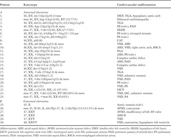
Table 1.
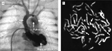
Clinical findings in a neonatal male with double outlet right ventricle, borderline hypoplasia of left heart, aortic coarctation, left radial hypoplasia, and an omphalocele. The angiogram (a) shows the ventricular anatomy and the aortic coarctation. In (b), we show the chromosomes as seen in metaphase after fluorescence banding. The karyotype is 48, XXYY, with the X and Y chromosomes marked by arrows.

This female neonate was found to have Williams–Beuren syndrome with an interatrial communication in the oval fossa, multiple pulmonary stenoses, developmental retardation, and typical facial dysmorphias. The angiography (a) demonstrates multiple and diffuse pulmonary stenoses. Fluorescence in situ hybridisation (b), using a DNA probe for the elastin (ELN) locus on the chromosomes in metaphase, shows the typical deletion (arrow) for the critical region 7q11.23 (green signal) giving the Williams–Beuren syndrome, and control hybridisation of the centromeric region (red signal). The karyotype is 46, XX. ish del (7)(q11.23)(ELN-).

Table 2.

This female patient has tetralogy of Fallot with double outlet ventriculo-arterial connection, a right aortic arch, persistence of the left superior caval vein, brachydactyly V, syndactyly of the feet, hypotonia and seizures. The angiogram (a) demonstrating the double outlet ventriculo-arterial connection from the right ventricle. Subtelomeric fluorescence in situ hybridisation (b), using a DNA probe on metaphase chromosomes, reveals cryptic deletion in the short arm of chromosome 1 (arrows). The karyotype is 46, XY.ish del (1)(p36.3) (CEB108-).
 You have
Access
- 11
- Cited by